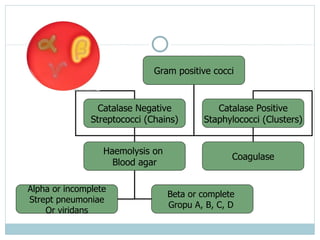
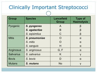
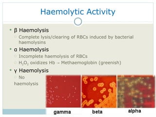
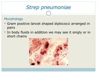
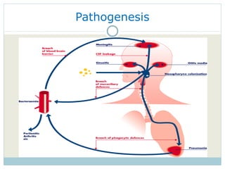
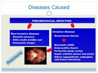
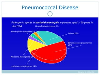
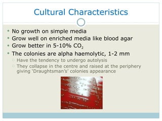
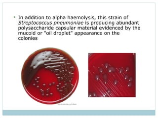
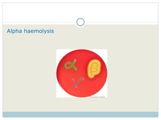
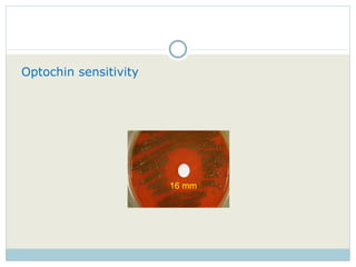
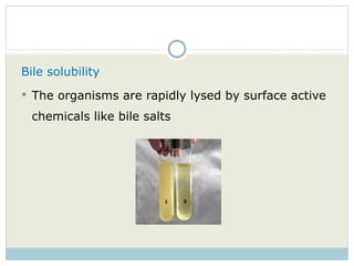
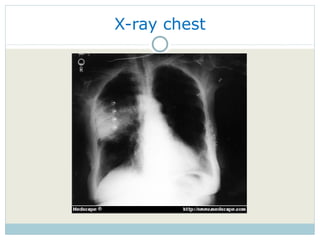
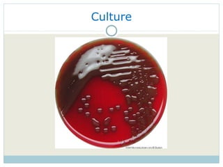
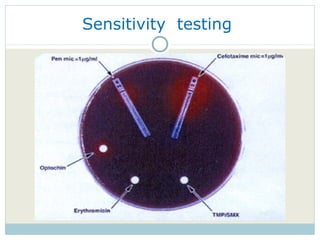
DR KAMRAN AFZAL ASST PROF MICROBIOLOGY Streptococcus pneumoniae
Gram positive cocci Catalase Negative Streptococci (Chains) Catalase Positive Staphylococci (Clusters) Haemolysis on  Blood agar Alpha or incomplete Strept pneumoniae Or viridans Beta or complete  Gropu A, B, C, D  Coagulase
The Genus Streptococcus FAMILY Streptococcaceae  Some important pathogens, commensals of mucous membranes Opportunistic pathogens 40 species, in 6 clusters Strong fermenters of carbohydrates  -> Lactic acid Facultative anaerobes Peptostreptococci are obligate anaerobe S
Clinically Important Streptococci Group Species Lancefield Group Type of Haemolysis Pyogenic S. pyogenes S. agalactiae S. eqisimilus A B C β β β Mitis S. pneumoniae S. mitis S. sanguis O O H α α α Anginosus S. anginosus G, F α Salivarius S. salivarius K γ Bovis S. bovis D α Mutans S. mutans No γ
Haemolytic Activity β  Haemolysis Complete lysis/clearing of RBCs induced by bacterial haemolysins α  Haemolysis Incomplete haemolysis of RBCs H 2 O 2  oxidizes Hb -> Methaemoglobin (greenish) γ  Haemolysis No  haemolysis
Strep pneumoniae Morphology Gram positive lancet shaped diplococci arranged in pairs  In body fluids in addition we may see it singly or in short chains
Antigenic Structure The organism is capsulated with    90 different capsular polysaccharide types Elicits humoral response - formation of antibodies The cell wall contains another polysaccharide that is common to all pneumococci C- Substance that binds to C-reactive protein
Virulence Factors Polysaccharide capsule - the major virulence factor It prevents phagocytosis by host immune cells Autolysin Activation of this protein lyses the bacteria  ->  Pneumolysin Pneumolysin Lyses host cells of immune system Facilitates colonization M-protein Antiphagocytic Lipoteichoic acid Activates complement IgA protease Helps in colonization
Predisposing Factors Alcohol or drug intoxication Immunocompromised Elderly Abnormality of the respiratory tract Viral infections, bronchial obstruction and respiratory tract injury Abnormal circulatory dynamics Splenectomy Chronic diseases Sickle cell anaemia
Transmission And Colonisation
Pathogenesis
Diseases Caused Non-invasive disease Sinusitis (sinuses) Otitis media (middle ear) Pneumonia (lungs) Invasive disease Bacteraemia (blood) Meningitis (CNS) Endocarditis (heart) Peritonitis (body cavity) Septic arthritis (bones and joints) Others (appendicitis, salpingitis, soft-tissue infections) PNEUMOCOCCAL INFECTION   Pneumonia
Pneumococcal Disease Wenger et al., J Infect Dis, 1990 Pathogenic agents in  bacterial meningitis  in persons aged   60 years in the USA Others 26% Streptococcus pneumoniae  49% Group B streptococcus 3% Haemophilus influenzae  4% Neisseria meningitidis  4% Listeria monocytogenes  14%
Clinical Features Septicemia Pneumonia Fever Hypotension Acute confusion Chills Dyspnea Tachypnea Headache Tachycardia Fever Dyspnea Lung consolidation Hemoptysis chills Chest pain Cyanosis malaise Rust colored sputum
Pneumonia Complications Bacteraemia in 15  20% of patients high mortality despite appropriate antibiotic therapy overall case fatality rate 15-20%  higher case fatality rates (30-40%) for elderly  Spread of pneumococci in the blood to other normally sterile sites can cause other invasive pneumococcal diseases (e.g. meningitis) Empyema (pus in the pleural cavity) in about 2% of cases
Lab Diagnosis Specimens Sputum  Blood CSF Swabs Urine Inoculation Blood, chocolate agar at 37 o C for 24-48 hrs
Cultural Characteristics No growth on simple media Grow well on enriched media like blood agar  Grow better in 5-10% CO 2 The colonies are alpha haemolytic, 1-2 mm  Have the tendency to undergo autolysis They collapse in the centre and raised at the periphery giving ‘Draughtsman’s’ colonies appearance
In addition to alpha haemolysis, this strain of  Streptococcus pneumoniae  is producing abundant polysaccharide capsular material evidenced by the mucoid or "oil droplet" appearance on the colonies
Identification Gram staining Catalase negative Alpha haemolysis Soluble in bile salts Sensitivity to optochin Capsular swelling (Quellung reaction) Antigen detection Animal pathogenicity Molecular typing
Gram Staining (Direct microscopy) Lancet (lanceolate) shaped diplococci having long axis in line with each other  Pus cells are also present
Alpha haemolysis
Optochin sensitivity   16 mm
Capsular Swelling Reaction If sputum or specimen mixed with a polyvalent antiserum and then stained, the capsule increases in size The Quellung test is used in pneumococcus typing
Bile solubility The organisms are rapidly lysed by surface active chemicals like bile salts
Antigen Detection Capsular polysaccharide antigen In serum, CSF or Urine  By latex agglutination method Rapid method Useful to diagnose bacteraemia and meningitis
Animal pathogenicity Intraperitoneal injection of the organisms The animal dies and the microorganisms can be isolated from blood
Molecular typing PCR RFLP DNA sequencing
Sensitivity testing
Treatment Penicillins, tetracylines and macrolides They have remained sensitive over the years Resistant strains are being reported Ceftriaxone for the resistant strains Vancomycin for ceftriaxone resistant strains
Pneumococcal Vaccine 90 different capsular types 23-valent pneumococcal vaccine contains purified capsular polysaccharides Serotype coverage 2,3 85-90% of serotypes responsible for all cases of invasive pneumococcal disease Vaccine includes major serotypes that have developed antimicrobial resistance
Case History A 53 year-old man with non-insulin-dependent diabetes Presents with symptoms of fever, chills and cough with rust colored sputum for 05 days  Pulse of 110/min, Fever 103  0 F Crackles in right lung field along with decreased air entry on respiratory examination
X-ray chest
Sputum Gram’s Stain
Culture
Susceptible to Optochin
Sensitivity  testing
Causative agent is  Streptococcus Pneumoniae It is the only strep that is susceptible to Optochin
Special technique..

Strep pneumoniae

  • 1.
    DR KAMRAN AFZALASST PROF MICROBIOLOGY Streptococcus pneumoniae
  • 2.
    Gram positive cocciCatalase Negative Streptococci (Chains) Catalase Positive Staphylococci (Clusters) Haemolysis on Blood agar Alpha or incomplete Strept pneumoniae Or viridans Beta or complete Gropu A, B, C, D Coagulase
  • 3.
    The Genus StreptococcusFAMILY Streptococcaceae Some important pathogens, commensals of mucous membranes Opportunistic pathogens 40 species, in 6 clusters Strong fermenters of carbohydrates -> Lactic acid Facultative anaerobes Peptostreptococci are obligate anaerobe S
  • 4.
    Clinically Important StreptococciGroup Species Lancefield Group Type of Haemolysis Pyogenic S. pyogenes S. agalactiae S. eqisimilus A B C β β β Mitis S. pneumoniae S. mitis S. sanguis O O H α α α Anginosus S. anginosus G, F α Salivarius S. salivarius K γ Bovis S. bovis D α Mutans S. mutans No γ
  • 5.
    Haemolytic Activity β Haemolysis Complete lysis/clearing of RBCs induced by bacterial haemolysins α Haemolysis Incomplete haemolysis of RBCs H 2 O 2 oxidizes Hb -> Methaemoglobin (greenish) γ Haemolysis No haemolysis
  • 6.
    Strep pneumoniae MorphologyGram positive lancet shaped diplococci arranged in pairs In body fluids in addition we may see it singly or in short chains
  • 7.
    Antigenic Structure Theorganism is capsulated with 90 different capsular polysaccharide types Elicits humoral response - formation of antibodies The cell wall contains another polysaccharide that is common to all pneumococci C- Substance that binds to C-reactive protein
  • 8.
    Virulence Factors Polysaccharidecapsule - the major virulence factor It prevents phagocytosis by host immune cells Autolysin Activation of this protein lyses the bacteria -> Pneumolysin Pneumolysin Lyses host cells of immune system Facilitates colonization M-protein Antiphagocytic Lipoteichoic acid Activates complement IgA protease Helps in colonization
  • 9.
    Predisposing Factors Alcoholor drug intoxication Immunocompromised Elderly Abnormality of the respiratory tract Viral infections, bronchial obstruction and respiratory tract injury Abnormal circulatory dynamics Splenectomy Chronic diseases Sickle cell anaemia
  • 10.
  • 11.
  • 12.
    Diseases Caused Non-invasivedisease Sinusitis (sinuses) Otitis media (middle ear) Pneumonia (lungs) Invasive disease Bacteraemia (blood) Meningitis (CNS) Endocarditis (heart) Peritonitis (body cavity) Septic arthritis (bones and joints) Others (appendicitis, salpingitis, soft-tissue infections) PNEUMOCOCCAL INFECTION Pneumonia
  • 13.
    Pneumococcal Disease Wengeret al., J Infect Dis, 1990 Pathogenic agents in bacterial meningitis in persons aged  60 years in the USA Others 26% Streptococcus pneumoniae 49% Group B streptococcus 3% Haemophilus influenzae 4% Neisseria meningitidis 4% Listeria monocytogenes 14%
  • 14.
    Clinical Features SepticemiaPneumonia Fever Hypotension Acute confusion Chills Dyspnea Tachypnea Headache Tachycardia Fever Dyspnea Lung consolidation Hemoptysis chills Chest pain Cyanosis malaise Rust colored sputum
  • 15.
    Pneumonia Complications Bacteraemiain 15  20% of patients high mortality despite appropriate antibiotic therapy overall case fatality rate 15-20% higher case fatality rates (30-40%) for elderly Spread of pneumococci in the blood to other normally sterile sites can cause other invasive pneumococcal diseases (e.g. meningitis) Empyema (pus in the pleural cavity) in about 2% of cases
  • 16.
    Lab Diagnosis SpecimensSputum Blood CSF Swabs Urine Inoculation Blood, chocolate agar at 37 o C for 24-48 hrs
  • 17.
    Cultural Characteristics Nogrowth on simple media Grow well on enriched media like blood agar Grow better in 5-10% CO 2 The colonies are alpha haemolytic, 1-2 mm Have the tendency to undergo autolysis They collapse in the centre and raised at the periphery giving ‘Draughtsman’s’ colonies appearance
  • 18.
    In addition toalpha haemolysis, this strain of Streptococcus pneumoniae is producing abundant polysaccharide capsular material evidenced by the mucoid or "oil droplet" appearance on the colonies
  • 19.
    Identification Gram stainingCatalase negative Alpha haemolysis Soluble in bile salts Sensitivity to optochin Capsular swelling (Quellung reaction) Antigen detection Animal pathogenicity Molecular typing
  • 20.
    Gram Staining (Directmicroscopy) Lancet (lanceolate) shaped diplococci having long axis in line with each other Pus cells are also present
  • 21.
  • 22.
  • 23.
    Capsular Swelling ReactionIf sputum or specimen mixed with a polyvalent antiserum and then stained, the capsule increases in size The Quellung test is used in pneumococcus typing
  • 24.
    Bile solubility Theorganisms are rapidly lysed by surface active chemicals like bile salts
  • 25.
    Antigen Detection Capsularpolysaccharide antigen In serum, CSF or Urine By latex agglutination method Rapid method Useful to diagnose bacteraemia and meningitis
  • 26.
    Animal pathogenicity Intraperitonealinjection of the organisms The animal dies and the microorganisms can be isolated from blood
  • 27.
    Molecular typing PCRRFLP DNA sequencing
  • 28.
  • 29.
    Treatment Penicillins, tetracylinesand macrolides They have remained sensitive over the years Resistant strains are being reported Ceftriaxone for the resistant strains Vancomycin for ceftriaxone resistant strains
  • 30.
    Pneumococcal Vaccine 90different capsular types 23-valent pneumococcal vaccine contains purified capsular polysaccharides Serotype coverage 2,3 85-90% of serotypes responsible for all cases of invasive pneumococcal disease Vaccine includes major serotypes that have developed antimicrobial resistance
  • 31.
    Case History A53 year-old man with non-insulin-dependent diabetes Presents with symptoms of fever, chills and cough with rust colored sputum for 05 days Pulse of 110/min, Fever 103 0 F Crackles in right lung field along with decreased air entry on respiratory examination
  • 32.
  • 33.
  • 34.
  • 35.
  • 36.
  • 37.
    Causative agent is Streptococcus Pneumoniae It is the only strep that is susceptible to Optochin
  • 38.

Editor's Notes