Downloaded 408 times
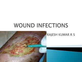
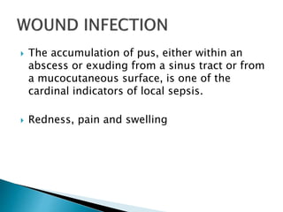


This document discusses wound infections, including the types (exogenous and endogenous), common causative organisms, and methods for diagnosis. It notes that pus accumulation is a sign of local infection and describes redness, pain, and swelling as additional indicators. Culture-based methods are outlined for identifying bacteria and determining pathogenicity from wound specimens. The importance of discussing isolated organisms with the physician is emphasized.


