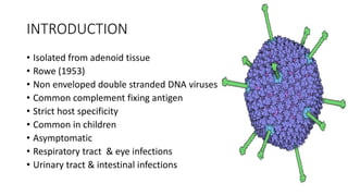
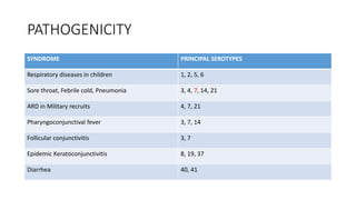
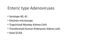
ADENOVIRUSES
DR.RAJESH KUMAR R S
INTRODUCTION
• Isolated from adenoid tissue
• Rowe (1953)
• Non enveloped double stranded DNA viruses
• Common complement fixing antigen
• Strict host specificity
• Common in children
• Asymptomatic
• Respiratory tract & eye infections
• Urinary tract & intestinal infections
MORPHOLOGY
RESISTANCE
• Relatively stable
• Viable for a week at 37˚ C
• Inactivated at 50˚ C
• Resistant to ether and bile salts
GROWTH AND HOST RANGE
• Host specific ( except type 12, 18 & 31)
• Tissue cultures - Human embryonic kidney
- HeLa
- Hep2
• Cytopathic changes – cell rounding
- aggregation into grape like clusters
- intranuclear inclusion bodies
CLASSIFICATION
• Family Adenoviridae
• 2 genera – Mastadenovirus
- Aviadenovirus
• 6 groups – A to F
- Haemagglutination, Fibre length, DNA fragment analysis &
oncogenic potential
• 50 serotypes
• AIDS patients
PATHOGENICITY
SYNDROME PRINCIPAL SEROTYPES
Respiratory diseases in children 1, 2, 5, 6
Sore throat, Febrile cold, Pneumonia 3, 4, 7, 14, 21
ARD in Military recruits 4, 7, 21
Pharyngoconjunctival fever 3, 7, 14
Follicular conjunctivitis 3, 7
Epidemic Keratoconjunctivitis 8, 19, 37
Diarrhea 40, 41
FOLLICULAR CONJUNCTIVITIS
EPIDEMIC KERATOCONJUNCTIVITIS
Enteric type Adenoviruses
• Serotype 40, 41
• Electron microscopy
• Trypsinised Monkey Kidney Cells
• Transformed Human Embryonic Kidney cells
• Stool ELISA
OTHER SYNDROMES
• Acute Hemorrhagic Cystitis – serotypes 11, 21
• Generalised Exanthem
• Mesenteric adenitis
• Intussusception
LABORATORY DIAGNOSIS
• Tissue culture – Cytopathic effects
- Complement Fixation Tests
- Haemagglutination
- Neutralisation
• Serodiagnosis
• Electron Microscopy
• Immunofluorescence
• PCR
PROPHYLAXIS
• Control of outbreaks in closed community
• ARD – Killed & Live vaccines
• No vaccine available for general use
• Gene therapy
ADENO ASSOCIATED VIRUSES
• 20 – 25 nm icosahedral viral particles
• Defective viruses
• Adenosatellite viruses
• Dependovirus
• Parvoviridae
• Electron microscopy, CFT or Immunofluorescence
• Type 1, 2, 3, 4
Adenoviruses

Adenoviruses

  • 1.
  • 2.
    INTRODUCTION • Isolated fromadenoid tissue • Rowe (1953) • Non enveloped double stranded DNA viruses • Common complement fixing antigen • Strict host specificity • Common in children • Asymptomatic • Respiratory tract & eye infections • Urinary tract & intestinal infections
  • 3.
  • 4.
    RESISTANCE • Relatively stable •Viable for a week at 37˚ C • Inactivated at 50˚ C • Resistant to ether and bile salts
  • 5.
    GROWTH AND HOSTRANGE • Host specific ( except type 12, 18 & 31) • Tissue cultures - Human embryonic kidney - HeLa - Hep2 • Cytopathic changes – cell rounding - aggregation into grape like clusters - intranuclear inclusion bodies
  • 6.
    CLASSIFICATION • Family Adenoviridae •2 genera – Mastadenovirus - Aviadenovirus • 6 groups – A to F - Haemagglutination, Fibre length, DNA fragment analysis & oncogenic potential • 50 serotypes • AIDS patients
  • 7.
    PATHOGENICITY SYNDROME PRINCIPAL SEROTYPES Respiratorydiseases in children 1, 2, 5, 6 Sore throat, Febrile cold, Pneumonia 3, 4, 7, 14, 21 ARD in Military recruits 4, 7, 21 Pharyngoconjunctival fever 3, 7, 14 Follicular conjunctivitis 3, 7 Epidemic Keratoconjunctivitis 8, 19, 37 Diarrhea 40, 41
  • 8.
  • 9.
  • 10.
    Enteric type Adenoviruses •Serotype 40, 41 • Electron microscopy • Trypsinised Monkey Kidney Cells • Transformed Human Embryonic Kidney cells • Stool ELISA
  • 11.
    OTHER SYNDROMES • AcuteHemorrhagic Cystitis – serotypes 11, 21 • Generalised Exanthem • Mesenteric adenitis • Intussusception
  • 12.
    LABORATORY DIAGNOSIS • Tissueculture – Cytopathic effects - Complement Fixation Tests - Haemagglutination - Neutralisation • Serodiagnosis • Electron Microscopy • Immunofluorescence • PCR
  • 13.
    PROPHYLAXIS • Control ofoutbreaks in closed community • ARD – Killed & Live vaccines • No vaccine available for general use • Gene therapy
  • 14.
    ADENO ASSOCIATED VIRUSES •20 – 25 nm icosahedral viral particles • Defective viruses • Adenosatellite viruses • Dependovirus • Parvoviridae • Electron microscopy, CFT or Immunofluorescence • Type 1, 2, 3, 4