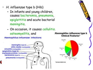
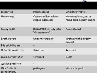
StreptococcusStreptococcus
pneumoniaepneumoniae
HemophilusHemophilus
influenzaeinfluenzae
StreptococcusStreptococcus
pneumoniaepneumoniae
Introduction
• gram positive lanceolate diplococci
• Resemble viridans streptococci
• Are normal inhabitants of upper RT
• Cause pneumonia & otitis media in children
• was first isolated simultaneously and independently
by the U.S. Army physician George Sternberg and
the French chemist Louis Pasteur
Morphology
• Gram positive diplococci
• alpha-hemolytic, facultative anaerobic member
of the genus Streptococcus.
• Individual cocci
– 1 um
– Lanceolate in appearance
• Capsulated
• Nonmotile
• Non sporing
• Non acid fast
Culture characteristics
• Aerobe & facultative
• Optimum temp. 37 o
C ( 25 – 42 o
C )
• Optimum PH – 7.6 ( 6.5 – 8.3)
• Grow only in enriched media
• Growth is improved by 5- 10 % co2
Characteristics ( contd..)
• Glucose/ serum broth:
– Produce uniform turbidity
– No pellicle formation
– After 36 hours autolysis occurs
• Blood Agar: after 18 hours at 37o
C
• Small (0.5 to 1)
• Round
• Entire
• Convex
• With an area of greenish discolouration around them
• On further incubation the colonies become flat with
raised edges and central depression, so that concentric
rings are seen on the surface when viewed from
above(draughtsman or carrom coin appearance)
“draughtsman” apperance
Biochemical reactions
• Catalase- -ve
• Oxidase- -ve
• Ferment several sugars
• Ferment inulin
• Are bile soluble
Serotypes
• Based on capsular polysaccharides
• More than 90 different serotypes are known
• these types differ in virulence, prevalence, and
extent of drug resistance
• Toxins:Hemolysin (Pneumolysin O) and leucocidin
• Capsular polysaccharide
– Protects the cocci from phagocytosis
virulence factors
Other virulence factors
• Ig A1 protease
• Protein adhesin
• Cell wall constituents
transmission
• methods include
– sneezing,
– coughing, and
– direct contact with an infected person.
Diseases
• Pneumonia
– Lobar pneumonia (localised in the lower lobes of
the lung) (more than 80% of cases)
– Broncho pneumonia (patchy involvment of the lung)
• bacteremia/septicaemia
• meningitis
• Conjuctivitis
• Suppurative lesions:
– Otitis media
– Paranasal Sinusitis
Diseases contd
• osteomyelitis,
• septic arthritis,
• endocarditis,
• peritonitis,
• cellulitis and
• brain abscesses.
• currently the leading cause of invasive bacterial
disease in children and the elderly.
• Pneumonia and otitis media are the most common
infections
Symptoms of pneumococcal disease
•depend on the part of the body that is infected.
•include fever, cough, shortness of breath, chest
pain, stiff neck, disorientation, sensitivity to light,
joint pain, chills, ear pain, sleeplessness, and
irritability.
• In severe cases, pneumococcal disease can cause
hearing loss, brain damage, and death.
Diagnosis
• Laboratory diagnosis
• Imaging
Laboratory diagnosis
• Specimens
• Microscopic Examination
• Culture
• Serological test
• Animal pathogenicity test
Potential specimen
• Blood
• Cerebrospinal fluid
• Sputum
• Pleural fluid or lung aspirate
• Joint fluid
• Bone
• Other abscess or tissue specimens
Gram Stain of a film of sputum from
a case of lobar pneumonia
Culture
• Culture in enriched media
• Routinely in Blood agar with 5-10 %
Carbondioxide
• Alphahemolytic colony noted
Identification: Optochin
sensitivity test
Quellung (capsular swelling) reaction/
serotyping
Bile solubility test
Animal pathogenecity
• Highly pathogenic for mice and rabbits but less
for guinea pig.
• Intraperitoneal inoculation (0.5) of homogenised
specimen of sputum or others leads to fatal
infection
• Mice-intraperitoneally when injected die within
24 hrs
• Mice-intracerebally when injected die within 6
hrs
• Organisms found in predominance in peritoneal
exudate and heart blood
• Other laboratory values that may be helpful in
diagnosis and treatment include the following:
– Complete blood count and differential
– Erythrocyte sedimentation rate (ESR)
– C-reactive protein (CRP)
Imaging studies
• Chest radiography
• Ultrasonography of the chest
• Computed tomography (CT) of the chest, sinuses,
face, or affected bones or joints
• Magnetic resonance imaging (MRI) of the brain (in
meningitis) or affected bones and joints
Griffith Tranformation
Prophylaxis
• A polysaccharide vaccine containing 23
serotypes is available
• Useful
• for children above 2 years of age
• Elderly , debilited & immunosuppressed
• Contraindicated in pts with Lymphomas &
children under 2 year
Treatment
• Penicillin:
– the drug of choice for many years
– Until recently 10 -15 % resistant strains
– Therefore AST should be done
• Other antibiotics:
– Cephalosporins
– Erythromycin
– Chloramphenicol
– Vancomycin –for highly resistant strains
Pneumococcus Vs other
Streptococci
• Morphology
• Quellung test
• Colony
characteristics
• Growth in liquid
media
• Bile solubility
• Inulin fermentation
• Optochin
sensitivity
• Intraperitoneal
inoculation in mice
(Mouse
pathogenicity test)
properties Pneumococcus Viridians strepto.
Morphology Capsulated,lanceolate-
shaped diplococci
Non-capsulated,oval or
round cells in short chains
Colony on BA Raised/flat initially later
“draughtsman”
Dome-shaped
Broth culture Uniform turbidity granularwith powdery
deposit
Bile solubility test + -
Optochin sensitivity Sensitive Resistant
Inulin fermentation ferments Donot
Quellung reaction + -
Mice/rabbbit
pathogenicity
pathogenic Non -pathogenic
Hemophilus influenzaeHemophilus influenzae
Definition
• Haemophilus influenzae (formerly called
Pfeiffer's bacillus or Bacillus influenzae) is a
Gram-negative, coccobacillary,
facultatively anaerobic pathogenic bacterium
belonging to the Pasteurellaceae family.
• H. influenzae was first described in 1892 by
Richard Pfeiffer during an influenza pandemic
• Greek haemal, blood; philos, friendly)
• present in the nasopharynx of approximately 75
percent of healthy children and adults.
Haemophilus influenzae
• gram-negative coccobacillus(short rod) and long
filamentous form in CSF in meningitis.
• 0.2 to 0.3 to 0.5 to 0.8 um
• Nonmotile, nonsporing pleomorphic
• Oxidase positive
• aerobic and facultative anaerobe.
• In vitro growth requires accessory growth factors,
including “X” factor (hemin) and “V” factor
(nicotinamide adenine dinucleotide [NAD]).
• Chocolate agar media are used for isolation. H.
influenzae will generally not grow on blood agar, which
lacks NAD.
Special media
• Levanthal agar
• Filde’s peptic digest
• Brain and Heart Infusion broth with
hemin and nicotinamide supplements
Satellitism
• H. influenzae will grow in the hemolytic zone of
Staphylococcus aureus on blood agar plates; the
hemolysis of cells by S. aureus releases factor V
which is needed for its growth. H. influenzae will
not grow outside the hemolytic zone of S. aureus
due to the lack of nutrients such as factor V in
these areas.
Classification/Serotypes
• In 1930, two major categories of H. influenzae
were defined: the unencapsulated strains and the
encapsulated strains.
• Encapsulated strains (typeable)
– The outermost structure of encapsulated H.
influenzae is composed of polyribosyl-
ribitolphosphate (PRP), a polysaccharide that is
responsible for virulence and immunity.
– on the basis of distinct capsular antigens six
types : a, b, c, d, e, and f.
– 95% of invasive disease caused by type b
(prevaccine)
• Unencapsulated strains (also termed nontypeable
(NTHi) lack capsular serotypes
– less invasive; they can, however, produce an
inflammatory response in humans
Diseases
• Most strains usually live in host without causing
disease
• but other factors (such as a viral infection,
mycoplasma, reduced immune function or
chronically inflamed tissues, e.g. from allergies)
create an opportunity.
• infect the host by sticking to the host cell using
trimeric autotransporter adhesins
• H. influenzae type b (Hib)
– In infants and young children,
causes bacteremia, pneumonia,
epiglottitis and acute bacterial
meningitis.
– On occasion, it causes cellulitis,
osteomyelitis, and
infectious arthritis.
• Unencapsulated H. influenzae strains:
– cause invasive disease similar to type b
infections but less virulent than encapsulated
strains
– are unaffected by the Hib vaccine
– cause ear infections (otitis media), eye
infections (conjunctivitis), and sinusitis in
children, and are associated with pneumonia.
– bronchitis in adults.
Virulence 
• no exotoxins
• neuraminidase and an IgA protease
•  Fimbriae adherence to human mucosal cells 
• capsular polysaccharide, (polyribosyl ribitol
phosphate (PRP) capsule )
– antiphagocytic, ineffective complement-mediated bacteriolysis.
Diagnosis
• Laboratory testing
• Imaging studies
Laboratory diagnosis
• Specimens:
– CSF, blood, pleural fluid, joint fluid, and
middle ear aspirates should be cultured
on Chocolate agar
Gram staining of body fluids from
various sites of infection
Culture
• chocolate agar, with added X (
hemin) and V (
nicotinamide adenine dinucleotide)
factors at 37 °C in a CO2-enriched
incubator
– convex, smooth, pale, grey or
transparent colonies.
• Fildes agar is best for isolation.
• In Levinthal medium, capsulated
strains show distinctive iridescence
.
X and V factors requirements
• Haemophilus influenzae requires X and V factors for
growth. In this culture haemophilus has only grown around
the paper disc that has been impregnated with X and V
factors. There is no bacterial growth around the discs that
only contain either X or V factor.
Immunological test
– Detection of the polyribosyl ribitol phosphate
(PRP) polysaccharide capsule
• countercurrent immunoelectrophoresis,
• latex particle agglutination,
• co-agglutination, and enzyme-linked
immunosorbent assay
– The most confirmatory method of establishing
the diagnosis; slide agglutination with type-
specific antisera
Laboratory testing
• Others
• Acute phase reactants:
– Characteristic elevated erythrocyte sedimentation
rates (ESRs) and C-reactive protein (CRP) levels in
patients with septic arthritis
Molecular methods
– Polymerase chain reaction (PCR)
– Countercurrent immunoelectrophoresis
•
Imaging studies
• Computed tomography (CT) scanning of the head
•
• Chest radiography: For suspected pulmonary
disease (eg, pneumonia)
• Lateral neck radiography: To confirm epiglottitis
•
• Echocardiography: For suspected pericarditis
Treatment
• A combined therapy with ampicillin plus
chloramphenicol or third generations
cephlosporine(Cefotaxime/ceftriaxone)
• Fluoroquinolones
• Macrolide antibiotics (e.g., clarithromycin) may be
used in patients with a history of allergy to beta-
lactam antibiotics
Prevention
• Effective vaccines for Haemophilus influenzae Type B
have been available since the early 1990s, and is
recommended for children under age 5 and asplenic
patients.
• World Health Organization recommends a pentavalent
vaccine, combining vaccines against diphtheria,
tetanus, pertussis, hepatitis B and Hib.
• Doses of Hib vaccine are usually recommended at
these ages:
– First Dose: 2 months of age
– Second Dose: 4 months of age
– Third Dose: 6 months of age (if needed,
depending on brand of vaccine)
– Final/Booster Dose: 12–15 months of age
– Children over 5 years old and adults usually do
not need Hib vaccine.
Haemophilus influenzae
type b Epidemiology
• Occurrence: worldwide
• Reservoir : human –asymptomatic carriers
• Transmission : commonly respiratory droplets
– neonates
• aspiration of amniotic fluid
• genital track secretions during delivery
• Temporal pattern : peaks in Sept-Dec and
March-May
• Communicability - generally limited but higher
in some circumstances
Thank You

Streptococcus pneumoniae mbbs

  • 1.
  • 2.
  • 3.
    Introduction • gram positivelanceolate diplococci • Resemble viridans streptococci • Are normal inhabitants of upper RT • Cause pneumonia & otitis media in children • was first isolated simultaneously and independently by the U.S. Army physician George Sternberg and the French chemist Louis Pasteur
  • 4.
    Morphology • Gram positivediplococci • alpha-hemolytic, facultative anaerobic member of the genus Streptococcus. • Individual cocci – 1 um – Lanceolate in appearance • Capsulated • Nonmotile • Non sporing • Non acid fast
  • 6.
    Culture characteristics • Aerobe& facultative • Optimum temp. 37 o C ( 25 – 42 o C ) • Optimum PH – 7.6 ( 6.5 – 8.3) • Grow only in enriched media • Growth is improved by 5- 10 % co2
  • 7.
    Characteristics ( contd..) •Glucose/ serum broth: – Produce uniform turbidity – No pellicle formation – After 36 hours autolysis occurs • Blood Agar: after 18 hours at 37o C • Small (0.5 to 1) • Round • Entire • Convex • With an area of greenish discolouration around them • On further incubation the colonies become flat with raised edges and central depression, so that concentric rings are seen on the surface when viewed from above(draughtsman or carrom coin appearance)
  • 9.
  • 10.
    Biochemical reactions • Catalase--ve • Oxidase- -ve • Ferment several sugars • Ferment inulin • Are bile soluble
  • 11.
    Serotypes • Based oncapsular polysaccharides • More than 90 different serotypes are known • these types differ in virulence, prevalence, and extent of drug resistance
  • 12.
    • Toxins:Hemolysin (PneumolysinO) and leucocidin • Capsular polysaccharide – Protects the cocci from phagocytosis virulence factors
  • 13.
    Other virulence factors •Ig A1 protease • Protein adhesin • Cell wall constituents
  • 14.
    transmission • methods include –sneezing, – coughing, and – direct contact with an infected person.
  • 15.
    Diseases • Pneumonia – Lobarpneumonia (localised in the lower lobes of the lung) (more than 80% of cases) – Broncho pneumonia (patchy involvment of the lung) • bacteremia/septicaemia • meningitis • Conjuctivitis • Suppurative lesions: – Otitis media – Paranasal Sinusitis
  • 16.
    Diseases contd • osteomyelitis, •septic arthritis, • endocarditis, • peritonitis, • cellulitis and • brain abscesses. • currently the leading cause of invasive bacterial disease in children and the elderly. • Pneumonia and otitis media are the most common infections
  • 18.
    Symptoms of pneumococcaldisease •depend on the part of the body that is infected. •include fever, cough, shortness of breath, chest pain, stiff neck, disorientation, sensitivity to light, joint pain, chills, ear pain, sleeplessness, and irritability. • In severe cases, pneumococcal disease can cause hearing loss, brain damage, and death.
  • 19.
  • 20.
    Laboratory diagnosis • Specimens •Microscopic Examination • Culture • Serological test • Animal pathogenicity test
  • 21.
    Potential specimen • Blood •Cerebrospinal fluid • Sputum • Pleural fluid or lung aspirate • Joint fluid • Bone • Other abscess or tissue specimens
  • 22.
    Gram Stain ofa film of sputum from a case of lobar pneumonia
  • 23.
    Culture • Culture inenriched media • Routinely in Blood agar with 5-10 % Carbondioxide • Alphahemolytic colony noted
  • 24.
  • 25.
    Quellung (capsular swelling)reaction/ serotyping
  • 26.
  • 27.
    Animal pathogenecity • Highlypathogenic for mice and rabbits but less for guinea pig. • Intraperitoneal inoculation (0.5) of homogenised specimen of sputum or others leads to fatal infection • Mice-intraperitoneally when injected die within 24 hrs • Mice-intracerebally when injected die within 6 hrs • Organisms found in predominance in peritoneal exudate and heart blood
  • 28.
    • Other laboratoryvalues that may be helpful in diagnosis and treatment include the following: – Complete blood count and differential – Erythrocyte sedimentation rate (ESR) – C-reactive protein (CRP)
  • 29.
    Imaging studies • Chestradiography • Ultrasonography of the chest • Computed tomography (CT) of the chest, sinuses, face, or affected bones or joints • Magnetic resonance imaging (MRI) of the brain (in meningitis) or affected bones and joints
  • 30.
  • 31.
    Prophylaxis • A polysaccharidevaccine containing 23 serotypes is available • Useful • for children above 2 years of age • Elderly , debilited & immunosuppressed • Contraindicated in pts with Lymphomas & children under 2 year
  • 32.
    Treatment • Penicillin: – thedrug of choice for many years – Until recently 10 -15 % resistant strains – Therefore AST should be done • Other antibiotics: – Cephalosporins – Erythromycin – Chloramphenicol – Vancomycin –for highly resistant strains
  • 33.
    Pneumococcus Vs other Streptococci •Morphology • Quellung test • Colony characteristics • Growth in liquid media • Bile solubility • Inulin fermentation • Optochin sensitivity • Intraperitoneal inoculation in mice (Mouse pathogenicity test)
  • 34.
    properties Pneumococcus Viridiansstrepto. Morphology Capsulated,lanceolate- shaped diplococci Non-capsulated,oval or round cells in short chains Colony on BA Raised/flat initially later “draughtsman” Dome-shaped Broth culture Uniform turbidity granularwith powdery deposit Bile solubility test + - Optochin sensitivity Sensitive Resistant Inulin fermentation ferments Donot Quellung reaction + - Mice/rabbbit pathogenicity pathogenic Non -pathogenic
  • 35.
  • 36.
    Definition • Haemophilus influenzae(formerly called Pfeiffer's bacillus or Bacillus influenzae) is a Gram-negative, coccobacillary, facultatively anaerobic pathogenic bacterium belonging to the Pasteurellaceae family. • H. influenzae was first described in 1892 by Richard Pfeiffer during an influenza pandemic • Greek haemal, blood; philos, friendly) • present in the nasopharynx of approximately 75 percent of healthy children and adults.
  • 37.
    Haemophilus influenzae • gram-negativecoccobacillus(short rod) and long filamentous form in CSF in meningitis. • 0.2 to 0.3 to 0.5 to 0.8 um • Nonmotile, nonsporing pleomorphic • Oxidase positive • aerobic and facultative anaerobe. • In vitro growth requires accessory growth factors, including “X” factor (hemin) and “V” factor (nicotinamide adenine dinucleotide [NAD]). • Chocolate agar media are used for isolation. H. influenzae will generally not grow on blood agar, which lacks NAD.
  • 38.
    Special media • Levanthalagar • Filde’s peptic digest • Brain and Heart Infusion broth with hemin and nicotinamide supplements
  • 39.
    Satellitism • H. influenzaewill grow in the hemolytic zone of Staphylococcus aureus on blood agar plates; the hemolysis of cells by S. aureus releases factor V which is needed for its growth. H. influenzae will not grow outside the hemolytic zone of S. aureus due to the lack of nutrients such as factor V in these areas.
  • 40.
    Classification/Serotypes • In 1930,two major categories of H. influenzae were defined: the unencapsulated strains and the encapsulated strains. • Encapsulated strains (typeable) – The outermost structure of encapsulated H. influenzae is composed of polyribosyl- ribitolphosphate (PRP), a polysaccharide that is responsible for virulence and immunity. – on the basis of distinct capsular antigens six types : a, b, c, d, e, and f. – 95% of invasive disease caused by type b (prevaccine)
  • 41.
    • Unencapsulated strains(also termed nontypeable (NTHi) lack capsular serotypes – less invasive; they can, however, produce an inflammatory response in humans
  • 42.
    Diseases • Most strainsusually live in host without causing disease • but other factors (such as a viral infection, mycoplasma, reduced immune function or chronically inflamed tissues, e.g. from allergies) create an opportunity. • infect the host by sticking to the host cell using trimeric autotransporter adhesins
  • 43.
    • H. influenzaetype b (Hib) – In infants and young children, causes bacteremia, pneumonia, epiglottitis and acute bacterial meningitis. – On occasion, it causes cellulitis, osteomyelitis, and infectious arthritis.
  • 44.
    • Unencapsulated H.influenzae strains: – cause invasive disease similar to type b infections but less virulent than encapsulated strains – are unaffected by the Hib vaccine – cause ear infections (otitis media), eye infections (conjunctivitis), and sinusitis in children, and are associated with pneumonia. – bronchitis in adults.
  • 45.
    Virulence  • no exotoxins •neuraminidase and an IgA protease •  Fimbriae adherence to human mucosal cells  • capsular polysaccharide, (polyribosyl ribitol phosphate (PRP) capsule ) – antiphagocytic, ineffective complement-mediated bacteriolysis.
  • 46.
  • 47.
    Laboratory diagnosis • Specimens: –CSF, blood, pleural fluid, joint fluid, and middle ear aspirates should be cultured on Chocolate agar
  • 48.
    Gram staining ofbody fluids from various sites of infection
  • 49.
    Culture • chocolate agar,with added X ( hemin) and V ( nicotinamide adenine dinucleotide) factors at 37 °C in a CO2-enriched incubator – convex, smooth, pale, grey or transparent colonies. • Fildes agar is best for isolation. • In Levinthal medium, capsulated strains show distinctive iridescence .
  • 50.
    X and Vfactors requirements • Haemophilus influenzae requires X and V factors for growth. In this culture haemophilus has only grown around the paper disc that has been impregnated with X and V factors. There is no bacterial growth around the discs that only contain either X or V factor.
  • 51.
    Immunological test – Detectionof the polyribosyl ribitol phosphate (PRP) polysaccharide capsule • countercurrent immunoelectrophoresis, • latex particle agglutination, • co-agglutination, and enzyme-linked immunosorbent assay – The most confirmatory method of establishing the diagnosis; slide agglutination with type- specific antisera
  • 52.
    Laboratory testing • Others •Acute phase reactants: – Characteristic elevated erythrocyte sedimentation rates (ESRs) and C-reactive protein (CRP) levels in patients with septic arthritis
  • 53.
    Molecular methods – Polymerasechain reaction (PCR) – Countercurrent immunoelectrophoresis •
  • 54.
    Imaging studies • Computedtomography (CT) scanning of the head • • Chest radiography: For suspected pulmonary disease (eg, pneumonia) • Lateral neck radiography: To confirm epiglottitis • • Echocardiography: For suspected pericarditis
  • 55.
    Treatment • A combinedtherapy with ampicillin plus chloramphenicol or third generations cephlosporine(Cefotaxime/ceftriaxone) • Fluoroquinolones • Macrolide antibiotics (e.g., clarithromycin) may be used in patients with a history of allergy to beta- lactam antibiotics
  • 56.
    Prevention • Effective vaccinesfor Haemophilus influenzae Type B have been available since the early 1990s, and is recommended for children under age 5 and asplenic patients. • World Health Organization recommends a pentavalent vaccine, combining vaccines against diphtheria, tetanus, pertussis, hepatitis B and Hib.
  • 57.
    • Doses ofHib vaccine are usually recommended at these ages: – First Dose: 2 months of age – Second Dose: 4 months of age – Third Dose: 6 months of age (if needed, depending on brand of vaccine) – Final/Booster Dose: 12–15 months of age – Children over 5 years old and adults usually do not need Hib vaccine.
  • 58.
    Haemophilus influenzae type bEpidemiology • Occurrence: worldwide • Reservoir : human –asymptomatic carriers • Transmission : commonly respiratory droplets – neonates • aspiration of amniotic fluid • genital track secretions during delivery • Temporal pattern : peaks in Sept-Dec and March-May • Communicability - generally limited but higher in some circumstances
  • 59.