Downloaded 81 times


![Laboratory Diagnosis (contd....)
B.Bacteriological
Investigation:
• Specimens:
– Pus: from wound or abscess
or burns]
– Nasal Swab: from
suspected carrier
– Food: to diagnose staphylococcal
intoxication
– Blood: to diagnose
endocarditis and bacteremia
– Sputum: to diagnose lower
respiratory tract infection](https://image.slidesharecdn.com/lecture4staphylococcus-230321150017-bc2910b8/85/Staphylococcus-30-320.jpg)


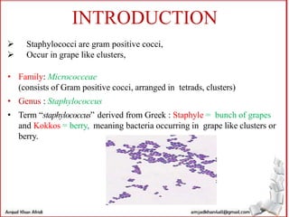
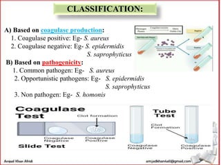
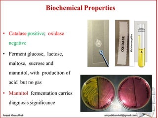
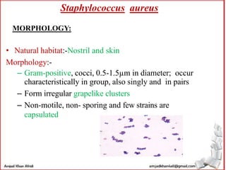
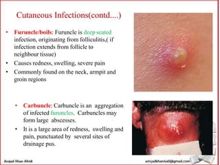
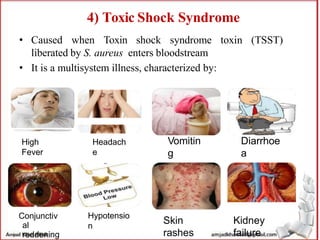
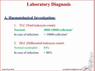
The document provides a comprehensive overview of staphylococci, highlighting their classification, historical background, morphology, growth characteristics, and pathogenicity. It details various infections caused by Staphylococcus aureus, including cutaneous and deep infections, exfoliative diseases, and food poisoning, along with modes of transmission and prevention strategies. It concludes with diagnostic methods for detecting staphylococcal infections, emphasizing laboratory investigations such as culture and biochemical tests.