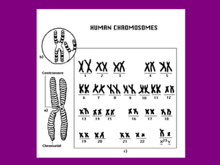
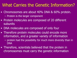
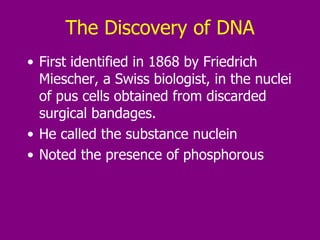
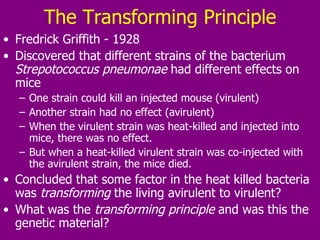
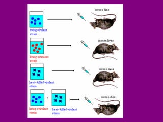
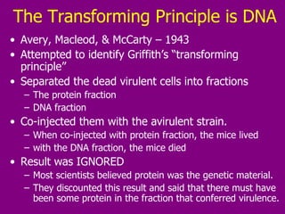
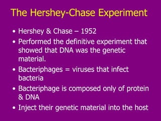
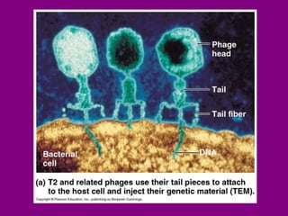
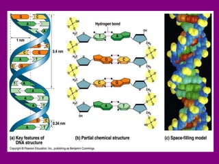
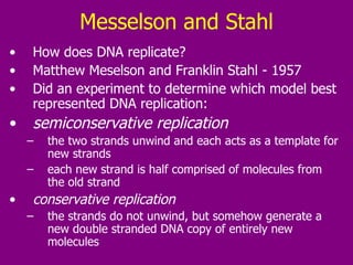
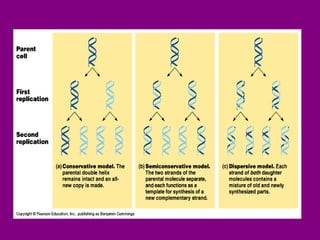
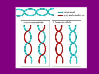
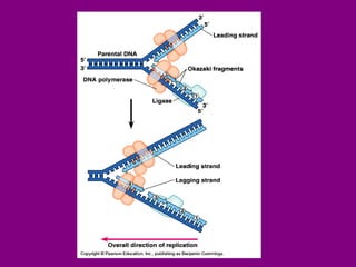
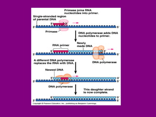
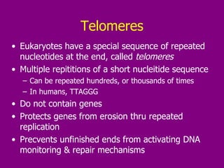
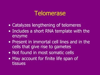
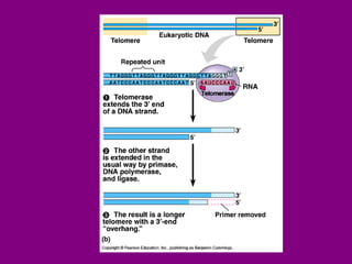
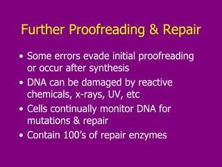
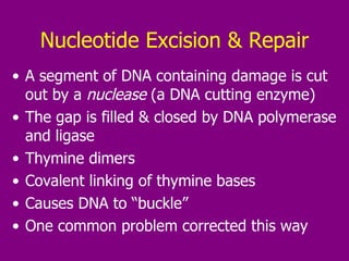
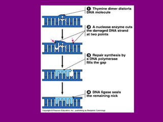
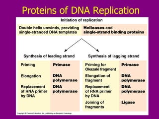
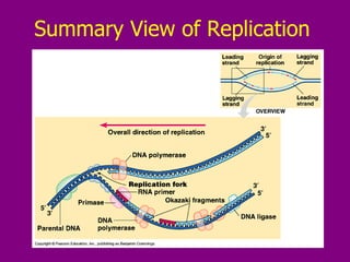
Molecular Genetics The Central Dogma of Biology
What is a Genetic “Factor”? From Mendel: we now accepted that there was genetic transmission of traits. Traits are transmitted by “factors” Organisms carry 2 copies of each “factor” The question now was: what is the factor that carries the genetic information?
Requirements of Genetic Material Must be able to  replicate , so it is reproduced in each cell of a growing organism.  Must be able to  control expression of traits Traits are determined by the proteins that act within us Proteins are determined by their sequences   Therefore, the genetic material must be able to  encode the sequence of proteins It must be able to  change  in a controlled way, to allow variation, adaptation, thus survival in a changing environment.
Chromosomes – The First Clue First ability to visualize chromosomes in the nucleus came at the turn of the century construction of increasingly powerful microscopes  the discovery of dyes that selectively colored various components of the cell   Scientists examined cellular nuclei and observed nuclear structures, which they called chromosomes Observation of these structures suggested their role in genetic transmission
Chromosome Observations Variety of chromosome types found in the nucleus  2 copies of each type of chromosome in most cells. All of the cells of an organism, except gametes  and rbc’s, have the same number of chromosomes.  All organisms of the same species have the same number of chromosomes.  The number of chromosomes in a cell doubles immediately prior to cell division  Gametes have exactly half of the number of chromosomes as the somatic cells of any organism. Gametes have just one copy of each chromosome type. Fertilization produces a diploid cell (a zygote), with the same number of chromosomes as somatic cells of that organism.
Implications Chromosomes behaved like Mendel’s “factors” Mendel's hereditary factors were either located on the chromosomes or were the chromosomes themselves.   Proof chromosomes were hereditary factors – 1905: The first physical trait was linked to the  presence of specific chromosomal material  conversely, the absence of that chromosome meant the absence of the particular physical trait.  Discovery of the sex chromosomes "X" and "Y."  distinguished from other chromosomes and from each other
Importance of Sex Chromosomes Somatic cells taken from female donors always contained 2 copies of the X chromosome   Somatic cells taken from male donors always contained one copy of the X chromosome and one copy of the Y chromosome   All of the other chromosomes in the nucleated cells of both male and female donors appeared identical Thus, gender was shown to be the direct result of a specific combination of chromosomal material The first phenotype (physical characteristic) to be assigned a chromosomal location  Specifically the X and Y chromosomes.
 
What Carries the Genetic Information? Chromosomes are about 40% DNA & 60% protein.  Protein is the larger component Protein molecules are composed of 20 different subunits DNA molecules are composed of only four Therefore protein molecules could encode more information, and a greater variety of information protein had the possibility for much more diversity than in DNA  Therefore, scientists believed that the protein in chromosomes must carry the genetic information
The Discovery of DNA First identified in 1868 by Friedrich Miescher, a Swiss biologist, in the nuclei of pus cells obtained from discarded surgical bandages.  He called the substance nuclein Noted the presence of phosphorous
The Transforming Principle Fredrick Griffith - 1928 Discovered that different strains of the bacterium  Strepotococcus pneumonae  had different effects on mice One strain could kill an injected mouse (virulent) Another strain had no effect (avirulent)  When the virulent strain was heat-killed and injected into mice, there was no effect.  But when a heat-killed virulent strain was co-injected with the avirulent strain, the mice died.  Concluded that some factor in the heat killed bacteria was  transforming  the living avirulent to virulent? What was the  transforming principle  and was this the genetic material?
 
The Transforming Principle is DNA Avery, Macleod, & McCarty – 1943 Attempted to identify Griffith’s “transforming principle” Separated the dead virulent cells into fractions The protein fraction DNA fraction Co-injected them with the avirulent strain. When co-injected with protein fraction, the mice lived with the DNA fraction, the mice died Result was IGNORED Most scientists believed protein was the genetic material.  They discounted this result and said that there must have been some protein in the fraction that conferred virulence.
The Hershey-Chase Experiment  Hershey & Chase – 1952 Performed the definitive experiment that showed that DNA was the genetic material.  Bacteriphages = viruses that infect bacteria Bacteriphage is composed only of protein & DNA Inject their genetic material into the host
 
The Experiment Prepared 2 cultures of bacteriophages Radiolabeled sulphur in one culture  there is sulphur in proteins, in the amino acids methionine and cysteine  there is no sulphur in DNA  Radiolabeled phosphorous in the second culture there is phosphorous in the phosphate backbone of DNA  none in any of the amino acids.  So this one culture in which only the phage protein was labeled, and one culture in which only the phage DNA was labeled.
Experiment Summary Performed side by side experiments with separate phage cultures in which either the protein capsule was labeled with radioactive sulfur or the DNA core was labeled with radioactive phosphorus.  The radioactively labeled phages were allowed to infect bacteria.  Agitation in a blender dislodged phage particles from bacterial cells.  Centrifugation pelleted cells, separating them from the phage particles left in the supernatant.
Results Summary Radioactive sulfur was found predominantly in the supernatant.  Radioactive phosphorus was found predominantly in the cell fraction, from which a new generation of infective phage was generated.  Thus, it was shown that the genetic material that encoded the growth of a new generation of phage was in the phosphorous-containing DNA.
 
Chargaff’s Rule   Once  DNA was recognized as the genetic material, scientists began investigating its mechanism and structure.  Erwin Chargaff – 1950  discovered the % content of the 4 nucleotides was the same in all tissues of the same species percentages could vary from species to species. He also found that in all animals (Chargaff’s rule):  %G = %C %A = %T This suggested that the structure of the DNA was specific and conserved in each organism.  The significance of these results was initially overlooked
Base Pairing in DNA Not understood ‘til Watson & Crick described double helix Adenine & guanine are  purines 2 organic rings Cytosine & guanine are  pyrimidines 1 organic ring Pairing a purine & a pyrimidine creates the correct 2 nm distance in the double helix A – T joined by 2 hydrogen bonds G – C joined by 3 hydrogen bonds
The Double Helix   James Watson and Francis Crick – 1953 Presented a model of the structure of DNA.  It was already known from chemical studies that DNA was a polymer of nucleotide (sugar, base and phosphate) units.  X-ray crystallographic data obtained by Rosalind Franklin, combined with the previous results from Chargaff and others, were fitted together by Watson and Crick into the double helix model.
Watson and Crick shared the 1962 Nobel Prize for Physiology and Medicine with Maurice Wilkins.  Rosalind Franklin died before this date.
DNA Structure The double helix is formed from two strands of DNA  DNA strands run in opposite directions  They are  complementary attached by hydrogen bonds between complimentary base pairs:  A - T and G - C  This complementary pairing of the bases suggests that, when DNA replicates, an exact duplicate of the parental genetic information is made.  The polymerization of a new complementary strand takes place using each of the old strands as a template.
 
Messelson and Stahl How does DNA replicate? Matthew Meselson and Franklin Stahl - 1957 Did an experiment to determine which model best represented DNA replication:  semiconservative replication   the two strands unwind and each acts as a template for new strands each new strand is half comprised of molecules from the old strand conservative replication   the strands do not unwind, but somehow generate a new double stranded DNA copy of entirely new molecules
 
 
The Experiment The original DNA strand was labeled with the heavy isotope of nitrogen, N-15.  This DNA was allowed to go through one round of replication with N-14 (non-labeled) the mixture was centrifuged so that the heavier DNA would form a band lower in the tube the intermediate (one N-15 strand and one N-14 strand) and light DNA (all N-14) would appear as a band higher in the tube
The expected results for each model were:
The actual results were as expected for the semiconservative model and thus Watson and Crick's suspicion was borne out.
Enzymes & Replication DNA replication is not a passive, spontaneous process.  More than a dozen enzymes & other proteins are required to unwind the double helix & to synthesize & finalize a new strand of DNA.  The molecular mechanism of DNA replication can best be understood from the point of view of the machinery required to accomplish it.  The unwound helix, with each strand being synthesized into a new double helix, is called the  replication fork .
The Enzymes of DNA Replication Topoisomerase  Responsible for initiation of unwinding of DNA. The tension holding the helix in its coiled and supercoiled structure is broken by nicking a single strand of DNA.   Helicase  Accomplishes unwinding of the original double strand, once supercoiling is eliminated by topoisomerase. The two strands want to bind together because of hydrogen bonding affinity for each other, so helicase requires energy (ATP) to break the strands apart.
Single-stranded Binding Proteins Important to maintain the stability of the replication fork.  Line up along unpaired DNA strans, holding them apart Single-stranded DNA is very unstable, so these proteins bind bind to it while it remains single stranded and keep it from being degraded.
Beginning: Origins of Replication Replication begins at specific sites called  origins of replication In prokaryotes, the bacterial chromosome has a specific  origin In eukaryotes, replication begins at many sites on the large molecule 100’s of origins Proteins that begin replication recognize the  origin  sequence These enzymes attach to DNA, separating the strands, and opening a  replication bubble The end of the  replication bubble  is the “Y” shaped  replication fork , where new strands of DNA elongate
 
Elongation Elongation of the new DNA strands is catalyzed by DNA polymerase Nucleotides align with complimentary bases on the template strand, and are added by the polymerase, one by one, to the growing chain  DNA polymerase proceeds along a single-stranded DNA molecule, recruiting free nucleotides to H-bond with the complementary nucleotides on the single strand  Forms a covalent phosphodiester bond with the previous nucleotide of the same strand  The energy stored in the triphosphate is used to covalently bind each new nucleotide to the growing second strand Replication proceeds in both directions
 
The Replication Fork
DNA Polymerase There are different forms of DNA polymerase DNA polymerase III is responsible for the synthesis of new DNA strands  DNA polymerase is actually an aggregate of several different protein subunits, so it is often called a  holoenzyme . Primary job is adding nucleotides to the growing chain  Also has proofreading activities
Proofreading & Repair DNA polymerase proofreads each nucleotide added against its’ template as it is added Removes incorrectly paired nucleotides & corrects
DNA Strands are Anti-parallel The sugar phosphate backbones of the 2 parent strands run in opposite directions “ upside down” to each other DNA is polar There is a 3’ end and a 5’ end At the 3’ end, a OH is attached to the 3’ C of the last deoxyribose At the other end a phosphate group is attached to the 5’ C of the last nucleotide DNA polymerase adds nucleotides only to the 3’ end of the growing chain So new DNA strand elongates only in the 5’   3” direction
 
Why 5’    3’? Why can DNA polymerase only  add nucleotides to the 3’ end ?  Needs a triphosphate to provide energy for the bond between an added nucleotide & the growing DNA strand.  This triphosphate is very unstable  can easily break into a monophosphate and an inorganic pyrophosphate At the 5' end, this triphosphate can easily break It is not be able to attach new nucleotides to the 5' end once the phosphate has broken off  The 3' end only has a hydroxyl group As long as new nucleotide triphosphates are brought by DNA polymerase, synthesis of a new strand continues, no matter how long the 3' end has remained free.
Leading & Lagging Strands The new strand made by adding to the 3’ end =  leading strand   Parent strand is 3’    5’ New strand is 5’    3’ How can DNA polymerase synthesize new copies of the 5'    3' strand, if it can only travel in one direction? To elongate in the other direction, the process must work  away  from the replication fork  The new strand formed on the 5’    3’ parent strand is called the  lagging strand
Building the Lagging Strand DNA polymerase makes a second copy in an overall 3’    5’ direction First, it produces short segments,  called  Okazaki fragments These are built in a 5’ –3’ direction Okazaki fragments are joined together by  ligase  to produce the new 3’  5’ lagging strand
Ligase Catalyzes the formation of a phosphodiester bond given an unattached adjacent 3‘ OH and 5‘ phosphate.  This can join Okazaki fragments This can also fill in the unattached gap left when an RNA primer is removed  DNA polymerase can organize the bond on the 5' end of the primer, but ligase is needed to make the bond on the 3' end.
 
Primers DNA polymerase cannot start synthesizing on a bare single strand.  It only adds to an existing chain It needs a  primer  with a 3'OH group on which to attach a nucleotide.  The start of a new chain is not DNA, but a short RNA primer Only one primer is needed for the leading strand One primer for each Okazaki fragment on the lagging strand
Primase Part of an aggregate of proteins called the  primeosome. Attaches the small RNA primer to the single-stranded DNA which acts as a substitute 3'OH so DNA polymerase can begin synthesis This RNA primer is eventually removed by RNase H  the gap is filled in by DNA polymerase I.
 
Ending the Strand DNA polymerase only adds to the 3’ end There is no way to complete the 5’ end of the new strand A small gap would be left at the 5’ end of each new strand Repeated replication would then make the strand shorter and shorter, eventually losing genes Not a problem in prokaryotes, because the DNA is circular There are no “ends”
Telomeres Eukaryotes have a special sequence of repeated nucleotides at the end, called  telomeres Multiple repititions of a short nucleitide sequence Can be repeated hundreds, or thousands of times In humans, TTAGGG Do not contain genes Protects genes from erosion thru repeated replication Precvents unfinished ends from activating DNA monitoring & repair mechanisms
Telomerase Catalyzes lengthening of telomeres Includes a short RNA template with the enzyme Present in immortal cell lines and in the cells that give rise to gametes Not found in most somatic cells May account for finite life span of tissues
 
Further Proofreading & Repair Some errors evade initial proofreading or occur after synthesis DNA can be damaged by reactive chemicals, x-rays, UV, etc Cells continually monitor DNA for mutations & repair Contain 100’s of repair enzymes
Nucleotide Excision & Repair A segment of DNA containing damage is cut out by a  nuclease  (a DNA cutting enzyme) The gap is filled & closed by DNA polymerase and ligase Thymine dimers Covalent linking of thymine bases Causes DNA to “buckle” One common problem corrected this way
 
Proteins of DNA Replication
Summary View of Replication

Molecular Genetics

  • 1.
    Molecular Genetics TheCentral Dogma of Biology
  • 2.
    What is aGenetic “Factor”? From Mendel: we now accepted that there was genetic transmission of traits. Traits are transmitted by “factors” Organisms carry 2 copies of each “factor” The question now was: what is the factor that carries the genetic information?
  • 3.
    Requirements of GeneticMaterial Must be able to replicate , so it is reproduced in each cell of a growing organism. Must be able to control expression of traits Traits are determined by the proteins that act within us Proteins are determined by their sequences Therefore, the genetic material must be able to encode the sequence of proteins It must be able to change in a controlled way, to allow variation, adaptation, thus survival in a changing environment.
  • 4.
    Chromosomes – TheFirst Clue First ability to visualize chromosomes in the nucleus came at the turn of the century construction of increasingly powerful microscopes the discovery of dyes that selectively colored various components of the cell Scientists examined cellular nuclei and observed nuclear structures, which they called chromosomes Observation of these structures suggested their role in genetic transmission
  • 5.
    Chromosome Observations Varietyof chromosome types found in the nucleus 2 copies of each type of chromosome in most cells. All of the cells of an organism, except gametes and rbc’s, have the same number of chromosomes. All organisms of the same species have the same number of chromosomes. The number of chromosomes in a cell doubles immediately prior to cell division Gametes have exactly half of the number of chromosomes as the somatic cells of any organism. Gametes have just one copy of each chromosome type. Fertilization produces a diploid cell (a zygote), with the same number of chromosomes as somatic cells of that organism.
  • 6.
    Implications Chromosomes behavedlike Mendel’s “factors” Mendel's hereditary factors were either located on the chromosomes or were the chromosomes themselves. Proof chromosomes were hereditary factors – 1905: The first physical trait was linked to the presence of specific chromosomal material conversely, the absence of that chromosome meant the absence of the particular physical trait. Discovery of the sex chromosomes "X" and "Y." distinguished from other chromosomes and from each other
  • 7.
    Importance of SexChromosomes Somatic cells taken from female donors always contained 2 copies of the X chromosome Somatic cells taken from male donors always contained one copy of the X chromosome and one copy of the Y chromosome All of the other chromosomes in the nucleated cells of both male and female donors appeared identical Thus, gender was shown to be the direct result of a specific combination of chromosomal material The first phenotype (physical characteristic) to be assigned a chromosomal location Specifically the X and Y chromosomes.
  • 8.
  • 9.
    What Carries theGenetic Information? Chromosomes are about 40% DNA & 60% protein. Protein is the larger component Protein molecules are composed of 20 different subunits DNA molecules are composed of only four Therefore protein molecules could encode more information, and a greater variety of information protein had the possibility for much more diversity than in DNA Therefore, scientists believed that the protein in chromosomes must carry the genetic information
  • 10.
    The Discovery ofDNA First identified in 1868 by Friedrich Miescher, a Swiss biologist, in the nuclei of pus cells obtained from discarded surgical bandages. He called the substance nuclein Noted the presence of phosphorous
  • 11.
    The Transforming PrincipleFredrick Griffith - 1928 Discovered that different strains of the bacterium Strepotococcus pneumonae had different effects on mice One strain could kill an injected mouse (virulent) Another strain had no effect (avirulent) When the virulent strain was heat-killed and injected into mice, there was no effect. But when a heat-killed virulent strain was co-injected with the avirulent strain, the mice died. Concluded that some factor in the heat killed bacteria was transforming the living avirulent to virulent? What was the transforming principle and was this the genetic material?
  • 12.
  • 13.
    The Transforming Principleis DNA Avery, Macleod, & McCarty – 1943 Attempted to identify Griffith’s “transforming principle” Separated the dead virulent cells into fractions The protein fraction DNA fraction Co-injected them with the avirulent strain. When co-injected with protein fraction, the mice lived with the DNA fraction, the mice died Result was IGNORED Most scientists believed protein was the genetic material. They discounted this result and said that there must have been some protein in the fraction that conferred virulence.
  • 14.
    The Hershey-Chase Experiment Hershey & Chase – 1952 Performed the definitive experiment that showed that DNA was the genetic material. Bacteriphages = viruses that infect bacteria Bacteriphage is composed only of protein & DNA Inject their genetic material into the host
  • 15.
  • 16.
    The Experiment Prepared2 cultures of bacteriophages Radiolabeled sulphur in one culture there is sulphur in proteins, in the amino acids methionine and cysteine there is no sulphur in DNA Radiolabeled phosphorous in the second culture there is phosphorous in the phosphate backbone of DNA none in any of the amino acids. So this one culture in which only the phage protein was labeled, and one culture in which only the phage DNA was labeled.
  • 17.
    Experiment Summary Performedside by side experiments with separate phage cultures in which either the protein capsule was labeled with radioactive sulfur or the DNA core was labeled with radioactive phosphorus. The radioactively labeled phages were allowed to infect bacteria. Agitation in a blender dislodged phage particles from bacterial cells. Centrifugation pelleted cells, separating them from the phage particles left in the supernatant.
  • 18.
    Results Summary Radioactivesulfur was found predominantly in the supernatant. Radioactive phosphorus was found predominantly in the cell fraction, from which a new generation of infective phage was generated. Thus, it was shown that the genetic material that encoded the growth of a new generation of phage was in the phosphorous-containing DNA.
  • 19.
  • 20.
    Chargaff’s Rule Once DNA was recognized as the genetic material, scientists began investigating its mechanism and structure. Erwin Chargaff – 1950 discovered the % content of the 4 nucleotides was the same in all tissues of the same species percentages could vary from species to species. He also found that in all animals (Chargaff’s rule): %G = %C %A = %T This suggested that the structure of the DNA was specific and conserved in each organism. The significance of these results was initially overlooked
  • 21.
    Base Pairing inDNA Not understood ‘til Watson & Crick described double helix Adenine & guanine are purines 2 organic rings Cytosine & guanine are pyrimidines 1 organic ring Pairing a purine & a pyrimidine creates the correct 2 nm distance in the double helix A – T joined by 2 hydrogen bonds G – C joined by 3 hydrogen bonds
  • 22.
    The Double Helix James Watson and Francis Crick – 1953 Presented a model of the structure of DNA. It was already known from chemical studies that DNA was a polymer of nucleotide (sugar, base and phosphate) units. X-ray crystallographic data obtained by Rosalind Franklin, combined with the previous results from Chargaff and others, were fitted together by Watson and Crick into the double helix model.
  • 23.
    Watson and Crickshared the 1962 Nobel Prize for Physiology and Medicine with Maurice Wilkins. Rosalind Franklin died before this date.
  • 24.
    DNA Structure Thedouble helix is formed from two strands of DNA DNA strands run in opposite directions They are complementary attached by hydrogen bonds between complimentary base pairs: A - T and G - C This complementary pairing of the bases suggests that, when DNA replicates, an exact duplicate of the parental genetic information is made. The polymerization of a new complementary strand takes place using each of the old strands as a template.
  • 25.
  • 26.
    Messelson and StahlHow does DNA replicate? Matthew Meselson and Franklin Stahl - 1957 Did an experiment to determine which model best represented DNA replication: semiconservative replication the two strands unwind and each acts as a template for new strands each new strand is half comprised of molecules from the old strand conservative replication the strands do not unwind, but somehow generate a new double stranded DNA copy of entirely new molecules
  • 27.
  • 28.
  • 29.
    The Experiment Theoriginal DNA strand was labeled with the heavy isotope of nitrogen, N-15. This DNA was allowed to go through one round of replication with N-14 (non-labeled) the mixture was centrifuged so that the heavier DNA would form a band lower in the tube the intermediate (one N-15 strand and one N-14 strand) and light DNA (all N-14) would appear as a band higher in the tube
  • 30.
    The expected resultsfor each model were:
  • 31.
    The actual resultswere as expected for the semiconservative model and thus Watson and Crick's suspicion was borne out.
  • 32.
    Enzymes & ReplicationDNA replication is not a passive, spontaneous process. More than a dozen enzymes & other proteins are required to unwind the double helix & to synthesize & finalize a new strand of DNA. The molecular mechanism of DNA replication can best be understood from the point of view of the machinery required to accomplish it. The unwound helix, with each strand being synthesized into a new double helix, is called the replication fork .
  • 33.
    The Enzymes ofDNA Replication Topoisomerase Responsible for initiation of unwinding of DNA. The tension holding the helix in its coiled and supercoiled structure is broken by nicking a single strand of DNA. Helicase Accomplishes unwinding of the original double strand, once supercoiling is eliminated by topoisomerase. The two strands want to bind together because of hydrogen bonding affinity for each other, so helicase requires energy (ATP) to break the strands apart.
  • 34.
    Single-stranded Binding ProteinsImportant to maintain the stability of the replication fork. Line up along unpaired DNA strans, holding them apart Single-stranded DNA is very unstable, so these proteins bind bind to it while it remains single stranded and keep it from being degraded.
  • 35.
    Beginning: Origins ofReplication Replication begins at specific sites called origins of replication In prokaryotes, the bacterial chromosome has a specific origin In eukaryotes, replication begins at many sites on the large molecule 100’s of origins Proteins that begin replication recognize the origin sequence These enzymes attach to DNA, separating the strands, and opening a replication bubble The end of the replication bubble is the “Y” shaped replication fork , where new strands of DNA elongate
  • 36.
  • 37.
    Elongation Elongation ofthe new DNA strands is catalyzed by DNA polymerase Nucleotides align with complimentary bases on the template strand, and are added by the polymerase, one by one, to the growing chain DNA polymerase proceeds along a single-stranded DNA molecule, recruiting free nucleotides to H-bond with the complementary nucleotides on the single strand Forms a covalent phosphodiester bond with the previous nucleotide of the same strand The energy stored in the triphosphate is used to covalently bind each new nucleotide to the growing second strand Replication proceeds in both directions
  • 38.
  • 39.
  • 40.
    DNA Polymerase Thereare different forms of DNA polymerase DNA polymerase III is responsible for the synthesis of new DNA strands DNA polymerase is actually an aggregate of several different protein subunits, so it is often called a holoenzyme . Primary job is adding nucleotides to the growing chain Also has proofreading activities
  • 41.
    Proofreading & RepairDNA polymerase proofreads each nucleotide added against its’ template as it is added Removes incorrectly paired nucleotides & corrects
  • 42.
    DNA Strands areAnti-parallel The sugar phosphate backbones of the 2 parent strands run in opposite directions “ upside down” to each other DNA is polar There is a 3’ end and a 5’ end At the 3’ end, a OH is attached to the 3’ C of the last deoxyribose At the other end a phosphate group is attached to the 5’ C of the last nucleotide DNA polymerase adds nucleotides only to the 3’ end of the growing chain So new DNA strand elongates only in the 5’  3” direction
  • 43.
  • 44.
    Why 5’  3’? Why can DNA polymerase only add nucleotides to the 3’ end ? Needs a triphosphate to provide energy for the bond between an added nucleotide & the growing DNA strand. This triphosphate is very unstable can easily break into a monophosphate and an inorganic pyrophosphate At the 5' end, this triphosphate can easily break It is not be able to attach new nucleotides to the 5' end once the phosphate has broken off The 3' end only has a hydroxyl group As long as new nucleotide triphosphates are brought by DNA polymerase, synthesis of a new strand continues, no matter how long the 3' end has remained free.
  • 45.
    Leading & LaggingStrands The new strand made by adding to the 3’ end = leading strand Parent strand is 3’  5’ New strand is 5’  3’ How can DNA polymerase synthesize new copies of the 5'  3' strand, if it can only travel in one direction? To elongate in the other direction, the process must work away from the replication fork The new strand formed on the 5’  3’ parent strand is called the lagging strand
  • 46.
    Building the LaggingStrand DNA polymerase makes a second copy in an overall 3’  5’ direction First, it produces short segments, called Okazaki fragments These are built in a 5’ –3’ direction Okazaki fragments are joined together by ligase to produce the new 3’  5’ lagging strand
  • 47.
    Ligase Catalyzes theformation of a phosphodiester bond given an unattached adjacent 3‘ OH and 5‘ phosphate. This can join Okazaki fragments This can also fill in the unattached gap left when an RNA primer is removed DNA polymerase can organize the bond on the 5' end of the primer, but ligase is needed to make the bond on the 3' end.
  • 48.
  • 49.
    Primers DNA polymerasecannot start synthesizing on a bare single strand. It only adds to an existing chain It needs a primer with a 3'OH group on which to attach a nucleotide. The start of a new chain is not DNA, but a short RNA primer Only one primer is needed for the leading strand One primer for each Okazaki fragment on the lagging strand
  • 50.
    Primase Part ofan aggregate of proteins called the primeosome. Attaches the small RNA primer to the single-stranded DNA which acts as a substitute 3'OH so DNA polymerase can begin synthesis This RNA primer is eventually removed by RNase H the gap is filled in by DNA polymerase I.
  • 51.
  • 52.
    Ending the StrandDNA polymerase only adds to the 3’ end There is no way to complete the 5’ end of the new strand A small gap would be left at the 5’ end of each new strand Repeated replication would then make the strand shorter and shorter, eventually losing genes Not a problem in prokaryotes, because the DNA is circular There are no “ends”
  • 53.
    Telomeres Eukaryotes havea special sequence of repeated nucleotides at the end, called telomeres Multiple repititions of a short nucleitide sequence Can be repeated hundreds, or thousands of times In humans, TTAGGG Do not contain genes Protects genes from erosion thru repeated replication Precvents unfinished ends from activating DNA monitoring & repair mechanisms
  • 54.
    Telomerase Catalyzes lengtheningof telomeres Includes a short RNA template with the enzyme Present in immortal cell lines and in the cells that give rise to gametes Not found in most somatic cells May account for finite life span of tissues
  • 55.
  • 56.
    Further Proofreading &Repair Some errors evade initial proofreading or occur after synthesis DNA can be damaged by reactive chemicals, x-rays, UV, etc Cells continually monitor DNA for mutations & repair Contain 100’s of repair enzymes
  • 57.
    Nucleotide Excision &Repair A segment of DNA containing damage is cut out by a nuclease (a DNA cutting enzyme) The gap is filled & closed by DNA polymerase and ligase Thymine dimers Covalent linking of thymine bases Causes DNA to “buckle” One common problem corrected this way
  • 58.
  • 59.
    Proteins of DNAReplication
  • 60.
    Summary View ofReplication