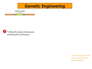
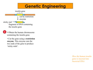
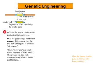
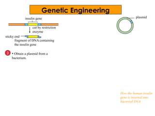
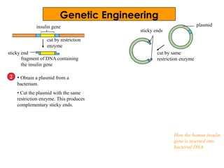
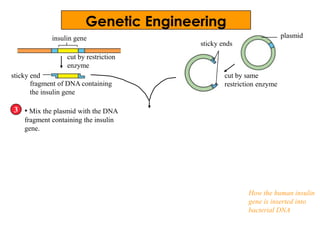
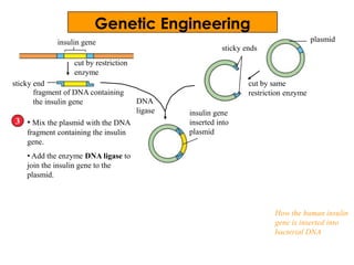
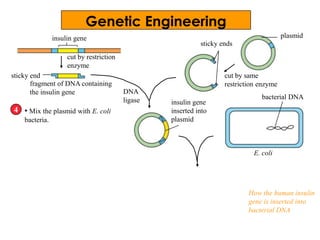
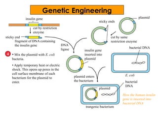
Chapter 20
Molecular
Genetics
Deoxyribonucleic acid (DNA)
• Deoxyribonucleic acid (DNA) is a
molecule that carries genetic
information.
• These genetic information is important
for all cellular functions, such as cell
division and cell differentiation.
• Almost all cells in our body contain
DNA inside their nuclei.
DNA
Each DNA molecule
consists of two parallel
strands twisted around
each other to form a
double helix.
A molecule of DNA
is wrapped around
proteins to form a
single chromatin
thread.
During cell
division, the
chromatin threads
coil more tightly to
form chromosomes
inside the cell
nucleus.
proteins
nuclear pore
nucleus
nuclear envelope
What is DNA made of?
Double helix ‘untwisted’
One strand of nucleotides
Components of a single nucleotide
DNA molecule: a long double helix
basedeoxyribose
sugar
phosphate
group
Sugar phosphate ‘backbone’
Basic units of DNA
adenine cytosine guanine thymine deoxyribose
sugar
phosphate
group
Bases
Nucleotides
base joins with the
phosphate group and
deoxyribose sugar group
Basic units of DNA
• The basic unit of DNA is a
nucleotide.
• Each nucleotide is made
of
- a sugar called
deoxyribose;
- a phosphate group; and
- a nitrogen-containing
base, all joined together
• The four bases of
nitrogen-containing bases
are
- adenine (A)
- thymine (T)
- cytosine (C)
adenine cytosine guanine thymine deoxyribose
sugar
phosphate
group
Bases
Nucleotides
adenine nucleotide thymine nucleotide
guanine nucleotide cytosine nucleotide
base joins with the
phosphate and deoxyribose
sugar group
The building blocks of DNA
• Nucleotides are joined together to form long chains
called polynucleotides.
• Each gene is made up of a sequence of nucleotides. This
sequence can vary.
bases
sugar-phosphate backbone
polynucleotide
Rule of base pairing
• The bases of one strand form bonds with bases of the
other strand according to the rule of base pairing.
• Adenine (A) bonds with thymine (T), while
cytosine (C) always bonds with guanine (G).
• Bases that bond with each other are known as
complementary base pairs.
base pair
Guide to be a God!
1) Colour each of the individual structures on
the worksheet with a different colour:
Example:
adenine = red
thymine = green
guanine = blue
cytosine = yellow
phosphate = brown
deoxyribose = purple
Guide to be a God!
2) Cut out each structure.
3) Using the small symbols (squares, circles and
stars) on the structures as guides, line up the
bases, phosphates and sugars.
4) Glue the appropriate pairs together to form
nucleotides.
Example:
Guide to be a God!
5) Construct the right side of your DNA molecule
by putting together in sequence a
cytosine, thymine, guanine and adenine
nucleotide.
6) Complete the left side of the DNA ladder by
adding complementary nucleotides or
nucleotides that fit. Your finished model should
resemble a ladder.
Guide to be a God!
7) To show replication of your model, separate
the left side from the right side on your desk,
leaving a space of about 15 to 20 cm.
8) Using the remaining nucleotides, add to the
left side of the model to build a new DNA
molecule. Do the same with the separated right
side.
Questions?!?!
1) When constructing the DNA molecule, what
did you notice about the orientation of the
two strands?
2) What DNA strand would bond opposite?
3) What is a similarity and a difference in DNA
between Homo sapien and Blattaria?
The DNA double helix
The DNA molecule has a spiral structure known as the
double helix. Both strands of DNA that run in opposite
directions are twisted to form this double helix.
a base pair
sugar-phosphate backbone
coiling of DNA
double helix
structure of DNA
Genes
• A DNA molecule contains many genes along its
length.
• A gene is a small segment of DNA which controls
the formation of a protein, such as an enzyme.
gene
DNA
molecule
Genes
• Each gene stores a message that determines how a
protein should be made in a cell.
• The message stored by a gene is known as the
genetic code.
• Proteins are responsible for the development of
certain characteristics in the body.
gene
DNA molecule
part of a DNA molecule unzipped to show a gene
M E S S A G E
a gene is a segment of DNA
protein coded
by the gene
Structure of a gene
• Each gene consists of two polynucleotide chains. One of the
chains determines the type of protein made. This chain is
called the template.
• The template is made up of a sequence of nucleotide bases.
• Three sequential bases code for one amino acid. This is
known as the triplet code or codon.
DNA template
Process of decoding and protein synthesis
polypeptide made of five
amino acids
triplet code/ codon
How are proteins made?
• Proteins in the cell are made through a two-step
process — transcription and translation.
• Transcription occurs when the message in the template
has to be copied into an RNA molecule called messenger
RNA (mRNA).
• Transcription occurs in the nucleus.
• Three bases in the mRNA made up a codon.
DNA template
TranscriptionmRNA
- RNA contains U
(uracil) instead of T
(thymine)
codon
How are proteins made?
• The mRNA moves out of the nucleus and carries the
message to the cytoplasm.
• A ribosome helps to translate the sequence of codons on
the mRNA into a protein molecule.
mRNA
polypeptide
Translation
Comparing DNA and RNA
DNA (double helix) RNA
Sugar unit is deoxyribose. Sugar unit is ribose.
Nitrogen-containing bases
are adenine (A), thymine
(T), cytosine (C) and
guanine (G).
Nitrogen-containing bases
are adenine (A), uracil (U),
cytosine (C) and guanine
(G).
Permanent molecule in
the nucleus
Temporary molecule that
is made when needed
Found only in nucleus Found in nucleus and
cytoplasm
1 part of a gene
Transcription and Translation
First, the gene unzips.
1 part of a gene
Transcription and Translation
template
mRNA molecule is
made
One of the strands in the gene is
used as the template to make
mRNA. This is transcription. The
mRNA molecule copies the genetic
code in the DNA template,
following the rule of base pairing.
1
Note that mRNA does not contain
T (thymine). A (adenine) in DNA
pairs with U (uracil) in mRNA.
Transcription and Translation
mRNA molecule is
made
ribosome
mRNA
nuclear
envelope
The mRNA leaves the nucleus
and attaches to a ribosome in
the cytoplasm.
2
nuclear pore
Transcription and Translation
tRNA • In the cytoplasm are amino acids
and transfer RNA (tRNA). Transfer
RNA or tRNA is another RNA
molecule also needed for protein
synthesis.
• tRNA molecules have amino acids
attached to one end of their
structure.Each tRNA is very specific
and attaches only to its own amino
acid For example, a tRNA with the
anticodon UAC always attaches to
the amino acid M.
• Each tRNA also has three bases at
one end. This is an anticodon that
can bind to complementary codons
on mRNA.
cytoplasm
amino
acids
3
anticodon
Transcription and Translation
codon
The anticodons on tRNA
bind with their respective
codons on mRNA.
tRNA
amino acid
attached to
tRNA
peptide bond
ribosome
4 • Translation starts with mRNA attaching
to a ribosome.
• The first two tRNAs together with their
amino acids also fit into the ribosome.
They attach to the codons on the mRNA
according to the rule of base pairing.
• A peptide bond is formed between the
two amino acids.
Transcription and Translation
5
peptide bond between
amino acids
first tRNA
is released
a new tRNA
fits into the
ribosome
• Once the peptide bond is formed
between the first two amino
acids, the ribosome moves along
one codon to the right of the
mRNA.
• As the ribosome moves to this
position, the first tRNA is released.
• At the same time, the third tRNA
and its amino acid slots into the
ribosome.
codon ribosome moves along
the mRNA strand
Transcription and Translation
6
another amino
acid is attached to
the chain
direction of movement
of ribosome
• Another amino acid is attached
to the chain.
Transcription and Translation
polypeptide formed
• The process continues as
the ribosome moves along
the mRNA.
• At the end of the mRNA
is a stop codon such as
UGA, UAA or UAG. A
stop codon does not have
any tRNA with
complementary codons.
This means that
anticodons ACU, AUU or
AUC do not exist.
• Eventually, the whole
chain of polypeptide is
produced. The ribosome
leaves the mRNA.
7
Transcription and Translation
polypeptide formed
• The ribosome may
attach to the same
mRNA for another
round of translation.
8
insulin gene
• Obtain the human chromosome
containing the insulin gene.
1
How the human insulin
gene is inserted into
bacterial DNA
Genetic Engineering
insulin gene
• Obtain the human chromosome
containing the insulin gene.
• Cut the gene using a restriction
enzyme. This enzyme cuts the
two ends of the gene to produce
‘sticky ends’.
1
cut by restriction
enzyme
How the human insulin
gene is inserted into
bacterial DNA
fragment of DNA containing
the insulin gene
sticky end
Genetic Engineering
insulin gene
• Obtain the human chromosome
containing the insulin gene.
• Cut the gene using a restriction
enzyme. This enzyme cuts the
two ends of the gene to produce
‘sticky ends’.
• Each ‘sticky end’ is a single
strand sequence of DNA bases.
These bases can pair with
complementary bases to form a
double strand.
1
cut by restriction
enzyme
fragment of DNA containing
the insulin gene
sticky end
How the human insulin
gene is inserted into
bacterial DNA
Genetic Engineering
insulin gene
• Obtain a plasmid from a
bacterium.
2
cut by restriction
enzyme
fragment of DNA containing
the insulin gene
sticky end
plasmid
How the human insulin
gene is inserted into
bacterial DNA
Genetic Engineering
insulin gene
• Obtain a plasmid from a
bacterium.
• Cut the plasmid with the same
restriction enzyme. This produces
complementary sticky ends.
2
cut by restriction
enzyme
fragment of DNA containing
the insulin gene
sticky end
plasmid
cut by same
restriction enzyme
sticky ends
How the human insulin
gene is inserted into
bacterial DNA
Genetic Engineering
insulin gene
• Mix the plasmid with the DNA
fragment containing the insulin
gene.
3
cut by restriction
enzyme
fragment of DNA containing
the insulin gene
sticky end
plasmid
cut by same
restriction enzyme
sticky ends
How the human insulin
gene is inserted into
bacterial DNA
Genetic Engineering
insulin gene
• Mix the plasmid with the DNA
fragment containing the insulin
gene.
• Add the enzyme DNA ligase to
join the insulin gene to the
plasmid.
3
cut by restriction
enzyme
fragment of DNA containing
the insulin gene
sticky end
plasmid
cut by same
restriction enzyme
sticky ends
insulin gene
inserted into
plasmid
How the human insulin
gene is inserted into
bacterial DNA
DNA
ligase
Genetic Engineering
insulin gene
• Mix the plasmid with E. coli
bacteria.
4
cut by restriction
enzyme
fragment of DNA containing
the insulin gene
sticky end
plasmid
cut by same
restriction enzyme
sticky ends
insulin gene
inserted into
plasmid
E. coli
bacterial DNA
How the human insulin
gene is inserted into
bacterial DNA
DNA
ligase
Genetic Engineering
Genetic Engineering
insulin gene
• Mix the plasmid with E. coli
bacteria.
• Apply temporary heat or electric
shock. This opens up pores in the
cell surface membrane of each
bacterium for the plasmid to
enter.
4
cut by restriction
enzyme
fragment of DNA containing
the insulin gene
sticky end
plasmid
cut by same
restriction enzyme
sticky ends
insulin gene
inserted into
plasmid
plasmid
bacterial
DNA
plasmid enters
the bacterium
trangenic bacterium
E. coli
bacterial DNA
How the human insulin
gene is inserted into
bacterial DNA
DNA
ligase

Molecular genetics 1 win q

  • 1.
  • 2.
    Deoxyribonucleic acid (DNA) •Deoxyribonucleic acid (DNA) is a molecule that carries genetic information. • These genetic information is important for all cellular functions, such as cell division and cell differentiation. • Almost all cells in our body contain DNA inside their nuclei.
  • 3.
    DNA Each DNA molecule consistsof two parallel strands twisted around each other to form a double helix. A molecule of DNA is wrapped around proteins to form a single chromatin thread. During cell division, the chromatin threads coil more tightly to form chromosomes inside the cell nucleus. proteins nuclear pore nucleus nuclear envelope
  • 4.
    What is DNAmade of? Double helix ‘untwisted’ One strand of nucleotides Components of a single nucleotide DNA molecule: a long double helix basedeoxyribose sugar phosphate group Sugar phosphate ‘backbone’
  • 5.
    Basic units ofDNA adenine cytosine guanine thymine deoxyribose sugar phosphate group Bases Nucleotides base joins with the phosphate group and deoxyribose sugar group
  • 6.
    Basic units ofDNA • The basic unit of DNA is a nucleotide. • Each nucleotide is made of - a sugar called deoxyribose; - a phosphate group; and - a nitrogen-containing base, all joined together • The four bases of nitrogen-containing bases are - adenine (A) - thymine (T) - cytosine (C) adenine cytosine guanine thymine deoxyribose sugar phosphate group Bases Nucleotides adenine nucleotide thymine nucleotide guanine nucleotide cytosine nucleotide base joins with the phosphate and deoxyribose sugar group
  • 7.
    The building blocksof DNA • Nucleotides are joined together to form long chains called polynucleotides. • Each gene is made up of a sequence of nucleotides. This sequence can vary. bases sugar-phosphate backbone polynucleotide
  • 8.
    Rule of basepairing • The bases of one strand form bonds with bases of the other strand according to the rule of base pairing. • Adenine (A) bonds with thymine (T), while cytosine (C) always bonds with guanine (G). • Bases that bond with each other are known as complementary base pairs. base pair
  • 10.
    Guide to bea God! 1) Colour each of the individual structures on the worksheet with a different colour: Example: adenine = red thymine = green guanine = blue cytosine = yellow phosphate = brown deoxyribose = purple
  • 11.
    Guide to bea God! 2) Cut out each structure. 3) Using the small symbols (squares, circles and stars) on the structures as guides, line up the bases, phosphates and sugars. 4) Glue the appropriate pairs together to form nucleotides.
  • 12.
  • 13.
    Guide to bea God! 5) Construct the right side of your DNA molecule by putting together in sequence a cytosine, thymine, guanine and adenine nucleotide. 6) Complete the left side of the DNA ladder by adding complementary nucleotides or nucleotides that fit. Your finished model should resemble a ladder.
  • 14.
    Guide to bea God! 7) To show replication of your model, separate the left side from the right side on your desk, leaving a space of about 15 to 20 cm. 8) Using the remaining nucleotides, add to the left side of the model to build a new DNA molecule. Do the same with the separated right side.
  • 15.
    Questions?!?! 1) When constructingthe DNA molecule, what did you notice about the orientation of the two strands? 2) What DNA strand would bond opposite? 3) What is a similarity and a difference in DNA between Homo sapien and Blattaria?
  • 16.
    The DNA doublehelix The DNA molecule has a spiral structure known as the double helix. Both strands of DNA that run in opposite directions are twisted to form this double helix. a base pair sugar-phosphate backbone coiling of DNA double helix structure of DNA
  • 17.
    Genes • A DNAmolecule contains many genes along its length. • A gene is a small segment of DNA which controls the formation of a protein, such as an enzyme. gene DNA molecule
  • 18.
    Genes • Each genestores a message that determines how a protein should be made in a cell. • The message stored by a gene is known as the genetic code. • Proteins are responsible for the development of certain characteristics in the body. gene DNA molecule part of a DNA molecule unzipped to show a gene M E S S A G E a gene is a segment of DNA protein coded by the gene
  • 19.
    Structure of agene • Each gene consists of two polynucleotide chains. One of the chains determines the type of protein made. This chain is called the template. • The template is made up of a sequence of nucleotide bases. • Three sequential bases code for one amino acid. This is known as the triplet code or codon. DNA template Process of decoding and protein synthesis polypeptide made of five amino acids triplet code/ codon
  • 20.
    How are proteinsmade? • Proteins in the cell are made through a two-step process — transcription and translation. • Transcription occurs when the message in the template has to be copied into an RNA molecule called messenger RNA (mRNA). • Transcription occurs in the nucleus. • Three bases in the mRNA made up a codon. DNA template TranscriptionmRNA - RNA contains U (uracil) instead of T (thymine) codon
  • 21.
    How are proteinsmade? • The mRNA moves out of the nucleus and carries the message to the cytoplasm. • A ribosome helps to translate the sequence of codons on the mRNA into a protein molecule. mRNA polypeptide Translation
  • 22.
    Comparing DNA andRNA DNA (double helix) RNA Sugar unit is deoxyribose. Sugar unit is ribose. Nitrogen-containing bases are adenine (A), thymine (T), cytosine (C) and guanine (G). Nitrogen-containing bases are adenine (A), uracil (U), cytosine (C) and guanine (G). Permanent molecule in the nucleus Temporary molecule that is made when needed Found only in nucleus Found in nucleus and cytoplasm
  • 23.
    1 part ofa gene Transcription and Translation
  • 24.
    First, the geneunzips. 1 part of a gene Transcription and Translation
  • 25.
    template mRNA molecule is made Oneof the strands in the gene is used as the template to make mRNA. This is transcription. The mRNA molecule copies the genetic code in the DNA template, following the rule of base pairing. 1 Note that mRNA does not contain T (thymine). A (adenine) in DNA pairs with U (uracil) in mRNA. Transcription and Translation
  • 26.
    mRNA molecule is made ribosome mRNA nuclear envelope ThemRNA leaves the nucleus and attaches to a ribosome in the cytoplasm. 2 nuclear pore Transcription and Translation
  • 27.
    tRNA • Inthe cytoplasm are amino acids and transfer RNA (tRNA). Transfer RNA or tRNA is another RNA molecule also needed for protein synthesis. • tRNA molecules have amino acids attached to one end of their structure.Each tRNA is very specific and attaches only to its own amino acid For example, a tRNA with the anticodon UAC always attaches to the amino acid M. • Each tRNA also has three bases at one end. This is an anticodon that can bind to complementary codons on mRNA. cytoplasm amino acids 3 anticodon Transcription and Translation
  • 28.
    codon The anticodons ontRNA bind with their respective codons on mRNA. tRNA amino acid attached to tRNA peptide bond ribosome 4 • Translation starts with mRNA attaching to a ribosome. • The first two tRNAs together with their amino acids also fit into the ribosome. They attach to the codons on the mRNA according to the rule of base pairing. • A peptide bond is formed between the two amino acids. Transcription and Translation
  • 29.
    5 peptide bond between aminoacids first tRNA is released a new tRNA fits into the ribosome • Once the peptide bond is formed between the first two amino acids, the ribosome moves along one codon to the right of the mRNA. • As the ribosome moves to this position, the first tRNA is released. • At the same time, the third tRNA and its amino acid slots into the ribosome. codon ribosome moves along the mRNA strand Transcription and Translation
  • 30.
    6 another amino acid isattached to the chain direction of movement of ribosome • Another amino acid is attached to the chain. Transcription and Translation
  • 31.
    polypeptide formed • Theprocess continues as the ribosome moves along the mRNA. • At the end of the mRNA is a stop codon such as UGA, UAA or UAG. A stop codon does not have any tRNA with complementary codons. This means that anticodons ACU, AUU or AUC do not exist. • Eventually, the whole chain of polypeptide is produced. The ribosome leaves the mRNA. 7 Transcription and Translation
  • 32.
    Transcription and Translation polypeptideformed • The ribosome may attach to the same mRNA for another round of translation. 8
  • 33.
    insulin gene • Obtainthe human chromosome containing the insulin gene. 1 How the human insulin gene is inserted into bacterial DNA Genetic Engineering
  • 34.
    insulin gene • Obtainthe human chromosome containing the insulin gene. • Cut the gene using a restriction enzyme. This enzyme cuts the two ends of the gene to produce ‘sticky ends’. 1 cut by restriction enzyme How the human insulin gene is inserted into bacterial DNA fragment of DNA containing the insulin gene sticky end Genetic Engineering
  • 35.
    insulin gene • Obtainthe human chromosome containing the insulin gene. • Cut the gene using a restriction enzyme. This enzyme cuts the two ends of the gene to produce ‘sticky ends’. • Each ‘sticky end’ is a single strand sequence of DNA bases. These bases can pair with complementary bases to form a double strand. 1 cut by restriction enzyme fragment of DNA containing the insulin gene sticky end How the human insulin gene is inserted into bacterial DNA Genetic Engineering
  • 36.
    insulin gene • Obtaina plasmid from a bacterium. 2 cut by restriction enzyme fragment of DNA containing the insulin gene sticky end plasmid How the human insulin gene is inserted into bacterial DNA Genetic Engineering
  • 37.
    insulin gene • Obtaina plasmid from a bacterium. • Cut the plasmid with the same restriction enzyme. This produces complementary sticky ends. 2 cut by restriction enzyme fragment of DNA containing the insulin gene sticky end plasmid cut by same restriction enzyme sticky ends How the human insulin gene is inserted into bacterial DNA Genetic Engineering
  • 38.
    insulin gene • Mixthe plasmid with the DNA fragment containing the insulin gene. 3 cut by restriction enzyme fragment of DNA containing the insulin gene sticky end plasmid cut by same restriction enzyme sticky ends How the human insulin gene is inserted into bacterial DNA Genetic Engineering
  • 39.
    insulin gene • Mixthe plasmid with the DNA fragment containing the insulin gene. • Add the enzyme DNA ligase to join the insulin gene to the plasmid. 3 cut by restriction enzyme fragment of DNA containing the insulin gene sticky end plasmid cut by same restriction enzyme sticky ends insulin gene inserted into plasmid How the human insulin gene is inserted into bacterial DNA DNA ligase Genetic Engineering
  • 40.
    insulin gene • Mixthe plasmid with E. coli bacteria. 4 cut by restriction enzyme fragment of DNA containing the insulin gene sticky end plasmid cut by same restriction enzyme sticky ends insulin gene inserted into plasmid E. coli bacterial DNA How the human insulin gene is inserted into bacterial DNA DNA ligase Genetic Engineering
  • 41.
    Genetic Engineering insulin gene •Mix the plasmid with E. coli bacteria. • Apply temporary heat or electric shock. This opens up pores in the cell surface membrane of each bacterium for the plasmid to enter. 4 cut by restriction enzyme fragment of DNA containing the insulin gene sticky end plasmid cut by same restriction enzyme sticky ends insulin gene inserted into plasmid plasmid bacterial DNA plasmid enters the bacterium trangenic bacterium E. coli bacterial DNA How the human insulin gene is inserted into bacterial DNA DNA ligase