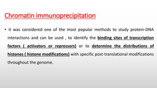
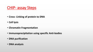
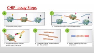
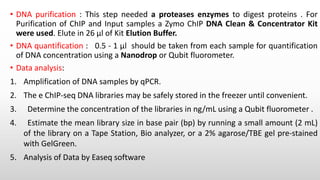
This document summarizes the steps of a ChIP-Seq (Chromatin Immunoprecipitation Sequencing) assay. The key steps are: 1) cross-linking proteins to DNA, 2) fragmenting chromatin, 3) immunoprecipitating the DNA-protein complex using antibodies specific to the protein of interest, 4) purifying and analyzing the DNA. ChIP-Seq allows researchers to identify the genomic binding sites of transcription factors and histone modifications genome-wide.
STS stands for sequence tagged site which is short DNA sequence, generally between 100 and 500 bp in length, that is easily recognizable and occurs only once in the chromosome or genome being studied.
BAC & YAC are artificially prepared chromosomes to clone DNA sequences.yeast artificial chromosome is capable of carrying upto 1000 kbp of inserted DNA sequence
Sanger sequencing is one of the DNA sequencing methods used to identify and determine the sequence (Nucleotide) of DNA .This is an enzymatic method of sequencing developed by Fred Sanger.
A physical map of a chromosome or a genome that shows the physical locations of genes and other DNA sequences of interest. Physical maps are used to help scientists identify and isolate genes by positional cloning.
According to the ICSM (Intergovernmental Committee on Surveying and Mapping), there are five different types of maps: General Reference, Topographical, Thematic, Navigation Charts and Cadastral Maps and Plans.
Introduction to RDT methods in genetic engineeringCollege
This presentation gives a small review about the RDT based methods used generally in genetic engineering. This presentation include various images about the technique, which will help the user in understanding the concepts easily.
In this slides the topic that which is discussed is "How PCR is involved in identification of Genotype"
I hope this will Help you in your presentation work.
"PCR can be used in identification of genotype."
In Situ Polymerase Chain Reaction (In situ PCR) is a powerful method that detects minute quantities of rare or single-copy number nucleic acid sequences in frozen or paraffin-embedded cells or tissue sections for the localization of those sequences within the cells. The principle of this method involves tissue fixing (to preserve the cell morphology) and subsequent treatment with proteolytic digestion (to provide access for the PCR reagents to the target DNA). The target sequences are amplified by those reagents and then detected by standard immunocytochemical protocols. In situ PCR combines the sensitivity of PCR or RT-PCR amplification along with the ability to perform morphological analysis on the same sample, and thus it is an attractive tool in diagnostic applications. One of the most prominent applications is the detection of infectious disease agents including HIV-1, HBV, HPV, HHV-6, CMV, and EBV.
Liquid biopsy: Overcome Challenges of Circulating DNA with Automated and Stan...QIAGEN
Circulating cell-free DNA (ccfDNA) originating from malignant tumors, a developing fetus and also from inflammatory tissues, is present in the cell-free nucleic acids in plasma, serum and other body fluids and is considered a “liquid biopsy”. Access to ccfDNA for analysis allows for specific detection of certain disease states based on a simple blood sample. Circulating cell-free DNA shows distinctive properties – it is present mostly as shorter fragments of less than 500 bp and the concentration of ccfDNA in a plasma or serum sample is low (approximately 1–100 ng/ml) compared to cellular materials and varies considerably between different individuals.
Because of their fragmented nature and low concentration, ccfDNA presents a particular challenge for efficient extraction / purification and quantification, such as by qPCR. We present data on solutions for the following critical problems concerning the purification of ccfDNA for research and molecular diagnostic applications:
• Pre-analytical workflow (blood processing) for analyzing ccfDNA
• Optimization of ccfDNA extraction from plasma samples: low target concentrations require efficient ccfDNA enrichment from larger sample volumes
• Novel automated extraction of ccfDNA using the QIAsymphony SP instrument for liquid biopsy diagnostic applications.
List of viruses causing Cancer:
Human papillomavirus (HPV)
Hepatitis C Virus
Hepatitis B Virus
Human Immunodeficiency Virus (HIV)
Human Herpes Virus 8 (HHV - 8)
Human T lymphocytic Virus (HTLV-1)
Epstein-Barr virus (EBV)
Merkel Cell Polyomavirus
Observation of Io’s Resurfacing via Plume Deposition Using Ground-based Adapt...Sérgio Sacani
Since volcanic activity was first discovered on Io from Voyager images in 1979, changes
on Io’s surface have been monitored from both spacecraft and ground-based telescopes.
Here, we present the highest spatial resolution images of Io ever obtained from a groundbased telescope. These images, acquired by the SHARK-VIS instrument on the Large
Binocular Telescope, show evidence of a major resurfacing event on Io’s trailing hemisphere. When compared to the most recent spacecraft images, the SHARK-VIS images
show that a plume deposit from a powerful eruption at Pillan Patera has covered part
of the long-lived Pele plume deposit. Although this type of resurfacing event may be common on Io, few have been detected due to the rarity of spacecraft visits and the previously low spatial resolution available from Earth-based telescopes. The SHARK-VIS instrument ushers in a new era of high resolution imaging of Io’s surface using adaptive
optics at visible wavelengths.
Slide 1: Title Slide
Extrachromosomal Inheritance
Slide 2: Introduction to Extrachromosomal Inheritance
Definition: Extrachromosomal inheritance refers to the transmission of genetic material that is not found within the nucleus.
Key Components: Involves genes located in mitochondria, chloroplasts, and plasmids.
Slide 3: Mitochondrial Inheritance
Mitochondria: Organelles responsible for energy production.
Mitochondrial DNA (mtDNA): Circular DNA molecule found in mitochondria.
Inheritance Pattern: Maternally inherited, meaning it is passed from mothers to all their offspring.
Diseases: Examples include Leber’s hereditary optic neuropathy (LHON) and mitochondrial myopathy.
Slide 4: Chloroplast Inheritance
Chloroplasts: Organelles responsible for photosynthesis in plants.
Chloroplast DNA (cpDNA): Circular DNA molecule found in chloroplasts.
Inheritance Pattern: Often maternally inherited in most plants, but can vary in some species.
Examples: Variegation in plants, where leaf color patterns are determined by chloroplast DNA.
Slide 5: Plasmid Inheritance
Plasmids: Small, circular DNA molecules found in bacteria and some eukaryotes.
Features: Can carry antibiotic resistance genes and can be transferred between cells through processes like conjugation.
Significance: Important in biotechnology for gene cloning and genetic engineering.
Slide 6: Mechanisms of Extrachromosomal Inheritance
Non-Mendelian Patterns: Do not follow Mendel’s laws of inheritance.
Cytoplasmic Segregation: During cell division, organelles like mitochondria and chloroplasts are randomly distributed to daughter cells.
Heteroplasmy: Presence of more than one type of organellar genome within a cell, leading to variation in expression.
Slide 7: Examples of Extrachromosomal Inheritance
Four O’clock Plant (Mirabilis jalapa): Shows variegated leaves due to different cpDNA in leaf cells.
Petite Mutants in Yeast: Result from mutations in mitochondrial DNA affecting respiration.
Slide 8: Importance of Extrachromosomal Inheritance
Evolution: Provides insight into the evolution of eukaryotic cells.
Medicine: Understanding mitochondrial inheritance helps in diagnosing and treating mitochondrial diseases.
Agriculture: Chloroplast inheritance can be used in plant breeding and genetic modification.
Slide 9: Recent Research and Advances
Gene Editing: Techniques like CRISPR-Cas9 are being used to edit mitochondrial and chloroplast DNA.
Therapies: Development of mitochondrial replacement therapy (MRT) for preventing mitochondrial diseases.
Slide 10: Conclusion
Summary: Extrachromosomal inheritance involves the transmission of genetic material outside the nucleus and plays a crucial role in genetics, medicine, and biotechnology.
Future Directions: Continued research and technological advancements hold promise for new treatments and applications.
Slide 11: Questions and Discussion
Invite Audience: Open the floor for any questions or further discussion on the topic.
Introduction:
RNA interference (RNAi) or Post-Transcriptional Gene Silencing (PTGS) is an important biological process for modulating eukaryotic gene expression.
It is highly conserved process of posttranscriptional gene silencing by which double stranded RNA (dsRNA) causes sequence-specific degradation of mRNA sequences.
dsRNA-induced gene silencing (RNAi) is reported in a wide range of eukaryotes ranging from worms, insects, mammals and plants.
This process mediates resistance to both endogenous parasitic and exogenous pathogenic nucleic acids, and regulates the expression of protein-coding genes.
What are small ncRNAs?
micro RNA (miRNA)
short interfering RNA (siRNA)
Properties of small non-coding RNA:
Involved in silencing mRNA transcripts.
Called “small” because they are usually only about 21-24 nucleotides long.
Synthesized by first cutting up longer precursor sequences (like the 61nt one that Lee discovered).
Silence an mRNA by base pairing with some sequence on the mRNA.
Discovery of siRNA?
The first small RNA:
In 1993 Rosalind Lee (Victor Ambros lab) was studying a non- coding gene in C. elegans, lin-4, that was involved in silencing of another gene, lin-14, at the appropriate time in the
development of the worm C. elegans.
Two small transcripts of lin-4 (22nt and 61nt) were found to be complementary to a sequence in the 3' UTR of lin-14.
Because lin-4 encoded no protein, she deduced that it must be these transcripts that are causing the silencing by RNA-RNA interactions.
Types of RNAi ( non coding RNA)
MiRNA
Length (23-25 nt)
Trans acting
Binds with target MRNA in mismatch
Translation inhibition
Si RNA
Length 21 nt.
Cis acting
Bind with target Mrna in perfect complementary sequence
Piwi-RNA
Length ; 25 to 36 nt.
Expressed in Germ Cells
Regulates trnasposomes activity
MECHANISM OF RNAI:
First the double-stranded RNA teams up with a protein complex named Dicer, which cuts the long RNA into short pieces.
Then another protein complex called RISC (RNA-induced silencing complex) discards one of the two RNA strands.
The RISC-docked, single-stranded RNA then pairs with the homologous mRNA and destroys it.
THE RISC COMPLEX:
RISC is large(>500kD) RNA multi- protein Binding complex which triggers MRNA degradation in response to MRNA
Unwinding of double stranded Si RNA by ATP independent Helicase
Active component of RISC is Ago proteins( ENDONUCLEASE) which cleave target MRNA.
DICER: endonuclease (RNase Family III)
Argonaute: Central Component of the RNA-Induced Silencing Complex (RISC)
One strand of the dsRNA produced by Dicer is retained in the RISC complex in association with Argonaute
ARGONAUTE PROTEIN :
1.PAZ(PIWI/Argonaute/ Zwille)- Recognition of target MRNA
2.PIWI (p-element induced wimpy Testis)- breaks Phosphodiester bond of mRNA.)RNAse H activity.
MiRNA:
The Double-stranded RNAs are naturally produced in eukaryotic cells during development, and they have a key role in regulating gene expression .
Richard's entangled aventures in wonderlandRichard Gill
Since the loophole-free Bell experiments of 2020 and the Nobel prizes in physics of 2022, critics of Bell's work have retreated to the fortress of super-determinism. Now, super-determinism is a derogatory word - it just means "determinism". Palmer, Hance and Hossenfelder argue that quantum mechanics and determinism are not incompatible, using a sophisticated mathematical construction based on a subtle thinning of allowed states and measurements in quantum mechanics, such that what is left appears to make Bell's argument fail, without altering the empirical predictions of quantum mechanics. I think however that it is a smoke screen, and the slogan "lost in math" comes to my mind. I will discuss some other recent disproofs of Bell's theorem using the language of causality based on causal graphs. Causal thinking is also central to law and justice. I will mention surprising connections to my work on serial killer nurse cases, in particular the Dutch case of Lucia de Berk and the current UK case of Lucy Letby.
Earliest Galaxies in the JADES Origins Field: Luminosity Function and Cosmic ...Sérgio Sacani
We characterize the earliest galaxy population in the JADES Origins Field (JOF), the deepest
imaging field observed with JWST. We make use of the ancillary Hubble optical images (5 filters
spanning 0.4−0.9µm) and novel JWST images with 14 filters spanning 0.8−5µm, including 7 mediumband filters, and reaching total exposure times of up to 46 hours per filter. We combine all our data
at > 2.3µm to construct an ultradeep image, reaching as deep as ≈ 31.4 AB mag in the stack and
30.3-31.0 AB mag (5σ, r = 0.1” circular aperture) in individual filters. We measure photometric
redshifts and use robust selection criteria to identify a sample of eight galaxy candidates at redshifts
z = 11.5 − 15. These objects show compact half-light radii of R1/2 ∼ 50 − 200pc, stellar masses of
M⋆ ∼ 107−108M⊙, and star-formation rates of SFR ∼ 0.1−1 M⊙ yr−1
. Our search finds no candidates
at 15 < z < 20, placing upper limits at these redshifts. We develop a forward modeling approach to
infer the properties of the evolving luminosity function without binning in redshift or luminosity that
marginalizes over the photometric redshift uncertainty of our candidate galaxies and incorporates the
impact of non-detections. We find a z = 12 luminosity function in good agreement with prior results,
and that the luminosity function normalization and UV luminosity density decline by a factor of ∼ 2.5
from z = 12 to z = 14. We discuss the possible implications of our results in the context of theoretical
models for evolution of the dark matter halo mass function.
2. Chromatin immunoprecipitation
• it was considered one of the most popular methods to study protein-DNA
interactions and can be used , to identify the binding sites of transcription
factors ( activators or repressors) or to determine the distributions of
histones ( histone modifications) with specific post-translational modifications
throughout the genome.
3. CHIP- assay Steps
• Cross- Linking of protein to DNA
• Cell lysis
• Chromatin Fragmentation
• Immunoprecipitation using specific Anti-bodies
• DNA purification
• DNA analysis
5. Cross-linking of protein to DNA : this step was performed using cross- linking
agent (e.g. 16% formaldehyde (W:V) (methanol free), glutaraldehyde,
disuccinimidyl glutarate [DSG](preferred) .
Chromatin fragmentation : The chromatin is fragmented or “sheared” to
mononucleosome sized fragments (150-300 bp), which is important for obtaining
high resolution sequencing data. Chromatin shearing is accomplished by
sonication or enzymatic digestion using micrococcal nuclease (MNase) and
monitored by gel (e.g. agarose) or capillary electrophoresis .
Immunoprecipitation using specific Anti-bodies : Selected Abs should be highly –
specific Abs and show highly effective binding with chromatin associated
proteins . The amount of primary antibody required for good signal in a ChIP is
usually 2–5 μg . The antibody is coupled to magnetic beads , and incubate at 4°C
for at least 12 h, with rotation such that the samples mix and the beads remain
suspended . the antibody-bound chromatin is isolated from bulk chromatin using
a magnet, followed by a series of stringent washes.
7. • DNA purification : This step needed a proteases enzymes to digest proteins . For
Purification of ChIP and Input samples a Zymo ChIP DNA Clean & Concentrator Kit
were used. Elute in 26 μl of Kit Elution Buffer.
• DNA quantification : 0.5 - 1 μl should be taken from each sample for quantification
of DNA concentration using a Nanodrop or Qubit fluorometer.
• Data analysis:
1. Amplification of DNA samples by qPCR.
2. The e ChIP-seq DNA libraries may be safely stored in the freezer until convenient.
3. Determine the concentration of the libraries in ng/mL using a Qubit fluorometer .
4. Estimate the mean library size in base pair (bp) by running a small amount (2 mL)
of the library on a Tape Station, Bio analyzer, or a 2% agarose/TBE gel pre-stained
with GelGreen.
5. Analysis of Data by Easeq software