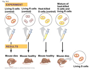
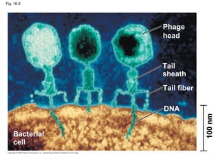
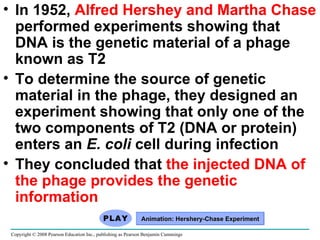
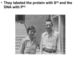
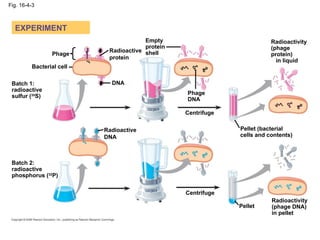
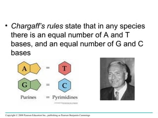
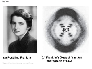
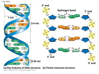
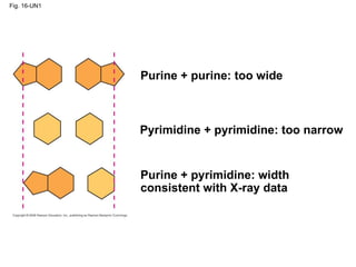
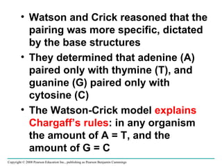
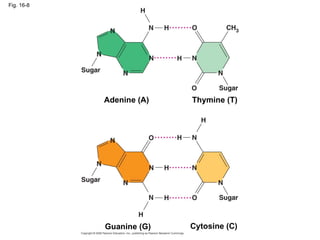
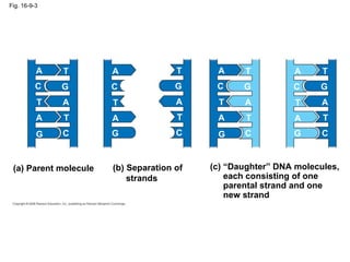
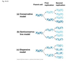
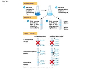
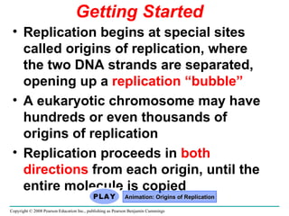
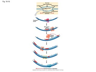
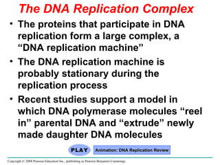
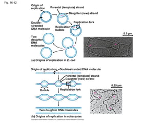
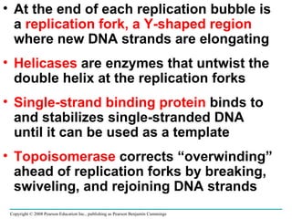
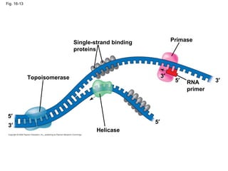
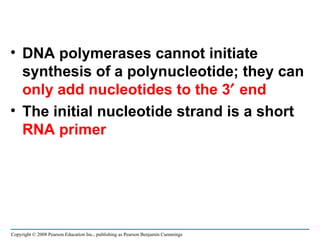
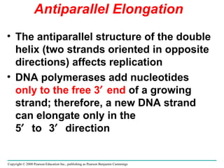
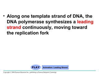
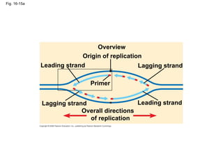
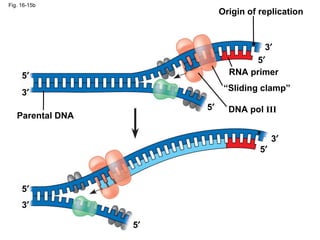
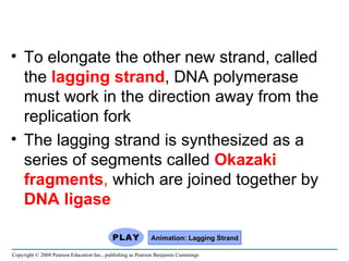
Chapter 16 The Molecular Basis of Inheritance
Concept 16.1: DNA is the genetic material Early in the 20th century, the identification of the molecules of inheritance loomed as a major challenge to biologists Copyright © 2008 Pearson Education Inc., publishing as Pearson Benjamin Cummings
When T. H. Morgan’s group showed that genes are located on chromosomes, the two components of chromosomes —DNA and   protein —became candidates for the genetic material. Was it the DNA or the protein that was the genetic material? The role of DNA in heredity was first discovered by studying bacteria and the viruses that infect them Copyright © 2008 Pearson Education Inc., publishing as Pearson Benjamin Cummings
Evidence That DNA Can Transform Bacteria The discovery of the genetic role of DNA began with research by  Frederick Griffith  in 1928 Griffith worked with two strains of a bacterium, one pathogenic and one harmless Copyright © 2008 Pearson Education Inc., publishing as Pearson Benjamin Cummings
Fig. 16-2 Living S cells (control) Living R cells (control) Heat-killed  S cells (control) Mixture of heat-killed  S cells and living R cells Mouse dies Mouse dies Mouse healthy Mouse healthy Living S cells RESULTS EXPERIMENT
What happened? When he mixed heat-killed remains of the pathogenic S strain with living cells of the harmless R strain, some living cells became pathogenic He called this phenomenon  transformation , now defined as a change in genotype and phenotype due  to assimilation of foreign DNA Copyright © 2008 Pearson Education Inc., publishing as Pearson Benjamin Cummings
In 1944, Oswald Avery, Maclyn McCarty, and Colin MacLeod announced that the  transforming substance was DNA Their conclusion was based on experimental evidence that  only DNA  worked in transforming harmless bacteria into pathogenic bacteria Many biologists remained skeptical, mainly because little was known about DNA Copyright © 2008 Pearson Education Inc., publishing as Pearson Benjamin Cummings
Evidence That Viral DNA Can Program Cells More evidence for DNA as the genetic material came from studies of viruses that infect bacteria Such viruses, called  bacteriophages  (or phages), are widely used in molecular genetics research Copyright © 2008 Pearson Education Inc., publishing as Pearson Benjamin Cummings Animation: Phage T2 Reproductive Cycle
Fig. 16-3 Bacterial  cell Phage head Tail sheath Tail fiber DNA 100 nm
In 1952,  Alfred Hershey and Martha Chase  performed experiments showing that DNA is the genetic material of a phage known as T2 To determine the source of genetic material in the phage, they designed an experiment showing that only one of the two components of T2 (DNA or protein) enters an  E. coli  cell during infection They concluded that  the injected DNA of the phage provides the genetic information  Copyright © 2008 Pearson Education Inc., publishing as Pearson Benjamin Cummings Animation: Hershery-Chase Experiment
They labeled the protein with S 35  and the DNA with P 32.
Fig. 16-4-3 EXPERIMENT Phage DNA Bacterial cell Radioactive protein Radioactive DNA Batch 1: radioactive sulfur ( 35 S) Batch 2: radioactive phosphorus ( 32 P) Empty protein shell Phage DNA Centrifuge Centrifuge Pellet Pellet (bacterial  cells and contents) Radioactivity (phage  protein)  in liquid Radioactivity (phage DNA)  in pellet
Additional Evidence That DNA Is the Genetic Material In 1950, Erwin Chargaff reported that DNA composition varies from one species to the next This evidence of diversity made DNA a more credible candidate for the genetic material Copyright © 2008 Pearson Education Inc., publishing as Pearson Benjamin Cummings Animation: DNA and RNA Structure
Chargaff’s rules  state that in any species there is an equal number of A and T bases, and an equal number of G and C bases Copyright © 2008 Pearson Education Inc., publishing as Pearson Benjamin Cummings
Building a Structural Model of DNA After most biologists became convinced that DNA was the genetic material, the challenge was to determine how its structure accounts for its role Maurice Wilkins and Rosalind Franklin  were using a technique called X-ray crystallography to study molecular structure Franklin produced a picture of the DNA molecule using this technique Copyright © 2008 Pearson Education Inc., publishing as Pearson Benjamin Cummings
Fig. 16-6 (a) Rosalind Franklin (b) Franklin’s X-ray diffraction    photograph of DNA
Franklin’s X-ray crystallographic images of DNA enabled Watson to deduce that DNA was helical  The X-ray images also enabled Watson to deduce the width of the helix and the spacing of the nitrogenous bases The width suggested that the DNA molecule was made up of two strands, forming a  double helix Copyright © 2008 Pearson Education Inc., publishing as Pearson Benjamin Cummings Animation: DNA Double Helix
Fig. 16-7b (c) Space-filling model
Fig. 16-7a Hydrogen bond 3   end 5   end 3.4 nm 0.34 nm 3   end 5   end (b) Partial chemical structure (a) Key features of DNA structure 1 nm
Watson and Crick built models of a double helix to conform to the X-rays and chemistry of DNA Franklin had concluded that there were two antiparallel sugar-phosphate backbones, with the nitrogenous bases paired in the molecule’s interior  At first, W and C thought that like bases paired. Copyright © 2008 Pearson Education Inc., publishing as Pearson Benjamin Cummings
Fig. 16-UN1 Purine + purine: too wide Pyrimidine + pyrimidine: too narrow Purine + pyrimidine: width consistent with X-ray data
 
Watson and Crick reasoned that the pairing was more specific, dictated by the base structures They determined that adenine (A) paired only with thymine (T), and guanine (G) paired only with cytosine (C) The Watson-Crick model  explains Chargaff’s rules : in any organism the amount of A = T, and the amount of G = C Copyright © 2008 Pearson Education Inc., publishing as Pearson Benjamin Cummings
 
Fig. 16-8 Cytosine (C) Adenine (A) Thymine (T) Guanine (G)
 
The relationship  between structure and   function  is manifest in the double helix Watson and Crick noted that the specific base pairing suggested a possible copying mechanism for genetic material Copyright © 2008 Pearson Education Inc., publishing as Pearson Benjamin Cummings
 
Fig. 16-9-3 A T G C T A T A G C (a) Parent molecule A T G C T A T A G C (c) “Daughter” DNA molecules, each consisting of one parental strand and one  new strand (b) Separation of strands A T G C T A T A G C A T G C T A T A G C
 
Watson and Crick’s  semiconservative model  of replication predicts that when a double helix replicates, each daughter molecule will have one old strand (derived or “conserved” from the parent molecule) and one newly made strand Competing models were the conservative model (the two parent strands rejoin) and the dispersive model (each strand is a mix of old and new) Copyright © 2008 Pearson Education Inc., publishing as Pearson Benjamin Cummings
 
Fig. 16-10 Parent cell First replication Second replication (a) Conservative model (b) Semiconserva- tive model (c) Dispersive model
 
Experiments by  Matthew Meselson and   Franklin Stahl  supported the semiconservative model  They labeled the nucleotides of the old strands with a heavy isotope of  nitrogen , while any new nucleotides were labeled with a lighter isotope Copyright © 2008 Pearson Education Inc., publishing as Pearson Benjamin Cummings
 
Fig. 16-11 EXPERIMENT RESULTS CONCLUSION 1 2 4 3 Conservative model Semiconservative model Dispersive  model Bacteria cultured in medium containing  15 N Bacteria transferred to  medium containing  14 N DNA sample centrifuged after 20 min (after first application) DNA sample centrifuged after 40 min (after second replication) More dense Less dense Second replication First replication
DNA Replication:  A Closer Look The copying of DNA is remarkable in its speed and accuracy More than a dozen  enzymes  and other proteins participate in DNA replication Copyright © 2008 Pearson Education Inc., publishing as Pearson Benjamin Cummings
Getting Started Replication begins at special sites called origins of replication, where the two DNA strands are separated, opening up a  replication “bubble” A eukaryotic chromosome may have hundreds or even thousands of origins of replication Replication proceeds in  both directions  from each origin, until the entire molecule is copied Copyright © 2008 Pearson Education Inc., publishing as Pearson Benjamin Cummings Animation: Origins of Replication
 
Fig. 16-16 Overview Origin of replication Leading strand Leading strand Lagging strand Lagging strand Overall directions of replication Template strand RNA primer Okazaki fragment Overall direction of replication 1 2 3  2 1 1 1 1 2 2 5  1 3  3  3  3  3  3  3  3  3  5  5  5  5  5  5  5  5  5  5  5  3  3 
 
The DNA Replication Complex The proteins that participate in DNA replication form a large complex, a “DNA replication machine” The DNA replication machine is probably stationary during the replication process Recent studies support a model in which DNA polymerase molecules “reel in” parental DNA and “extrude” newly made daughter DNA molecules Copyright © 2008 Pearson Education Inc., publishing as Pearson Benjamin Cummings Animation: DNA Replication Review
Fig. 16-12 Origin of replication Parental (template) strand Daughter (new) strand Replication fork Replication bubble Two daughter DNA molecules (a) Origins of replication in  E. coli Origin of replication Double-stranded DNA molecule Parental (template) strand Daughter (new) strand Bubble Replication fork Two daughter DNA molecules (b) Origins of replication in eukaryotes 0.5 µm 0.25 µm Double- stranded DNA molecule
At the end of each replication bubble is a  replication fork, a Y-shaped region  where new DNA strands are elongating Helicases  are enzymes that untwist the double helix at the replication forks Single-strand binding protein  binds to and stabilizes single-stranded DNA until it can be used as a template Topoisomerase  corrects “overwinding” ahead of replication forks by breaking, swiveling, and rejoining DNA strands Copyright © 2008 Pearson Education Inc., publishing as Pearson Benjamin Cummings
Fig. 16-13 Topoisomerase Helicase Primase Single-strand binding proteins RNA primer 5  5  5  3  3  3 
DNA polymerases cannot initiate synthesis of a polynucleotide; they can  only add nucleotides to the 3   end The initial nucleotide strand is a short  RNA primer Copyright © 2008 Pearson Education Inc., publishing as Pearson Benjamin Cummings
An enzyme called  primase  can start an RNA chain from scratch and adds RNA nucleotides one at a time using the parental DNA as a template The primer is short (5–10 nucleotides long), and the 3   end serves as the starting point for the new DNA strand Copyright © 2008 Pearson Education Inc., publishing as Pearson Benjamin Cummings
Synthesizing a New DNA Strand Enzymes called  DNA polymerases  catalyze the elongation of new DNA at a replication fork Most DNA polymerases require a primer and a DNA template strand The rate of elongation is about 500 nucleotides per second in bacteria and 50 per second in human cells Copyright © 2008 Pearson Education Inc., publishing as Pearson Benjamin Cummings
Each nucleotide that is added to a growing DNA strand is a  nucleoside triphosphate dATP supplies adenine to DNA and is similar to the ATP of energy metabolism The difference is in their sugars: dATP has deoxyribose while ATP has ribose As each monomer of dATP joins the DNA strand, it loses two phosphate groups as a molecule of pyrophosphate Copyright © 2008 Pearson Education Inc., publishing as Pearson Benjamin Cummings
Fig. 16-14 A C T G G G G C C C C C A A A T T T New strand 5   end Template strand  3   end 5   end 3   end 3   end 5   end 5   end 3   end Base Sugar Phosphate Nucleoside triphosphate Pyrophosphate DNA polymerase
Antiparallel Elongation The antiparallel structure of the double helix (two strands oriented in opposite directions) affects replication DNA polymerases add nucleotides  only to the free 3  end  of a growing strand; therefore, a new DNA strand can elongate only in the 5  to  3  direction Copyright © 2008 Pearson Education Inc., publishing as Pearson Benjamin Cummings
Along one template strand of DNA, the DNA polymerase synthesizes a  leading strand  continuously, moving toward the replication fork Copyright © 2008 Pearson Education Inc., publishing as Pearson Benjamin Cummings Animation: Leading Strand
Fig. 16-15a Overview Leading strand Leading strand Lagging strand Lagging strand Origin of replication Primer Overall directions  of replication
Fig. 16-15b Origin of replication RNA primer “ Sliding clamp” DNA pol  III Parental DNA 3  5  5  5  5  5  5  3  3  3 
To elongate the other new strand, called the  lagging strand , DNA polymerase must work in the direction away from the replication fork The lagging strand is synthesized as a series of segments called  Okazaki fragments ,  which are joined together by  DNA ligase Copyright © 2008 Pearson Education Inc., publishing as Pearson Benjamin Cummings Animation: Lagging Strand

Ap Chapter 16

  • 1.
    Chapter 16 TheMolecular Basis of Inheritance
  • 2.
    Concept 16.1: DNAis the genetic material Early in the 20th century, the identification of the molecules of inheritance loomed as a major challenge to biologists Copyright © 2008 Pearson Education Inc., publishing as Pearson Benjamin Cummings
  • 3.
    When T. H.Morgan’s group showed that genes are located on chromosomes, the two components of chromosomes —DNA and protein —became candidates for the genetic material. Was it the DNA or the protein that was the genetic material? The role of DNA in heredity was first discovered by studying bacteria and the viruses that infect them Copyright © 2008 Pearson Education Inc., publishing as Pearson Benjamin Cummings
  • 4.
    Evidence That DNACan Transform Bacteria The discovery of the genetic role of DNA began with research by Frederick Griffith in 1928 Griffith worked with two strains of a bacterium, one pathogenic and one harmless Copyright © 2008 Pearson Education Inc., publishing as Pearson Benjamin Cummings
  • 5.
    Fig. 16-2 LivingS cells (control) Living R cells (control) Heat-killed S cells (control) Mixture of heat-killed S cells and living R cells Mouse dies Mouse dies Mouse healthy Mouse healthy Living S cells RESULTS EXPERIMENT
  • 6.
    What happened? Whenhe mixed heat-killed remains of the pathogenic S strain with living cells of the harmless R strain, some living cells became pathogenic He called this phenomenon transformation , now defined as a change in genotype and phenotype due to assimilation of foreign DNA Copyright © 2008 Pearson Education Inc., publishing as Pearson Benjamin Cummings
  • 7.
    In 1944, OswaldAvery, Maclyn McCarty, and Colin MacLeod announced that the transforming substance was DNA Their conclusion was based on experimental evidence that only DNA worked in transforming harmless bacteria into pathogenic bacteria Many biologists remained skeptical, mainly because little was known about DNA Copyright © 2008 Pearson Education Inc., publishing as Pearson Benjamin Cummings
  • 8.
    Evidence That ViralDNA Can Program Cells More evidence for DNA as the genetic material came from studies of viruses that infect bacteria Such viruses, called bacteriophages (or phages), are widely used in molecular genetics research Copyright © 2008 Pearson Education Inc., publishing as Pearson Benjamin Cummings Animation: Phage T2 Reproductive Cycle
  • 9.
    Fig. 16-3 Bacterial cell Phage head Tail sheath Tail fiber DNA 100 nm
  • 10.
    In 1952, Alfred Hershey and Martha Chase performed experiments showing that DNA is the genetic material of a phage known as T2 To determine the source of genetic material in the phage, they designed an experiment showing that only one of the two components of T2 (DNA or protein) enters an E. coli cell during infection They concluded that the injected DNA of the phage provides the genetic information Copyright © 2008 Pearson Education Inc., publishing as Pearson Benjamin Cummings Animation: Hershery-Chase Experiment
  • 11.
    They labeled theprotein with S 35 and the DNA with P 32.
  • 12.
    Fig. 16-4-3 EXPERIMENTPhage DNA Bacterial cell Radioactive protein Radioactive DNA Batch 1: radioactive sulfur ( 35 S) Batch 2: radioactive phosphorus ( 32 P) Empty protein shell Phage DNA Centrifuge Centrifuge Pellet Pellet (bacterial cells and contents) Radioactivity (phage protein) in liquid Radioactivity (phage DNA) in pellet
  • 13.
    Additional Evidence ThatDNA Is the Genetic Material In 1950, Erwin Chargaff reported that DNA composition varies from one species to the next This evidence of diversity made DNA a more credible candidate for the genetic material Copyright © 2008 Pearson Education Inc., publishing as Pearson Benjamin Cummings Animation: DNA and RNA Structure
  • 14.
    Chargaff’s rules state that in any species there is an equal number of A and T bases, and an equal number of G and C bases Copyright © 2008 Pearson Education Inc., publishing as Pearson Benjamin Cummings
  • 15.
    Building a StructuralModel of DNA After most biologists became convinced that DNA was the genetic material, the challenge was to determine how its structure accounts for its role Maurice Wilkins and Rosalind Franklin were using a technique called X-ray crystallography to study molecular structure Franklin produced a picture of the DNA molecule using this technique Copyright © 2008 Pearson Education Inc., publishing as Pearson Benjamin Cummings
  • 16.
    Fig. 16-6 (a)Rosalind Franklin (b) Franklin’s X-ray diffraction photograph of DNA
  • 17.
    Franklin’s X-ray crystallographicimages of DNA enabled Watson to deduce that DNA was helical The X-ray images also enabled Watson to deduce the width of the helix and the spacing of the nitrogenous bases The width suggested that the DNA molecule was made up of two strands, forming a double helix Copyright © 2008 Pearson Education Inc., publishing as Pearson Benjamin Cummings Animation: DNA Double Helix
  • 18.
    Fig. 16-7b (c)Space-filling model
  • 19.
    Fig. 16-7a Hydrogenbond 3  end 5  end 3.4 nm 0.34 nm 3  end 5  end (b) Partial chemical structure (a) Key features of DNA structure 1 nm
  • 20.
    Watson and Crickbuilt models of a double helix to conform to the X-rays and chemistry of DNA Franklin had concluded that there were two antiparallel sugar-phosphate backbones, with the nitrogenous bases paired in the molecule’s interior At first, W and C thought that like bases paired. Copyright © 2008 Pearson Education Inc., publishing as Pearson Benjamin Cummings
  • 21.
    Fig. 16-UN1 Purine+ purine: too wide Pyrimidine + pyrimidine: too narrow Purine + pyrimidine: width consistent with X-ray data
  • 22.
  • 23.
    Watson and Crickreasoned that the pairing was more specific, dictated by the base structures They determined that adenine (A) paired only with thymine (T), and guanine (G) paired only with cytosine (C) The Watson-Crick model explains Chargaff’s rules : in any organism the amount of A = T, and the amount of G = C Copyright © 2008 Pearson Education Inc., publishing as Pearson Benjamin Cummings
  • 24.
  • 25.
    Fig. 16-8 Cytosine(C) Adenine (A) Thymine (T) Guanine (G)
  • 26.
  • 27.
    The relationship between structure and function is manifest in the double helix Watson and Crick noted that the specific base pairing suggested a possible copying mechanism for genetic material Copyright © 2008 Pearson Education Inc., publishing as Pearson Benjamin Cummings
  • 28.
  • 29.
    Fig. 16-9-3 AT G C T A T A G C (a) Parent molecule A T G C T A T A G C (c) “Daughter” DNA molecules, each consisting of one parental strand and one new strand (b) Separation of strands A T G C T A T A G C A T G C T A T A G C
  • 30.
  • 31.
    Watson and Crick’s semiconservative model of replication predicts that when a double helix replicates, each daughter molecule will have one old strand (derived or “conserved” from the parent molecule) and one newly made strand Competing models were the conservative model (the two parent strands rejoin) and the dispersive model (each strand is a mix of old and new) Copyright © 2008 Pearson Education Inc., publishing as Pearson Benjamin Cummings
  • 32.
  • 33.
    Fig. 16-10 Parentcell First replication Second replication (a) Conservative model (b) Semiconserva- tive model (c) Dispersive model
  • 34.
  • 35.
    Experiments by Matthew Meselson and Franklin Stahl supported the semiconservative model They labeled the nucleotides of the old strands with a heavy isotope of nitrogen , while any new nucleotides were labeled with a lighter isotope Copyright © 2008 Pearson Education Inc., publishing as Pearson Benjamin Cummings
  • 36.
  • 37.
    Fig. 16-11 EXPERIMENTRESULTS CONCLUSION 1 2 4 3 Conservative model Semiconservative model Dispersive model Bacteria cultured in medium containing 15 N Bacteria transferred to medium containing 14 N DNA sample centrifuged after 20 min (after first application) DNA sample centrifuged after 40 min (after second replication) More dense Less dense Second replication First replication
  • 38.
    DNA Replication: A Closer Look The copying of DNA is remarkable in its speed and accuracy More than a dozen enzymes and other proteins participate in DNA replication Copyright © 2008 Pearson Education Inc., publishing as Pearson Benjamin Cummings
  • 39.
    Getting Started Replicationbegins at special sites called origins of replication, where the two DNA strands are separated, opening up a replication “bubble” A eukaryotic chromosome may have hundreds or even thousands of origins of replication Replication proceeds in both directions from each origin, until the entire molecule is copied Copyright © 2008 Pearson Education Inc., publishing as Pearson Benjamin Cummings Animation: Origins of Replication
  • 40.
  • 41.
    Fig. 16-16 OverviewOrigin of replication Leading strand Leading strand Lagging strand Lagging strand Overall directions of replication Template strand RNA primer Okazaki fragment Overall direction of replication 1 2 3  2 1 1 1 1 2 2 5  1 3  3  3  3  3  3  3  3  3  5  5  5  5  5  5  5  5  5  5  5  3  3 
  • 42.
  • 43.
    The DNA ReplicationComplex The proteins that participate in DNA replication form a large complex, a “DNA replication machine” The DNA replication machine is probably stationary during the replication process Recent studies support a model in which DNA polymerase molecules “reel in” parental DNA and “extrude” newly made daughter DNA molecules Copyright © 2008 Pearson Education Inc., publishing as Pearson Benjamin Cummings Animation: DNA Replication Review
  • 44.
    Fig. 16-12 Originof replication Parental (template) strand Daughter (new) strand Replication fork Replication bubble Two daughter DNA molecules (a) Origins of replication in E. coli Origin of replication Double-stranded DNA molecule Parental (template) strand Daughter (new) strand Bubble Replication fork Two daughter DNA molecules (b) Origins of replication in eukaryotes 0.5 µm 0.25 µm Double- stranded DNA molecule
  • 45.
    At the endof each replication bubble is a replication fork, a Y-shaped region where new DNA strands are elongating Helicases are enzymes that untwist the double helix at the replication forks Single-strand binding protein binds to and stabilizes single-stranded DNA until it can be used as a template Topoisomerase corrects “overwinding” ahead of replication forks by breaking, swiveling, and rejoining DNA strands Copyright © 2008 Pearson Education Inc., publishing as Pearson Benjamin Cummings
  • 46.
    Fig. 16-13 TopoisomeraseHelicase Primase Single-strand binding proteins RNA primer 5  5  5  3  3  3 
  • 47.
    DNA polymerases cannotinitiate synthesis of a polynucleotide; they can only add nucleotides to the 3  end The initial nucleotide strand is a short RNA primer Copyright © 2008 Pearson Education Inc., publishing as Pearson Benjamin Cummings
  • 48.
    An enzyme called primase can start an RNA chain from scratch and adds RNA nucleotides one at a time using the parental DNA as a template The primer is short (5–10 nucleotides long), and the 3  end serves as the starting point for the new DNA strand Copyright © 2008 Pearson Education Inc., publishing as Pearson Benjamin Cummings
  • 49.
    Synthesizing a NewDNA Strand Enzymes called DNA polymerases catalyze the elongation of new DNA at a replication fork Most DNA polymerases require a primer and a DNA template strand The rate of elongation is about 500 nucleotides per second in bacteria and 50 per second in human cells Copyright © 2008 Pearson Education Inc., publishing as Pearson Benjamin Cummings
  • 50.
    Each nucleotide thatis added to a growing DNA strand is a nucleoside triphosphate dATP supplies adenine to DNA and is similar to the ATP of energy metabolism The difference is in their sugars: dATP has deoxyribose while ATP has ribose As each monomer of dATP joins the DNA strand, it loses two phosphate groups as a molecule of pyrophosphate Copyright © 2008 Pearson Education Inc., publishing as Pearson Benjamin Cummings
  • 51.
    Fig. 16-14 AC T G G G G C C C C C A A A T T T New strand 5  end Template strand 3  end 5  end 3  end 3  end 5  end 5  end 3  end Base Sugar Phosphate Nucleoside triphosphate Pyrophosphate DNA polymerase
  • 52.
    Antiparallel Elongation Theantiparallel structure of the double helix (two strands oriented in opposite directions) affects replication DNA polymerases add nucleotides only to the free 3  end of a growing strand; therefore, a new DNA strand can elongate only in the 5  to  3  direction Copyright © 2008 Pearson Education Inc., publishing as Pearson Benjamin Cummings
  • 53.
    Along one templatestrand of DNA, the DNA polymerase synthesizes a leading strand continuously, moving toward the replication fork Copyright © 2008 Pearson Education Inc., publishing as Pearson Benjamin Cummings Animation: Leading Strand
  • 54.
    Fig. 16-15a OverviewLeading strand Leading strand Lagging strand Lagging strand Origin of replication Primer Overall directions of replication
  • 55.
    Fig. 16-15b Originof replication RNA primer “ Sliding clamp” DNA pol III Parental DNA 3  5  5  5  5  5  5  3  3  3 
  • 56.
    To elongate theother new strand, called the lagging strand , DNA polymerase must work in the direction away from the replication fork The lagging strand is synthesized as a series of segments called Okazaki fragments , which are joined together by DNA ligase Copyright © 2008 Pearson Education Inc., publishing as Pearson Benjamin Cummings Animation: Lagging Strand