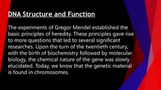
The experiments ofGregor Mendel established the
basic principles of heredity. These principles gave rise
to more questions that led to several significant
researches. Upon the turn of the twentieth century,
with the birth of biochemistry followed by molecular
biology, the chemical nature of the gene was slowly
elucidated. Today, we know that the genetic material
is found in chromosomes.
DNA Structure and Function
3.
'Griffith's Transformation Experiment
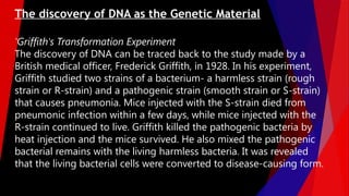
Thediscovery of DNA can be traced back to the study made by a
British medical officer, Frederick Griffith, in 1928. In his experiment,
Griffith studied two strains of a bacterium- a harmless strain (rough
strain or R-strain) and a pathogenic strain (smooth strain or S-strain)
that causes pneumonia. Mice injected with the S-strain died from
pneumonic infection within a few days, while mice injected with the
R-strain continued to live. Griffith killed the pathogenic bacteria by
heat injection and the mice survived. He also mixed the pathogenic
bacterial remains with the living harmless bacteria. It was revealed
that the living bacterial cells were converted to disease-causing form.
The discovery of DNA as the Genetic Material
4.
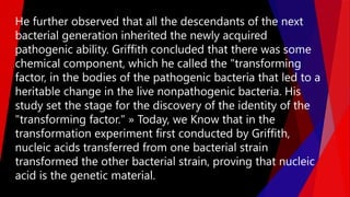
He further observedthat all the descendants of the next
bacterial generation inherited the newly acquired
pathogenic ability. Griffith concluded that there was some
chemical component, which he called the "transforming
factor, in the bodies of the pathogenic bacteria that led to a
heritable change in the live nonpathogenic bacteria. His
study set the stage for the discovery of the identity of the
"transforming factor." » Today, we Know that in the
transformation experiment first conducted by Griffith,
nucleic acids transferred from one bacterial strain
transformed the other bacterial strain, proving that nucleic
acid is the genetic material.
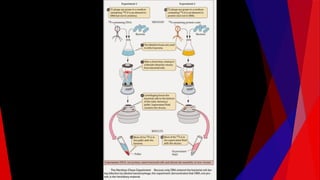
7.
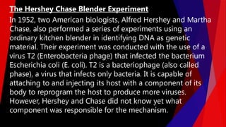
In 1952, twoAmerican biologists, Alfred Hershey and Martha
Chase, also performed a series of experiments using an
ordinary kitchen blender in identifying DNA as genetic
material. Their experiment was conducted with the use of a
virus T2 (Enterobacteria phage) that infected the bacterium
Escherichia coli (E. coli). T2 is a bacteriophage (also called
phase), a virus that infects only bacteria. It is capable of
attaching to and injecting its host with a component of its
body to reprogram the host to produce more viruses.
However, Hershey and Chase did not know yet what
component was responsible for the mechanism.
The Hershey Chase Blender Experiment
8.
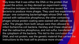
They knew thatit was either the DNA or the protein that
caused the action, so they devised an experiment using
radioactive isotopes to determine which one caused the
bacteria to produce more phages. They used two experimental
setups- one containing bacteriophages whose DNA were
stained with radioactive phosphorus; the other containing
phages whose protein coating were stained with radioactive
sulfur. The phages from both setups were then allowed to
infect Escherichia coli bacteria. Hershey and Chase observed
that the radioactive phosphorus, and not sulfur, transferred to
the cytoplasm of the bacteria. This led to the conclusion that
DNA, and not protein, was the genetic material that carried the
instructions to the host cell to produce more viruses.
10.
The series ofexperiments after Hershey and Chase
focused on the structure of DNA and what it looked
like. Several scientists laid the foundation before the
famous tandem of Watson and Crick won the Nobel
Prize for elucidating the structure of DNA.
The Elucidation of the DNA Structure
11.
In the 1920s,P. A. Levene (Phoebus Aaron Theodore Levene),
an American biochemist, analyzed the components of DNA.
He established the fact that DNA is composed of four
nitrogenous bases (cytosine, guanine, adenine, and
thymine), a deoxyribose sugar, and a phosphate group. He
also laid the groundwork for the building block of DNA,
known as a nucleotide, which consists of a base attached to
a sugar, and the phosphate is attached to another sugar
molecule. However, he was not able to establish the correct
proportions of the bases, which he assumed at the time to
be equal.
Levene's Nucleotides
12.
In the late1940s, Austrian biochemist Erwin Chargaff, analyzed the
proportion of the nitrogenous bases in the DNA of various species.
He found that each species had unique percentages of each type of
nucleotide. The human cell, for example, had 31% of its bases as
adenine, 31% as thymine, 19% as guanine, and 19% as cytosine.
What was common among species was that the amount of adenine
(A) was always equal to the amount of thymine (T), and the amount
of guanine (G) was always equal to the amount of cytosine (C). With
these data, he established the following relationships, collectively
known as the Chargaff rules:
1. DNA contains A, T, G, and C, which vary from species to species.
2. Within the species, the amount of base pairs are equal; that is,
A = T and G = C.
Chargaff Rules
13.
Rosalind Franklin, aresearcher from King's College in London,
studied the structure of DNA using a technique known as X-ray
crystallography. In November 1951, Franklin delivered a lecture
to a group of scientists, including biologist James Watson, on
the two forms of DNA, which she referred to as Type A (dry
form) and Type B (wet form). In the lecture, she mentioned that
phosphate units in a DNA are located in the external part of the
molecule, a model that likewise appeared in the DNA model of
Watson and Crick in 1953. Photo 51 is an X-ray diffraction
photograph of DNA taken by Franklin in 1952. In the
experiment, a minute amount of hydrated DNA was exposed to
an X-ray beam for more than 60 hours, which resulted in the
scattering of its component molecules to produce an image
useful in the elucidation of the 3-D structure of DNA.
Rosalind Franklin's X-ray Diffraction
Photo 51
14.
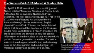
On April 25,1953, an article in the scientific journal
Nature entitled "Molecular Structure of Nucleic Acids:
A Structure for Deoxyribose Nucleic Acid" was
published. The two-page article (pages 737-738 in the
171st volume of Nature) was authored by the
American biologist James Watson and English
physicist Francis Crick. This was the first published
article that described the structure of the DNA as a
double helix. Considered as a "pearl" of science, the
article contained the answers to how the genetic
information inside the nucleus of cells was stored and
passed from one generation to another. The article
was considered a giant scientific leap and a turning
point in the development and rapid progress of
molecular biology and genetics as a science.
The Watson-Crick DNA Model: A Double Helix
Francis Crick (left) and James Watson
(right) with their model of the famous
double-stranded helical DNA structure
15.
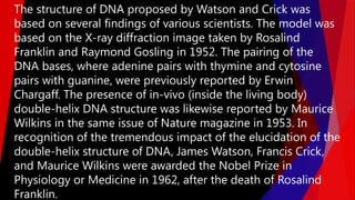
The structure ofDNA proposed by Watson and Crick was
based on several findings of various scientists. The model was
based on the X-ray diffraction image taken by Rosalind
Franklin and Raymond Gosling in 1952. The pairing of the
DNA bases, where adenine pairs with thymine and cytosine
pairs with guanine, were previously reported by Erwin
Chargaff. The presence of in-vivo (inside the living body)
double-helix DNA structure was likewise reported by Maurice
Wilkins in the same issue of Nature magazine in 1953. In
recognition of the tremendous impact of the elucidation of the
double-helix structure of DNA, James Watson, Francis Crick,
and Maurice Wilkins were awarded the Nobel Prize in
Physiology or Medicine in 1962, after the death of Rosalind
Franklin.
16.
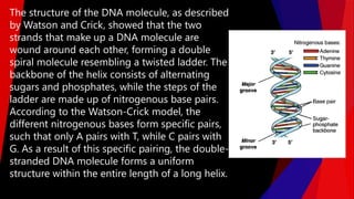
The structure ofthe DNA molecule, as described
by Watson and Crick, showed that the two
strands that make up a DNA molecule are
wound around each other, forming a double
spiral molecule resembling a twisted ladder. The
backbone of the helix consists of alternating
sugars and phosphates, while the steps of the
ladder are made up of nitrogenous base pairs.
According to the Watson-Crick model, the
different nitrogenous bases form specific pairs,
such that only A pairs with T, while C pairs with
G. As a result of this specific pairing, the double-
stranded DNA molecule forms a uniform
structure within the entire length of a long helix.
17.
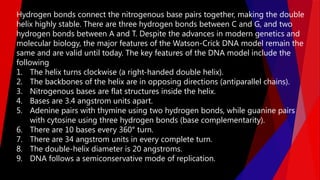
Hydrogen bonds connectthe nitrogenous base pairs together, making the double
helix highly stable. There are three hydrogen bonds between C and G, and two
hydrogen bonds between A and T. Despite the advances in modern genetics and
molecular biology, the major features of the Watson-Crick DNA model remain the
same and are valid until today. The key features of the DNA model include the
following
1. The helix turns clockwise (a right-handed double helix).
2. The backbones of the helix are in opposing directions (antiparallel chains).
3. Nitrogenous bases are flat structures inside the helix.
4. Bases are 3.4 angstrom units apart.
5. Adenine pairs with thymine using two hydrogen bonds, while guanine pairs
with cytosine using three hydrogen bonds (base complementarity).
6. There are 10 bases every 360° turn.
7. There are 34 angstrom units in every complete turn.
8. The double-helix diameter is 20 angstroms.
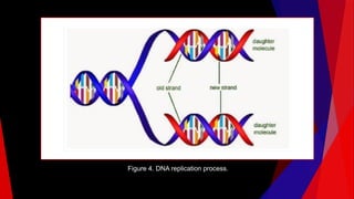
9. DNA follows a semiconservative mode of replication.
18.
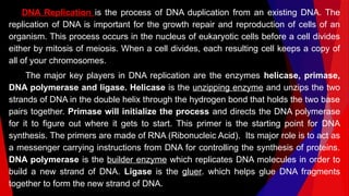
DNA Replication isthe process of DNA duplication from an existing DNA. The
replication of DNA is important for the growth repair and reproduction of cells of an
organism. This process occurs in the nucleus of eukaryotic cells before a cell divides
either by mitosis of meiosis. When a cell divides, each resulting cell keeps a copy of
all of your chromosomes.
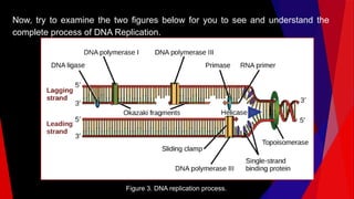
The major key players in DNA replication are the enzymes helicase, primase,
DNA polymerase and ligase. Helicase is the unzipping enzyme and unzips the two
strands of DNA in the double helix through the hydrogen bond that holds the two base
pairs together. Primase will initialize the process and directs the DNA polymerase
for it to figure out where it gets to start. This primer is the starting point for DNA
synthesis. The primers are made of RNA (Ribonucleic Acid). Its major role is to act as
a messenger carrying instructions from DNA for controlling the synthesis of proteins.
DNA polymerase is the builder enzyme which replicates DNA molecules in order to
build a new strand of DNA. Ligase is the gluer. which helps glue DNA fragments
together to form the new strand of DNA.
19.
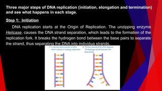
Three major stepsof DNA replication (initiation, elongation and termination)
and see what happens in each stage.
Step 1: Initiation
DNA replication starts at the Origin of Replication. The unzipping enzyme
Helicase, causes the DNA strand separation, which leads to the formation of the
replication fork. It breaks the hydrogen bond between the base pairs to separate
the strand, thus separating the DNA into individua strands.
20.
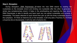
Step 2: Elongation
Duringelongation, DNA Polymerase III makes the new DNA strand by reading the
nucleotides on the template strand and binding one nucleotide after the other to generate a
whole new complementary strand. It helps in the proofreading and repairing the new strand.
DNA Polymerase is able to identify and back track any mis paired nucleotides and corrects it
immediately. The bases attached to each strand then pair up with the three nucleotides found in
the cytoplasm. If it finds an Adenine (A) on the template, it will only add a Thymine (T). If it finds
a Guanine (G) on the template, it will only add a Cytosine (C).
21.
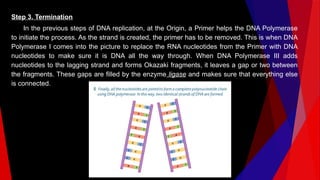
Step 3. Termination
Inthe previous steps of DNA replication, at the Origin, a Primer helps the DNA Polymerase
to initiate the process. As the strand is created, the primer has to be removed. This is when DNA
Polymerase I comes into the picture to replace the RNA nucleotides from the Primer with DNA
nucleotides to make sure it is DNA all the way through. When DNA Polymerase III adds
nucleotides to the lagging strand and forms Okazaki fragments, it leaves a gap or two between
the fragments. These gaps are filled by the enzyme ligase and makes sure that everything else
is connected.
22.
Now, try toexamine the two figures below for you to see and understand the
complete process of DNA Replication.
Figure 3. DNA replication process.
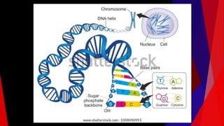
Ever wonder whywe are so complex? Our bodies are able to function in growth,
cell repair, or development even without our conscious effort. How does the
body know when to perform such bodily activities? The answer lies in specific
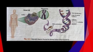
sets of instructions inside our DNA. To appreciate what DNA is or where it is
located in our body, one must untangle the chromosome to reveal its
components. Every human body cell contains 23 pairs of chromosomes, or a
total of 46 chromosomes. A single chromosome contains many genes joined
together like beads on a string. The genes are packed in bundles of these
chromosomes. Our body cells contain about 20000-25 000 genes. A gene, as
shown in figure 13-6, is a distinct portion of the DNA responsible for an inherited
trait. Genes are coded instructions for everything that must happen in the body,
including how we function and how we look. Normally, one gene controls one
trait; but some traits are coded by more than one gene.
The Nucleic Acids and Their Connection With Inheritance
25.
Collectively, DNA isa type of the nucleic acids contained in our cells.
Nuclei acids are organic compounds that function as storage of
genetic information, which is transmitted from one generation to the
next in all living organisms. It is the physical carrier of inheritance
that is passed from parents to offspring. Nucleic acids also function
in protein synthesis as they carry the code needed in the formation
of specific proteins. There are two types of nucleic acids found in
living organisms - deoxyribonucleic acid (DNA) and ribonucleic acid
(RNA). Both types are made up of basic building blocks called
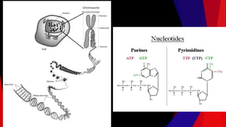
nucleotides. A nucleotide is made up of a five-carbon sugar, a
phosphate group, and a nitrogenous base. The nitrogenous bases are
either double-ringed purines (guanine G] and adenine [A) or single-
ringed pyrimidines (cytocine [C], thymine [T], and uracil (U). These
bases and the nucleic acids in the body will be discussed further in
Chapter 18.
27.
James Watson andFrancis Crick described the structure of DNA as a
double helix of repeating nucleotides that are made up of a sugar
(deoxyribose), a nitrogenous base of either purines (adenine and
guanine) or pyrimidines (thy mine and cytosine), and a phosphate
group. The pairing of nitrogenous bases is so specific that only
adenine pairs with thymine, while only cytosine pairs with guanine.
Thus, a gene refers to a specific sequence of nitrogenous bases that
codes for a specific protein. DNA can be compared to a blueprint of
guidelines that the body must follow to exist and function properly.
RNA, on the other hand, helps to carry out the blueprint's guidelines.
RNA is able to perform a variety of functions and is thus more
diverse, while DNA is able to carry complex information for longer
periods of time and is thus more stable.
28.
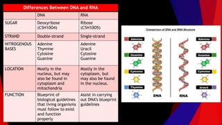
Differences Between DNAand RNA
DNA RNA
SUGAR Deoxyribose
(C5H10O4)
Ribose
(C5H10O5)
STRAND Double-strand Single-strand
NITROGENOUS
BASES
Adenine
Thymine
Cytosine
Guanine
Adenine
Uracil
Cytosine
Guanine
LOCATION Mostly in the
nucleus, but may
also be found in
cytoplasm and
mitochondria
Mostly in the
cytoplasm, but
may also be found
in the nucleus.
FUNCTION Blueprint of
biological guidelines
that living organisms
must follow to exist
and function
properly
Assist in carrying
out DNA’s blueprint
guidelines
29.
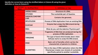
Identify the correctterm using the shuffled letters in Column B using the given
definition or clue on Column C.
A. Correct
Word
B. Shuffled Word C. Definition or Clue
1 UOEBLD XHILE Structure of a DNA.
2 OTSYINEC The nucleotide pair of Guanine.
3
XOEDCIELCUNOYBIR
CADI
Contains the genomes.
4 RPCATILEOIN Process of DNA duplication from an existing DNA
5 ELIHCSAE
Enzyme that unzips the DNA strand during
replication.
6 BSEA What do you call the Adenine-Thymine pair?
7 OKAKIZA
Fragments of DNA that are produced during the
process of DNA replication
8 YDRGOHNE
The type of bond which breaks down when
helicase starts to unzip the DNA strand.
9 ENGE
Basic unit of heredity which carries the
characteristic of parents to children.
10 OLAIONTNEG
This is the step of DNA replication where the DNA
Polymerase creates new strands of nucleotide
specifically paired to another nucleotide.


![Collectively, DNA is a type of the nucleic acids contained in our cells.
Nuclei acids are organic compounds that function as storage of
genetic information, which is transmitted from one generation to the
next in all living organisms. It is the physical carrier of inheritance
that is passed from parents to offspring. Nucleic acids also function
in protein synthesis as they carry the code needed in the formation
of specific proteins. There are two types of nucleic acids found in
living organisms - deoxyribonucleic acid (DNA) and ribonucleic acid
(RNA). Both types are made up of basic building blocks called
nucleotides. A nucleotide is made up of a five-carbon sugar, a
phosphate group, and a nitrogenous base. The nitrogenous bases are
either double-ringed purines (guanine G] and adenine [A) or single-
ringed pyrimidines (cytocine [C], thymine [T], and uracil (U). These
bases and the nucleic acids in the body will be discussed further in
Chapter 18.](https://image.slidesharecdn.com/dna-260128173349-daa8493f/85/DNA-structure-function-history-of-each-scientist-25-320.jpg)