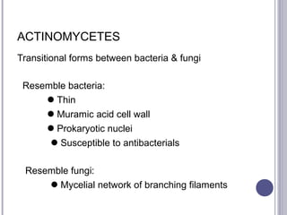
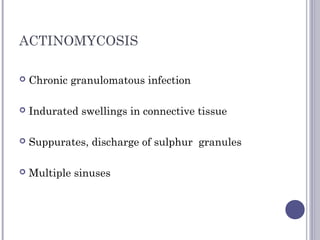
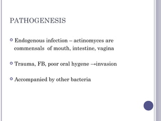
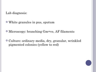
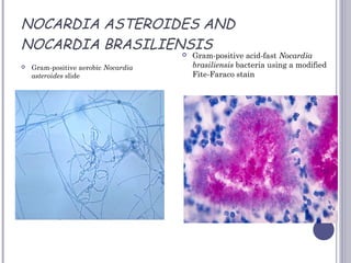
ACTINOMYCETES & NOCARDIA
Dr.S.Pramodhini,
Associate Professor,
Department of
Microbiology,
MGMC& RI
ACTINOMYCETES
Transitional forms between bacteria & fungi
Resemble bacteria:
Thin
Muramic acid cell wall
Prokaryotic nuclei
 Susceptible to antibacterials
Resemble fungi:
 Mycelial network of branching filaments
 Higher bacteria with superficial resemblance to fungi
Related to mycobacteria & corynebacteria
Gram +ve,
Non Motile,
Non Sporing,
Non Capsulated
Filaments break up → bacillary & coccoid
3 medically important genera
 Actinomyces – anaerobic/microaerophilic
non acidfast
 Nocardia – aerobic, acid fast (weakly)
 Streptomyces – aerobic, non acidfast
HISTORY
• Bollinger (1887) from ‘lumpy jaw’ of cattle
• Wolf & Israel (1891) from human lesions
• ‘‘Ray fungus’ – ray like appearance in granules from
lesions
Human pathogen
• A. israelii- most common
• A. eriksoni
• A. naeslundii
• A. odontolyticus
• A. meyeri
• A. viscosus
In cattle
• A. bovis
ACTINOMYCOSIS
 Chronic granulomatous infection
 Indurated swellings in connective tissue
 Suppurates, discharge of sulphur granules
 Multiple sinuses
PATHOGENESIS
 Endogenous infection – actinomyces are
commensals of mouth, intestine, vagina
 Trauma, FB, poor oral hygene →invasion
 Accompanied by other bacteria
CLINICAL DISEASE
• Cervicofacial – cheek & submaxillary
discharge from lower jaw
• Thoracic – lungs, pleura, pericardium
Discharge thro’ chest wall
• Abdominal – ileocaecal, abdominal wall,
Spread to liver
• Pelvic – intrauterine devices
CERVICOFACIAL
ACTINOMYCOSIS
THORACIC ACTINOMYCOSIS
CLINICAL DISEASE
Actinomyces are also responsible for
-Gingivitis, periodontitis
-Sublingual plaques, root caries
-Spread to CNS from primary focus
-Actinomycotic Mycetoma - tumour
with multiple discharging sinuses, mainly foot
LAB. DIAGNOSIS
Specimen –
pus, sputum, tissue biopsy
Methods –
microscopy, culture, IF & HPE
Microscopy:
 Dilute with saline, shake, allow to settle - Sulphur
granules sediment
 Sediments pipetted out
 Crushed b/w slides and examined
 Applying gauze pads on discharge
SULPHUR GRANULES IN
ACTINOMYCOSIS 
Sulphur granules :
 Minor specks to 5 mm in size
 Macroscopic bacterial colonies
 Crushed betn. slides & then gram stained
 Central gram +ve mycelial clump
 peripheral gram –ve radiating clubs
 sun ray appearance
 Clubs are Ag-Ab complexes
STAINING
Histopathologic changes due to the gram-positive organism, Actinomyces israelii.
Using a modified Fite-Faraco stain, a “sulphur granule” is shown in the middle of the image.
These granules actually represent colonies of A. israelii, a gram-positive, anaerobic
filamentous bacteria
Culture:
 Pus / granules washed in saline aseptically
 Crushed with sterile glass rod
Inoculated into:
 Thioglycolate broth
 Brain-heart infusion agar
 Incubate anaerobically
 In broth- fluffy balls at bottom (A. israelii)
- uniform turbidity (A. bovis)
 On agar- small spidery colonies in 48-72hrs
larger & heaped up in10 days
 Isolates identified by gram stain, biochem. tests &
confirmed by fluorescent Ab tests
CULTURAL CHARACTERISTICS
Immunofluorescence:
 Sulphur granules & mycelia in tissue sections using
fluorescent specific antisera
HPE:
 Central mycelial mass
 Peripheral pus cells & chronic inflam. cells
EPIDEMIOLOGY
 World wide
 Common in rural areas, agricultural workers
 Young males (10-30yrs)
 60% cervicofacial, 20% abdominal
 Pelvic actinomycosis in female (IUCD)
TREATMENT
 Large doses of antibiotics for prolonged periods
(months)
 Penicillins & tetracyclines
 Surgical drainage
NOCARDIA
Nocard first described
 Gram +ve
 Aerobic
 Acid fast (weakly – 1% H2SO4) )
(actinomyces are anaerobic, non AF)
 N. asteroides, N. brasiliensis, N. caviae
 Exogenous inf. - Nocardia are found in soil
GRAM STAIN- NOCARDIA
CLINICAL FEATURES
Cutaneous, S/C, systemic lesions
 Cutaneous – abscess, cellulitis, lymphatic
 S/C – actinomycotic mycetoma
 Systemic- immunodeficient (N. asteroides)
 Primary as pneumonia, lung abscess or TB like
illness
 Metastatic in brain, kidney etc
CLINICAL MANIFESTATIONS
Lab diagnosis:
 White granules in pus, sputum
 Microscopy: branching Gm+ve, AF filaments
 Culture: ordinary media, dry, granular, wrinkled
pigmented colonies (yellow to red)
NOCARDIA ACID FAST STAIN
NOCARDIA ASTEROIDES AND
NOCARDIA BRASILIENSIS
 Gram-positive aerobic Nocardia
asteroides slide
 Gram-positive acid-fast Nocardia
brasiliensis bacteria using a modified
Fite-Faraco stain
CULTURAL CHARACTERISTICS
TREATMENT
 Cotrimoxazole
 Minocycline
 Amikacin
 Cefotaxime
for several months
along with surgical drainage
STREPTOMYCOSIS
 The streptomyces species usually cause the disease
entity known as mycetoma (fungus tumor).
 These infections are usually subcutaneous, but they
can penetrate deeper and invade the bone.
 Some species produce a protease which inhibits
macrophages.
 Material sent to the lab is pus or skin biopsy.
 The streptomycetes are aerobic like Nocardia,
and can grow on both bacterial and fungal
(Sabouraud) media. They produce a chalky aerial
mycelium with much branching .
STREPTOMYCES GRANULES
Actinomycotic mycetomatous granule due to the bacteria Streptomyces somaliensis
STREPTOMYCOSIS
 The organisms are found world-wide.
 There are no serological tests
 The drugs of choice are the combination of
sulfamethoxazole/trimethoprim or amphotericinB.
 In the tropics, this disease may go undiagnosed or untreated for
so long that surgical amputation may be the only effective
treatment.
ACTINOMYCOTIC MYCETOMA
Mycetoma:
 Localised chronic granulomatous lesions of
subcut. & Deeper tissues
 Commonly of foot, less often hand & other parts
 Present as tumour with multiple sinuses
 Maduramycosis (gill in 1842 from madura)
MYCETOMA
 Mycetomas are usually caused by fungi
 Less commonly by bacteria
Bacterial agents:
 Actinomyces ( A. bovisA. israelii, A. bovis)
 N. brasiliensis, N. caviae, N. asteroides
 Steptomyces somaliensis
 Actinomadura (A. madurae, A. pelletieri)
 Even Staph. aureus (botryomycosis)
Aetiological diagnosis important in treatment
Colour of granule:
 Actinomycotic – white or yellow
 Eumycotic – black
Microscopy:
 Actinomycotic – thin filaments (1μm)
 Eumycotic – thick (4-5μm)
Cuture: isolation & identification

Actinomycetes

  • 1.
    ACTINOMYCETES & NOCARDIA Dr.S.Pramodhini, AssociateProfessor, Department of Microbiology, MGMC& RI
  • 2.
    ACTINOMYCETES Transitional forms betweenbacteria & fungi Resemble bacteria: Thin Muramic acid cell wall Prokaryotic nuclei  Susceptible to antibacterials Resemble fungi:  Mycelial network of branching filaments
  • 3.
     Higher bacteriawith superficial resemblance to fungi Related to mycobacteria & corynebacteria Gram +ve, Non Motile, Non Sporing, Non Capsulated Filaments break up → bacillary & coccoid
  • 4.
    3 medically importantgenera  Actinomyces – anaerobic/microaerophilic non acidfast  Nocardia – aerobic, acid fast (weakly)  Streptomyces – aerobic, non acidfast
  • 5.
    HISTORY • Bollinger (1887)from ‘lumpy jaw’ of cattle • Wolf & Israel (1891) from human lesions • ‘‘Ray fungus’ – ray like appearance in granules from lesions
  • 6.
    Human pathogen • A.israelii- most common • A. eriksoni • A. naeslundii • A. odontolyticus • A. meyeri • A. viscosus In cattle • A. bovis
  • 7.
    ACTINOMYCOSIS  Chronic granulomatousinfection  Indurated swellings in connective tissue  Suppurates, discharge of sulphur granules  Multiple sinuses
  • 8.
    PATHOGENESIS  Endogenous infection– actinomyces are commensals of mouth, intestine, vagina  Trauma, FB, poor oral hygene →invasion  Accompanied by other bacteria
  • 9.
    CLINICAL DISEASE • Cervicofacial– cheek & submaxillary discharge from lower jaw • Thoracic – lungs, pleura, pericardium Discharge thro’ chest wall • Abdominal – ileocaecal, abdominal wall, Spread to liver • Pelvic – intrauterine devices
  • 10.
  • 11.
  • 12.
    CLINICAL DISEASE Actinomyces arealso responsible for -Gingivitis, periodontitis -Sublingual plaques, root caries -Spread to CNS from primary focus -Actinomycotic Mycetoma - tumour with multiple discharging sinuses, mainly foot
  • 13.
    LAB. DIAGNOSIS Specimen – pus,sputum, tissue biopsy Methods – microscopy, culture, IF & HPE
  • 14.
    Microscopy:  Dilute withsaline, shake, allow to settle - Sulphur granules sediment  Sediments pipetted out  Crushed b/w slides and examined  Applying gauze pads on discharge
  • 15.
  • 17.
    Sulphur granules : Minor specks to 5 mm in size  Macroscopic bacterial colonies  Crushed betn. slides & then gram stained  Central gram +ve mycelial clump  peripheral gram –ve radiating clubs  sun ray appearance  Clubs are Ag-Ab complexes
  • 20.
    STAINING Histopathologic changes dueto the gram-positive organism, Actinomyces israelii. Using a modified Fite-Faraco stain, a “sulphur granule” is shown in the middle of the image. These granules actually represent colonies of A. israelii, a gram-positive, anaerobic filamentous bacteria
  • 21.
    Culture:  Pus /granules washed in saline aseptically  Crushed with sterile glass rod Inoculated into:  Thioglycolate broth  Brain-heart infusion agar  Incubate anaerobically
  • 22.
     In broth-fluffy balls at bottom (A. israelii) - uniform turbidity (A. bovis)  On agar- small spidery colonies in 48-72hrs larger & heaped up in10 days  Isolates identified by gram stain, biochem. tests & confirmed by fluorescent Ab tests
  • 23.
  • 24.
    Immunofluorescence:  Sulphur granules& mycelia in tissue sections using fluorescent specific antisera HPE:  Central mycelial mass  Peripheral pus cells & chronic inflam. cells
  • 25.
    EPIDEMIOLOGY  World wide Common in rural areas, agricultural workers  Young males (10-30yrs)  60% cervicofacial, 20% abdominal  Pelvic actinomycosis in female (IUCD)
  • 26.
    TREATMENT  Large dosesof antibiotics for prolonged periods (months)  Penicillins & tetracyclines  Surgical drainage
  • 27.
    NOCARDIA Nocard first described Gram +ve  Aerobic  Acid fast (weakly – 1% H2SO4) ) (actinomyces are anaerobic, non AF)  N. asteroides, N. brasiliensis, N. caviae  Exogenous inf. - Nocardia are found in soil
  • 28.
  • 29.
    CLINICAL FEATURES Cutaneous, S/C,systemic lesions  Cutaneous – abscess, cellulitis, lymphatic  S/C – actinomycotic mycetoma  Systemic- immunodeficient (N. asteroides)  Primary as pneumonia, lung abscess or TB like illness  Metastatic in brain, kidney etc
  • 30.
  • 31.
    Lab diagnosis:  Whitegranules in pus, sputum  Microscopy: branching Gm+ve, AF filaments  Culture: ordinary media, dry, granular, wrinkled pigmented colonies (yellow to red)
  • 33.
  • 34.
    NOCARDIA ASTEROIDES AND NOCARDIABRASILIENSIS  Gram-positive aerobic Nocardia asteroides slide  Gram-positive acid-fast Nocardia brasiliensis bacteria using a modified Fite-Faraco stain
  • 35.
  • 36.
    TREATMENT  Cotrimoxazole  Minocycline Amikacin  Cefotaxime for several months along with surgical drainage
  • 37.
    STREPTOMYCOSIS  The streptomycesspecies usually cause the disease entity known as mycetoma (fungus tumor).  These infections are usually subcutaneous, but they can penetrate deeper and invade the bone.  Some species produce a protease which inhibits macrophages.
  • 38.
     Material sentto the lab is pus or skin biopsy.  The streptomycetes are aerobic like Nocardia, and can grow on both bacterial and fungal (Sabouraud) media. They produce a chalky aerial mycelium with much branching .
  • 39.
    STREPTOMYCES GRANULES Actinomycotic mycetomatousgranule due to the bacteria Streptomyces somaliensis
  • 40.
    STREPTOMYCOSIS  The organismsare found world-wide.  There are no serological tests  The drugs of choice are the combination of sulfamethoxazole/trimethoprim or amphotericinB.  In the tropics, this disease may go undiagnosed or untreated for so long that surgical amputation may be the only effective treatment.
  • 41.
    ACTINOMYCOTIC MYCETOMA Mycetoma:  Localisedchronic granulomatous lesions of subcut. & Deeper tissues  Commonly of foot, less often hand & other parts  Present as tumour with multiple sinuses  Maduramycosis (gill in 1842 from madura)
  • 42.
  • 43.
     Mycetomas areusually caused by fungi  Less commonly by bacteria Bacterial agents:  Actinomyces ( A. bovisA. israelii, A. bovis)  N. brasiliensis, N. caviae, N. asteroides  Steptomyces somaliensis  Actinomadura (A. madurae, A. pelletieri)  Even Staph. aureus (botryomycosis)
  • 44.
    Aetiological diagnosis importantin treatment Colour of granule:  Actinomycotic – white or yellow  Eumycotic – black Microscopy:  Actinomycotic – thin filaments (1μm)  Eumycotic – thick (4-5μm) Cuture: isolation & identification