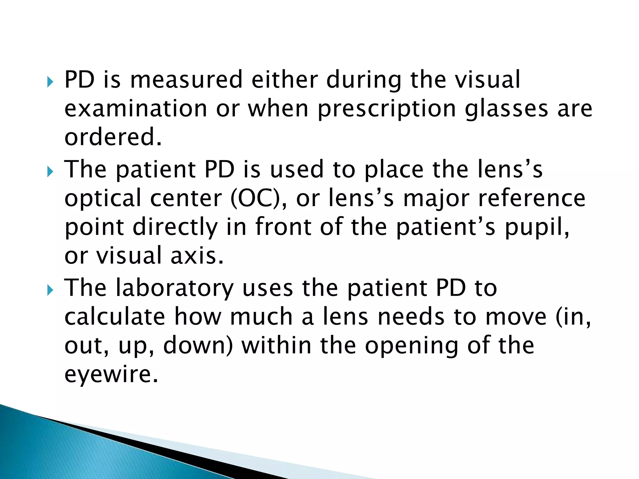
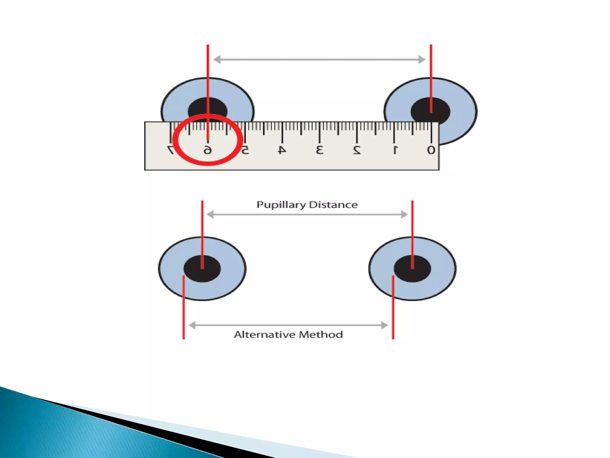
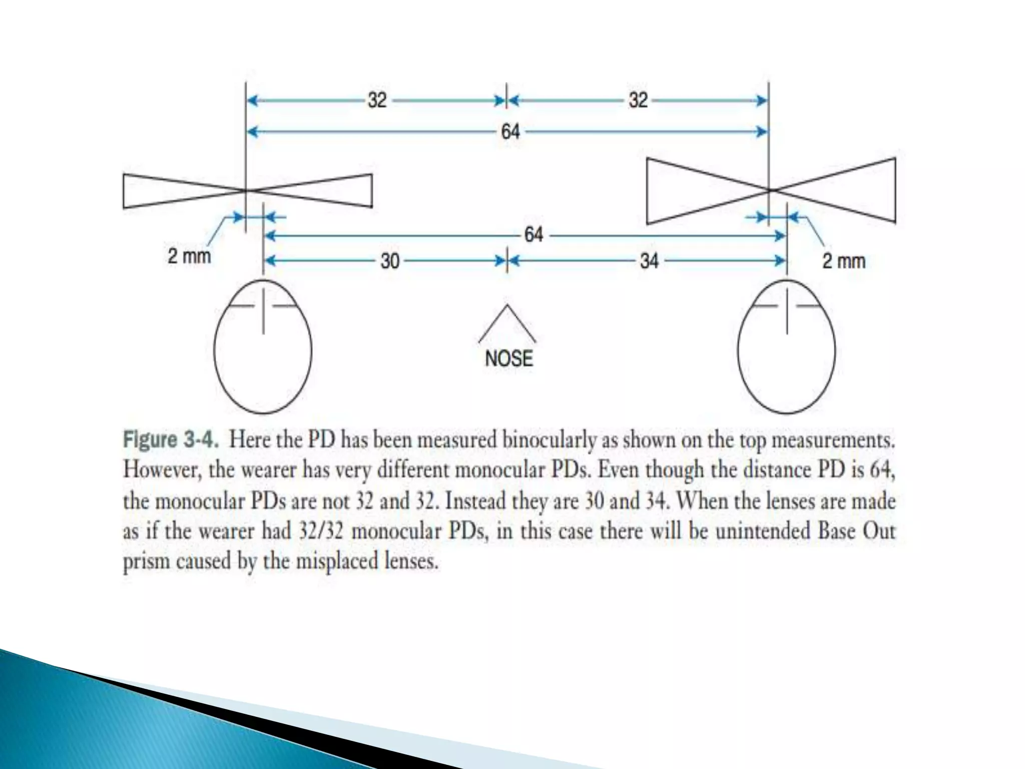
This document discusses pupil distance (PD), which is the distance between the centers of the pupils. It provides information on measuring binocular PD, monocular PD, and near PD. Binocular PD is measured from one pupil to the other using a ruler. Monocular PD measures each eye individually. Near PD is needed for reading glasses and is measured at a closer distance. Accurate PD measurement is important for properly positioning lenses in prescription glasses.
Contact lens for congenital aphakia and other eye conditions for infants and toddlers. The slide presentation encompasses indications for CL fitting in paediatric, contact lens options, fitting techniques, challenges and contact lens as myopia control.
Detailed instumentaion and use of manual Lensometer and just a outline of automated lensometer.
I have used the picture of manual lensometer with out the parts describtion because i have explained orally by showing the picture..
Hope u all like it and may help you in learning better. :)
SOFT CONTACT LENS FITTING
1. Alternative names of soft contact lens.
2. Need to know fitting requirement and performance requirements.
3. Centration and decentration of soft contact lens. -- There are cartesian system and binasal system.
4. what governs fitting of lens.
5. There are need to know about physical properties of soft contact lens.
6. Now, what is sag and sagital depth.
7. Finally, SAME SAG AND SAME DIAMETER but DIFFERENT DESIGN AND DIFFERENT BEHAVIOUR.
8. Parameters of soft contact lens -
total diameter
back optic zone radius
centre thickness
front optic zone radius
water content
9. There are two types of prescribing methods -
empirical prescribing
trial fit prescribing
10. Effect of a blink with soft contact lens - too flat and too steep.
11. Requirements of lens movement.
12. Lens lag position - primary gaze, up gaze and lateral gaze position.
13. Compulsory of lower lid push up test.
14. Ranges of fitting of soft contact lens - either too fit or too loose or ideal fitting.
15. All step of soft contact lens fitting is done.
Magnification is a method of increasing the size of the image
so that enough of the retina is stimulated to send an impulse
through the optic nerve allowing an object to be perceived .
you will get information and knowledge about the direct ophthalmic instrument known as ophthalmoscope.
its principle, parts, types, its different filters, techniques, uses, and its method is explained in these slides.
This is a guide for Visual function assessment in low vision. Useful for Optometrists in providing better care to Low vision Patients by assessing the conditions better.
Brückner, in 1962, published a paper in German describing a "trans-illumination" test extremely useful in the diagnosis of small angle deviations and amblyopia in young uncooperative children. A bright coaxial light source, such as a direct ophthalmoscope, is used.
Cold Sores: Causes, Treatments, and Prevention Strategies | The Lifesciences ...The Lifesciences Magazine
Cold Sores, medically known as herpes labialis, are caused by the herpes simplex virus (HSV). HSV-1 is primarily responsible for cold sores, although HSV-2 can also contribute in some cases.
International Cancer Survivors Day is celebrated during June, placing the spotlight not only on cancer survivors, but also their caregivers.
CANSA has compiled a list of tips and guidelines of support:
https://cansa.org.za/who-cares-for-cancer-patients-caregivers/
Deep Leg Vein Thrombosis (DVT): Meaning, Causes, Symptoms, Treatment, and Mor...The Lifesciences Magazine
Deep Leg Vein Thrombosis occurs when a blood clot forms in one or more of the deep veins in the legs. These clots can impede blood flow, leading to severe complications.
Empowering ACOs: Leveraging Quality Management Tools for MIPS and BeyondHealth Catalyst
Join us as we delve into the crucial realm of quality reporting for MSSP (Medicare Shared Savings Program) Accountable Care Organizations (ACOs).
In this session, we will explore how a robust quality management solution can empower your organization to meet regulatory requirements and improve processes for MIPS reporting and internal quality programs. Learn how our MeasureAble application enables compliance and fosters continuous improvement.
Navigating Challenges: Mental Health, Legislation, and the Prison System in B...Guillermo Rivera
This conference will delve into the intricate intersections between mental health, legal frameworks, and the prison system in Bolivia. It aims to provide a comprehensive overview of the current challenges faced by mental health professionals working within the legislative and correctional landscapes. Topics of discussion will include the prevalence and impact of mental health issues among the incarcerated population, the effectiveness of existing mental health policies and legislation, and potential reforms to enhance the mental health support system within prisons.
Under Pressure : Kenneth Kruk's StrategyKenneth Kruk
Kenneth Kruk's story of transforming challenges into opportunities by leading successful medical record transitions and bridging scientific knowledge gaps during COVID-19.
The dimensions of healthcare quality refer to various attributes or aspects that define the standard of healthcare services. These dimensions are used to evaluate, measure, and improve the quality of care provided to patients. A comprehensive understanding of these dimensions ensures that healthcare systems can address various aspects of patient care effectively and holistically. Dimensions of Healthcare Quality and Performance of care include the following; Appropriateness, Availability, Competence, Continuity, Effectiveness, Efficiency, Efficacy, Prevention, Respect and Care, Safety as well as Timeliness.
Health Education on prevention of hypertensionRadhika kulvi
Hypertension is a chronic condition of concern due to its role in the causation of coronary heart diseases. Hypertension is a worldwide epidemic and important risk factor for coronary artery disease, stroke and renal diseases. Blood pressure is the force exerted by the blood against the walls of the blood vessels and is sufficient to maintain tissue perfusion during activity and rest. Hypertension is sustained elevation of BP. In adults, HTN exists when systolic blood pressure is equal to or greater than 140mmHg or diastolic BP is equal to or greater than 90mmHg. The
1. Sahibzada Hakim Anjum Nadeem
Co-Incharge OTTC, Optician, Refractionist, COAVS
CEO Anjum Eye Care & Optical Company
Optometrist, Al-Khair Eye Hospital Lahore
Email: shanjum92@gmail.com
2. • The anatomic PD is the distance between the
centre of one pupil to the centre of the other
pupil, measured in millimeters.
• Prescription glasses are made so that the
distance between the optical centres of the
glasses' lenses, is the same as the PD.
• PD varies from person to person.
• But once an adult, PD does not change.
• Once PD is known, there is no need to measure
it every time you buy prescription glasses.
3. PD is measured either during the visual
examination or when prescription glasses are
ordered.
The patient PD is used to place the lens’s
optical center (OC), or lens’s major reference
point directly in front of the patient’s pupil,
or visual axis.
The laboratory uses the patient PD to
calculate how much a lens needs to move (in,
out, up, down) within the opening of the
eyewire.
4. For all prescriptions without prism, the lens’s
OC will be placed in front of the patient’s
pupil.
For all prescriptions with prism, the OC will
be shifted away from the wearer’s pupil,
forcing them to look through the prism area
of the lens.
5. Binocular PD:
A binocular PD is a single measurement taken
from the visual axis of the right eye to the
visual axis of the left eye.
The PD is measured with a millimeter ruler
commonly referred to as a PD ruler.
The resulting measurement is called the
binocular PD.
6.
7. Steps in measuring the Binocular Distance PD:
Dispenser positions at 40cm( 16 inches)
Dispenser closes right eye, subject fixates at
the dispenser’s left eye.
Dispenser lines up zero point on subject right
eye at the pupil centre, left pupillary border
or left limbus.
8. Dispenser closes left eye, open right eye,
subjects fixates right eye.
Dispenser reads off scale directly in line with
left pupil center, left pupillary border, or left
limbus.
Dispenser closes right eye, open left eye,
subjects fixates left eye.
Dispenser checks to make sure zero point is
correct.
9. Common difficulties are:
Dispenser cannot close one eye:
Occasionally the person doing the measuring is
unable to close one eye independent of the
other. This can be remedied by occluding
(covering) the eye with the free hand.
Occluding the eye with the hand held flat
appears to be a natural part of the test and does
not reveal a person’s inability to close only one
eye.
10. Dispenser visually impaired in one eye:
If the dispenser is blind in one eye, or has
visual acuity too poor to allow the ruler to
be read accurately, then the technique is
modified.
The dispenser places the good eye directly
in front of the subject’s right eye and at the
normal distance. The zero mark is lined up
as usual.
The dispenser then moves sideways until
the good eye is positioned in front of the
subject’s left eye and the measurement is
read.
11. Subject is strabismic:
To determine a true reading, simply cover the
subject’s eye not being observed. This ensures
that the subject is fixating with the eye under
observation and ensures that it is not turned
unless eccentric fixation is present.
Even if eccentric fixation is present, the PD
measurement is still correct, since the subject
never uses this eye in any other position
relative to the dominant eye.
12. Subject is uncooperative child:
If the subject is young or uncooperative,
making normal PD measurements impossible,
the dispenser may have to take a canthus-to-
canthus measurement.
13. Dispenser PD differs significantly from the
subject because the line of sight is not parallel.
The above error will be increased if the PD rule
is not tilted on the subject’s nose so that the
scale is in the most recessed area.
The parallactic error will also be increased even
more if the dispenser is too close to the subject.
Too close is closer than the normal 40 cm (16
inch) distance.
Dispenser and subject not at the same level.
14. An error can result if the subject’s head moves.
An error can result if the person measuring
moves his or her head.
An error will result if the person measuring
does not close or occlude one eye at a time to
ensure sighting from directly in front of the
subject’s eye under observation.
The subject may not look directly at the
measurer’s pupil during the test, as he or she
should, which will result in an error.
15. A separate measurement is taken from the
center of the patient’s nose to the visual axis
in the right eye and left eye.
Monocular PDs should be taken for every
single job except those that will be used for
lined-style multifocal lenses.
For progressive lenses, a monocular PD is
required.
16. Record monocular PDs on a patient record or
lab order form as two numbers,
Such as 32/31, 29/28, 28/28, or 33/34.
It is assumed that the first number provided
will be for the right eye.
17.
18. Steps in measuring the monocular PD Using Ruler:
The monocular PD is best taken using a
pupillometer. When a pupillometer is not
available, monocular PDs are taken by
measuring from the center of the nose to
the center of the pupils.
The procedure consists of the following three
steps:
19. Measure binocular PD. Use pupil center as
reference point.
Before moving the ruler, note the scale
reading on the ruler at the center of nose.
This is the right monocular PD.
Subtract this readings from the binocular PD
to obtain the left PD.
20.
21.
22.
23. A corneal reflex pupilometer, or CRP, is used
to measure the PD.
The CRP is a sophisticated tool that
accurately and repeatedly measures patient
PDs.
A CRP measures the distance from the center
of a patient’s nose to their visual axis
(roughly their pupil center).
24. A CRP has an optician’s side and a patient’s
side.
The patient’s side has a set of nosepads to
rest on the patient’s nose.
When the patient looks inside the CRP, they
will see a target.
A CPR can measure the both monocular and
binocular PD.
25.
26.
27. The near PD is required for single vision
reading glasses or for multifocals.
Opticals centers will be in line of sight when
eyes are converged for readings.
In bifocals, the distance portion is ground to
distance PD and near portion to the near PD.
28. Steps in measuring the near PD Using PD Ruler:
Dispenser places his dominant eye in front of
subjects nose at the subject near working
distance.
Normally distance = 40cm
Dispenser closes the poorer eye.
Subjects fixates dispenser’s open eye.
29. Dispenser places zero point of PD rule at the
center of subjects right pupil.
Dispenser reads scale marking at the center
of subjects left pupil.
The subjects is not required to shift gaze.
The dispenser is not required to change eyes
during the procedure.
30.
31. Taking Near PD Using a Pupillometer:
Usually a PD measuring instrument will allow
both distance and near PD to be measured.
This is done through the use of a movable
internal lens that changes the image
distance and convergence for the subject.
The near readings are carried out in the same
manner as the distance readings.
32. For the near “reading” area of a pair of glasses
to be used most comfortably, it must be
positioned accurately in the lens.
Horizontal placement of the near segment
viewing area is determined by the near PD.
The horizontal position of bifocal segments is
specified as the distance from the far point PD
that the segments are set in toward the bridge.
The total inset is the difference between the
distance PD and the near PD.
33. Because of the possibility of unequal
monocular PDs, segment inset is usually
specified individually for each eye.
Ordinarily segment inset is the difference
between the distance PD and the near PD,
divided by 2:
34. For example, if the distance PD is 68 and the
near PD is 64, then the segment (seg) inset
for each eye is 2 mm.