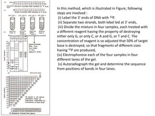
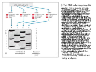
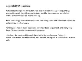
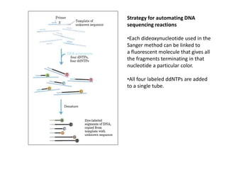
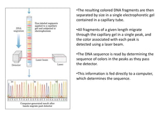
The document discusses techniques for DNA sequencing, including early methods developed in the 1970s by Maxam and Gilbert as well as Sanger. It provides details on how both methods work, such as using specific chemical or enzymatic reactions to generate labeled DNA fragments of different lengths corresponding to nucleotide positions in the sequence. The document also describes how these methods were later automated, using fluorescent tags on dideoxynucleotides and capillary electrophoresis to simultaneously sequence multiple samples in a single gel. This allowed rapid determination of thousands of nucleotides and enabled large genome sequencing projects such as the Human Genome Project.
Sanger sequencing is a method of DNA sequencing based on the selective incorporation of chain-terminating dideoxynucleotides by DNA polymerase during in vitro DNA replication.
Techniques based on the principle of selectively amplifying a subset of restriction fragments from a complex mixture of DNA fragments obtained after digestion of genomic DNA with restriction endonucleases.
Open reading frame is part of reading frame that contains no stop codons or region of amino acids coding triple codons.
ORF starts with start codon and ends at stop codon.
Deciphering DNA sequences is essential for virtually all branches of biological research. With the
advent of capillary electrophoresis (CE)-based Sanger sequencing, scientists gained the ability to
elucidate genetic information from any given biological system. This technology has become widely
adopted in laboratories around the world, yet has always been hampered by inherent limitations in
throughput, scalability, speed, and resolution that often preclude scientists from obtaining the essential
information they need for their course of study. To overcome these barriers, an entirely new technology
was required—Next-Generation Sequencing (NGS), a fundamentally different approach to sequencing
that triggered numerous ground-breaking discoveries and ignited a revolution in genomic science.
After sequencing of the genome has been done, the first thing that comes to mind is "Where are the genes?". Genome annotation is the process of attaching information to the biological sequences. It is an active area of research and it would help scientists a lot to undergo with their wet lab projects once they know the coding parts of a genome.
Sanger sequencing is one of the DNA sequencing methods used to identify and determine the sequence (Nucleotide) of DNA .This is an enzymatic method of sequencing developed by Fred Sanger.
Next Generation Sequencing (NGS) Is A Modern And Cost Effective Sequencing Technology Which Enables Scientists To Sequence Nucleic Acids At Much Faster Rate. In This Presentation, You Will Learn About What is NGS, Idea Behind NGS, Methodology And Protocol, Widely Adapted NGS Protocols, Applications And References For Further Study.
Next Gen Sequencing Technologies OverviewPatrick Merel
A presentation of the major nextgen sequencing technologies. Price indications were valid 1st quarter of 2009. Mac keynote file, half french half english. Hope this can help.
Autobiography of frederick sanger double nobel prize winnerrita martin
Autobiography of Frederick Sanger British biochemist double Noble prize winner as done major contributions to field of chemistry some of his works include structure of insulin and Recombinant DNA
DNA sequencing is the process of determining the nucleic acid sequence – the order of nucleotides in DNA. It includes any method or technology that is used to determine the order of the four bases: adenine, guanine, cytosine, and thymine. The advent of rapid DNA sequencing methods has greatly accelerated biological and medical research and discovery.
Knowledge of DNA sequences has become indispensable for basic biological research, DNA Genographic Projects and in numerous applied fields such as medical diagnosis, biotechnology, forensic biology, virology and biological systematics. Comparing healthy and mutated DNA sequences can diagnose different diseases including various cancers,characterize antibody repertoire, and can be used to guide patient treatment.[5Having a quick way to sequence DNA allows for faster and more individualized medical care to be administered, and for more organisms to be identified and cataloged.
The rapid speed of sequencing attained with modern DNA sequencing technology has been instrumental in the sequencing of complete DNA sequences, or genomes, of numerous types and species of life, including the human genome and other complete DNA sequences of many animal, plant, and microbial species.
The first DNA sequences were obtained in the early 1970s by academic researchers using laborious methods based on two-dimensional chromatography. Following the development of fluorescence-based sequencing methods with a DNA sequencer, DNA sequencing has become easier and orders of magnitude faster.
DNA sequencing refers to the general laboratory technique for determining the exact sequence of nucleotides, or bases, in a DNA molecule. The sequence of the bases (often referred to by the first letters of their chemical names: A, T, C, and G) encodes the biological information that cells use to develop and operate.Whole Genome Sequencing
•Allows doctors to closely analyze a patient's genes for mutations and health indicators.
•Can detect intellectual disabilities and developmental delays.
•WGS is currently available at Yale for patients in the NICU and PICU.
•Involves Genetics.Sequencing may be utilized to determine the order of nucleotides in small targeted genomic regions or entire genomes. Illumina sequencing enables a wide variety of applications, allowing researchers to ask virtually any question related to the genome, transcriptome, or epigenome of any organism.The spectrum of analysis of NGS can extend from a small number of genes to an entire genome, depending on the goal. Whole-genome sequencing (WGS) and whole-exome sequencing (WES) provide the sequence of DNA bases across the genome and exome, respectively.Capillary electrophoresis (CE) instruments are capable of performing both Sanger sequencing and fragment analysis. Fragment analysis is a method in which DNA fragments are fluorescently labeled, separated by CE, and sized by comparison to an internal standard. sanger and Maxam-Gilbert sequencing technologies were classified
Gene Sequencing, a tool to analyze the exact order of nucleotide sequence in the DNA -Deoxyribonucleic Acid.
Focuses on Two methods:
a. Maxam-Gilbert (Chemical Degradation) Method
b. Sanger's Method (Dideoxy Chain termination Method)
DNA consists of a linear string of nucleotides, or bases, for simplicity, referred to by the first letters of their chemical names--A, T, C and G. The process of deducing the order of nucleotides in DNA is called DNA sequencing. Since the DNA sequence confers information that the cell uses to make RNA molecules and proteins, establishing the sequence of DNA is key for understanding how genomes work. The technology for DNA sequencing was made faster and less expensive as a part of the Human Genome Project. And recent developments have profoundly increased the efficiency of DNA sequencing even further.
DNA sequencing is the process of determining the nucleic acid sequence – the order of nucleotides in DNA. It includes any method or technology that is used to determine the order of the four bases: adenine, guanine, cytosine, and thymine.
Sequencing DNA means determining the order of the four chemical building blocks - called "bases" - that make up the DNA molecule. The sequence tells scientists the kind of genetic information that is carried in a particular DNA segment. For example, scientists can use sequence information to determine which stretches of DNA contain genes and which stretches carry regulatory instructions, turning genes on or off. In addition, and importantly, sequence data can highlight changes in a gene that may cause disease.
Slide 1: Title Slide
Extrachromosomal Inheritance
Slide 2: Introduction to Extrachromosomal Inheritance
Definition: Extrachromosomal inheritance refers to the transmission of genetic material that is not found within the nucleus.
Key Components: Involves genes located in mitochondria, chloroplasts, and plasmids.
Slide 3: Mitochondrial Inheritance
Mitochondria: Organelles responsible for energy production.
Mitochondrial DNA (mtDNA): Circular DNA molecule found in mitochondria.
Inheritance Pattern: Maternally inherited, meaning it is passed from mothers to all their offspring.
Diseases: Examples include Leber’s hereditary optic neuropathy (LHON) and mitochondrial myopathy.
Slide 4: Chloroplast Inheritance
Chloroplasts: Organelles responsible for photosynthesis in plants.
Chloroplast DNA (cpDNA): Circular DNA molecule found in chloroplasts.
Inheritance Pattern: Often maternally inherited in most plants, but can vary in some species.
Examples: Variegation in plants, where leaf color patterns are determined by chloroplast DNA.
Slide 5: Plasmid Inheritance
Plasmids: Small, circular DNA molecules found in bacteria and some eukaryotes.
Features: Can carry antibiotic resistance genes and can be transferred between cells through processes like conjugation.
Significance: Important in biotechnology for gene cloning and genetic engineering.
Slide 6: Mechanisms of Extrachromosomal Inheritance
Non-Mendelian Patterns: Do not follow Mendel’s laws of inheritance.
Cytoplasmic Segregation: During cell division, organelles like mitochondria and chloroplasts are randomly distributed to daughter cells.
Heteroplasmy: Presence of more than one type of organellar genome within a cell, leading to variation in expression.
Slide 7: Examples of Extrachromosomal Inheritance
Four O’clock Plant (Mirabilis jalapa): Shows variegated leaves due to different cpDNA in leaf cells.
Petite Mutants in Yeast: Result from mutations in mitochondrial DNA affecting respiration.
Slide 8: Importance of Extrachromosomal Inheritance
Evolution: Provides insight into the evolution of eukaryotic cells.
Medicine: Understanding mitochondrial inheritance helps in diagnosing and treating mitochondrial diseases.
Agriculture: Chloroplast inheritance can be used in plant breeding and genetic modification.
Slide 9: Recent Research and Advances
Gene Editing: Techniques like CRISPR-Cas9 are being used to edit mitochondrial and chloroplast DNA.
Therapies: Development of mitochondrial replacement therapy (MRT) for preventing mitochondrial diseases.
Slide 10: Conclusion
Summary: Extrachromosomal inheritance involves the transmission of genetic material outside the nucleus and plays a crucial role in genetics, medicine, and biotechnology.
Future Directions: Continued research and technological advancements hold promise for new treatments and applications.
Slide 11: Questions and Discussion
Invite Audience: Open the floor for any questions or further discussion on the topic.
The increased availability of biomedical data, particularly in the public domain, offers the opportunity to better understand human health and to develop effective therapeutics for a wide range of unmet medical needs. However, data scientists remain stymied by the fact that data remain hard to find and to productively reuse because data and their metadata i) are wholly inaccessible, ii) are in non-standard or incompatible representations, iii) do not conform to community standards, and iv) have unclear or highly restricted terms and conditions that preclude legitimate reuse. These limitations require a rethink on data can be made machine and AI-ready - the key motivation behind the FAIR Guiding Principles. Concurrently, while recent efforts have explored the use of deep learning to fuse disparate data into predictive models for a wide range of biomedical applications, these models often fail even when the correct answer is already known, and fail to explain individual predictions in terms that data scientists can appreciate. These limitations suggest that new methods to produce practical artificial intelligence are still needed.
In this talk, I will discuss our work in (1) building an integrative knowledge infrastructure to prepare FAIR and "AI-ready" data and services along with (2) neurosymbolic AI methods to improve the quality of predictions and to generate plausible explanations. Attention is given to standards, platforms, and methods to wrangle knowledge into simple, but effective semantic and latent representations, and to make these available into standards-compliant and discoverable interfaces that can be used in model building, validation, and explanation. Our work, and those of others in the field, creates a baseline for building trustworthy and easy to deploy AI models in biomedicine.
Bio
Dr. Michel Dumontier is the Distinguished Professor of Data Science at Maastricht University, founder and executive director of the Institute of Data Science, and co-founder of the FAIR (Findable, Accessible, Interoperable and Reusable) data principles. His research explores socio-technological approaches for responsible discovery science, which includes collaborative multi-modal knowledge graphs, privacy-preserving distributed data mining, and AI methods for drug discovery and personalized medicine. His work is supported through the Dutch National Research Agenda, the Netherlands Organisation for Scientific Research, Horizon Europe, the European Open Science Cloud, the US National Institutes of Health, and a Marie-Curie Innovative Training Network. He is the editor-in-chief for the journal Data Science and is internationally recognized for his contributions in bioinformatics, biomedical informatics, and semantic technologies including ontologies and linked data.
Introduction:
RNA interference (RNAi) or Post-Transcriptional Gene Silencing (PTGS) is an important biological process for modulating eukaryotic gene expression.
It is highly conserved process of posttranscriptional gene silencing by which double stranded RNA (dsRNA) causes sequence-specific degradation of mRNA sequences.
dsRNA-induced gene silencing (RNAi) is reported in a wide range of eukaryotes ranging from worms, insects, mammals and plants.
This process mediates resistance to both endogenous parasitic and exogenous pathogenic nucleic acids, and regulates the expression of protein-coding genes.
What are small ncRNAs?
micro RNA (miRNA)
short interfering RNA (siRNA)
Properties of small non-coding RNA:
Involved in silencing mRNA transcripts.
Called “small” because they are usually only about 21-24 nucleotides long.
Synthesized by first cutting up longer precursor sequences (like the 61nt one that Lee discovered).
Silence an mRNA by base pairing with some sequence on the mRNA.
Discovery of siRNA?
The first small RNA:
In 1993 Rosalind Lee (Victor Ambros lab) was studying a non- coding gene in C. elegans, lin-4, that was involved in silencing of another gene, lin-14, at the appropriate time in the
development of the worm C. elegans.
Two small transcripts of lin-4 (22nt and 61nt) were found to be complementary to a sequence in the 3' UTR of lin-14.
Because lin-4 encoded no protein, she deduced that it must be these transcripts that are causing the silencing by RNA-RNA interactions.
Types of RNAi ( non coding RNA)
MiRNA
Length (23-25 nt)
Trans acting
Binds with target MRNA in mismatch
Translation inhibition
Si RNA
Length 21 nt.
Cis acting
Bind with target Mrna in perfect complementary sequence
Piwi-RNA
Length ; 25 to 36 nt.
Expressed in Germ Cells
Regulates trnasposomes activity
MECHANISM OF RNAI:
First the double-stranded RNA teams up with a protein complex named Dicer, which cuts the long RNA into short pieces.
Then another protein complex called RISC (RNA-induced silencing complex) discards one of the two RNA strands.
The RISC-docked, single-stranded RNA then pairs with the homologous mRNA and destroys it.
THE RISC COMPLEX:
RISC is large(>500kD) RNA multi- protein Binding complex which triggers MRNA degradation in response to MRNA
Unwinding of double stranded Si RNA by ATP independent Helicase
Active component of RISC is Ago proteins( ENDONUCLEASE) which cleave target MRNA.
DICER: endonuclease (RNase Family III)
Argonaute: Central Component of the RNA-Induced Silencing Complex (RISC)
One strand of the dsRNA produced by Dicer is retained in the RISC complex in association with Argonaute
ARGONAUTE PROTEIN :
1.PAZ(PIWI/Argonaute/ Zwille)- Recognition of target MRNA
2.PIWI (p-element induced wimpy Testis)- breaks Phosphodiester bond of mRNA.)RNAse H activity.
MiRNA:
The Double-stranded RNAs are naturally produced in eukaryotic cells during development, and they have a key role in regulating gene expression .
A brief information about the SCOP protein database used in bioinformatics.
The Structural Classification of Proteins (SCOP) database is a comprehensive and authoritative resource for the structural and evolutionary relationships of proteins. It provides a detailed and curated classification of protein structures, grouping them into families, superfamilies, and folds based on their structural and sequence similarities.
(May 29th, 2024) Advancements in Intravital Microscopy- Insights for Preclini...Scintica Instrumentation
Intravital microscopy (IVM) is a powerful tool utilized to study cellular behavior over time and space in vivo. Much of our understanding of cell biology has been accomplished using various in vitro and ex vivo methods; however, these studies do not necessarily reflect the natural dynamics of biological processes. Unlike traditional cell culture or fixed tissue imaging, IVM allows for the ultra-fast high-resolution imaging of cellular processes over time and space and were studied in its natural environment. Real-time visualization of biological processes in the context of an intact organism helps maintain physiological relevance and provide insights into the progression of disease, response to treatments or developmental processes.
In this webinar we give an overview of advanced applications of the IVM system in preclinical research. IVIM technology is a provider of all-in-one intravital microscopy systems and solutions optimized for in vivo imaging of live animal models at sub-micron resolution. The system’s unique features and user-friendly software enables researchers to probe fast dynamic biological processes such as immune cell tracking, cell-cell interaction as well as vascularization and tumor metastasis with exceptional detail. This webinar will also give an overview of IVM being utilized in drug development, offering a view into the intricate interaction between drugs/nanoparticles and tissues in vivo and allows for the evaluation of therapeutic intervention in a variety of tissues and organs. This interdisciplinary collaboration continues to drive the advancements of novel therapeutic strategies.
Observation of Io’s Resurfacing via Plume Deposition Using Ground-based Adapt...Sérgio Sacani
Since volcanic activity was first discovered on Io from Voyager images in 1979, changes
on Io’s surface have been monitored from both spacecraft and ground-based telescopes.
Here, we present the highest spatial resolution images of Io ever obtained from a groundbased telescope. These images, acquired by the SHARK-VIS instrument on the Large
Binocular Telescope, show evidence of a major resurfacing event on Io’s trailing hemisphere. When compared to the most recent spacecraft images, the SHARK-VIS images
show that a plume deposit from a powerful eruption at Pillan Patera has covered part
of the long-lived Pele plume deposit. Although this type of resurfacing event may be common on Io, few have been detected due to the rarity of spacecraft visits and the previously low spatial resolution available from Earth-based telescopes. The SHARK-VIS instrument ushers in a new era of high resolution imaging of Io’s surface using adaptive
optics at visible wavelengths.
2. •In its capacity as a repository of information, a DNA molecule’s most important
property is its nucleotide sequence.
•Until the late 1970s, determining the sequence of a nucleic acid containing even
five or ten nucleotides was difficult and very laborious.
•The development of two new techniques in 1977, one by Alan Maxam and Walter
Gilbert and the other by Frederick Sanger, has made possible the sequencing of
ever larger DNA molecules with an ease unimagined just a few decades ago.
3. •In both Sanger and Maxam-Gilbert sequencing, the general principle is to reduce the
DNA to four sets of labeled fragments.
•The reaction producing each set is base-specific, so the lengths of the fragments
correspond to positions in the DNA sequence where a certain base occurs.
• For example, for an oligonucleotide with the sequence pAATCGACT, labeled at the 5’
end (the left end), a reaction that breaks the DNA after each C residue will
generate two labeled fragments: a four-nucleotide and a seven-nucleotide fragment;
• a reaction that breaks the DNA after each G will produce only one labeled, five
nucleotide fragment.
•Because the fragments are radioactively labeled at their 5’ ends, only the fragment
to the 5’ side of the break is visualized. The fragment sizes correspond to the relative
positions of C and G residues in the sequence.
•When the sets of fragments corresponding to each of the four bases are
electrophoretically separated side by side, they produce a ladder of bands from
which the sequence can be read directly.
4. Maxam Gilbert’s chemical degradation methods
•Maxam–Gilbert sequencing is a method of DNA sequencing developed by Allan
Maxam and Walter Gilbert in 1976–1977.
• This method is based on nucleobase-specific partial chemical modification of
DNA and subsequent cleavage of the DNA backbone at sites adjacent to the
modified nucleotides
•Maxam–Gilbert sequencing was the first widely adopted method for DNA
sequencing, along with the Sanger dideoxy method.
5. In this method, which is illustrated in Figure, following
steps are involved :
(i) Lebel the 3' ends of DNA with 32P.
(ii) Separate two strands, both lebel led at 3' ends,
(iii) Divide the mixture in four samples, each treated with
a different reagent having the property of destroying
either only G, or only C, or A and G, or T and C. The
concentration of reagent is so adjusted that 50% of target
base is destroyed, so that fragments of different sizes
having 32P are produced,
(iv) Electrophorese each of the four samples in four
different lanes of the gel.
(v) Autoradiograph the gel and determine the sequence
from positions of bands in four lanes.
6. DNA sequencing by the Sanger
method
• This method makes use of the
mechanism of DNA synthesis by DNA
polymerase.
(a)DNA polymerases require both a
primer(a short oligonucleotide
strand),to which nucleotides are
added, and a template strand to
guide selection of each new
nucleotide.
In cells, the 3‘-hydroxyl group of the
primer reacts with an incoming
deoxynucleoside triphosphate
(dNTP)to form a new phosphodiester
bond.
(b) The Sanger sequencing
procedure uses dideoxynucleoside
triphosphate
(ddNTP) analogs to interrupt DNA
synthesis.
(The Sanger method is also known
as the dideoxy method.)
When a ddNTP is inserted in place
of a dNTP, strand elongation is
halted after the analog is added,
because it lacks the 3'-hydroxyl
group needed for the next step
7. (c)The DNA to be sequenced is
used as the template strand,
and a short primer,
radioactively or fluorescently
labeled, is annealed to it. By
addition of small amounts of a
single ddNTP, for example
ddCTP, to another wise normal
reaction system, the
synthesized strands will be
prematurely terminated at
some locations where dC
normally occurs. Given the
excess of dCTP over ddCTP the
chance that the analog will be
incorporated whenever a dC is
to be added is small.
However, ddCTP is present in
sufficient amounts to ensure
that each new strand has a
high probability of acquiring
atleast one ddC at somepoint
during synthesis. The result is
a solution containing a
mixture of labeled fragments,
each ending with a C residue.
Each C residue in the
sequence generates a set of
fragments of a particular
length, such that the different
sized fragments, separated by
electrophoresis, reveal the
location of C residues.
This procedure is repeated
separately for each of the
four ddNTPs, and the
sequence can be read directly
from an autoradiogram of the
gel. Because shorter DNA
fragments migrate faster, the
fragments near the bottom of
the gel represent the
nucleotide positions closest
to the primer(the5' end), and
the sequence is read (in the
5' to 3' direction) from
bottom to top. Note that the
sequence obtained is
that of the strand
complementary to the strand
being analyzed.
8. Automated DNA sequencing
•DNA sequencing is readily automated by a variation of Sanger’s sequencing
method in which the dideoxynucleotides used for each reaction are labeled
with a differently colored fluorescent tag.
•This technology allows DNA sequences containing thousands of nucleotides to be
determined in a few hours.
•Entire genomes of many organisms have now been sequenced, and many very
large DNA sequencing projects are in progress.
• Perhaps the most ambitious of these is the Human Genome Project, in
which researchers have sequenced all 3.2 billion base pairs of the DNA in a human
cell.
9. Strategy for automating DNA
sequencing reactions
•Each dideoxynucleotide used in the
Sanger method can be linked to
a fluorescent molecule that gives all
the fragments terminating in that
nucleotide a particular color.
•All four labeled ddNTPs are added
to a single tube.
10. •The resulting colored DNA fragments are then
separated by size in a single electrophoretic gel
contained in a capillary tube.
•All fragments of a given length migrate
through the capillary gel in a single peak, and
the color associated with each peak is
detected using a laser beam.
•The DNA sequence is read by determining the
sequence of colors in the peaks as they pass
the detector.
•This information is fed directly to a computer,
which determines the sequence.