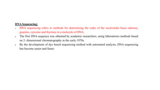
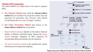
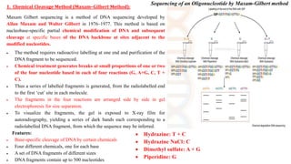
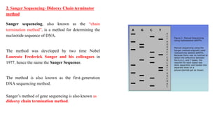
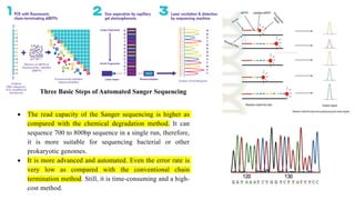
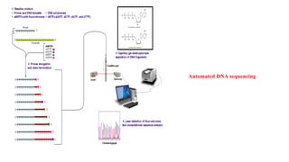
The document discusses different methods of DNA sequencing including the Maxam-Gilbert and Sanger chain termination methods as well as newer next generation sequencing techniques. It describes the principles, steps, and significance of the Maxam-Gilbert and Sanger methods and how next generation sequencing improved DNA sequencing by allowing millions of DNA molecules to be sequenced simultaneously in an automated process.
Sanger sequencing is one of the DNA sequencing methods used to identify and determine the sequence (Nucleotide) of DNA .This is an enzymatic method of sequencing developed by Fred Sanger.
Sanger sequencing is a method of DNA sequencing based on the selective incorporation of chain-terminating dideoxynucleotides by DNA polymerase during in vitro DNA replication.
The DNA Data Bank of Japan (DDBJ) is a biological database that collects DNA sequences. It is located at the National Institute of Genetics (NIG) in the Shizuoka prefecture of Japan. It is also a member of the International Nucleotide Sequence Database Collaboration or INSDC.
BAC & YAC are artificially prepared chromosomes to clone DNA sequences.yeast artificial chromosome is capable of carrying upto 1000 kbp of inserted DNA sequence
A physical map of a chromosome or a genome that shows the physical locations of genes and other DNA sequences of interest. Physical maps are used to help scientists identify and isolate genes by positional cloning.
According to the ICSM (Intergovernmental Committee on Surveying and Mapping), there are five different types of maps: General Reference, Topographical, Thematic, Navigation Charts and Cadastral Maps and Plans.
DNA consists of a linear string of nucleotides, or bases, for simplicity, referred to by the first letters of their chemical names--A, T, C and G. The process of deducing the order of nucleotides in DNA is called DNA sequencing. Since the DNA sequence confers information that the cell uses to make RNA molecules and proteins, establishing the sequence of DNA is key for understanding how genomes work. The technology for DNA sequencing was made faster and less expensive as a part of the Human Genome Project. And recent developments have profoundly increased the efficiency of DNA sequencing even further.
DNA sequencing is the process of determining the nucleic acid sequence – the order of nucleotides in DNA. It includes any method or technology that is used to determine the order of the four bases: adenine, guanine, cytosine, and thymine. The advent of rapid DNA sequencing methods has greatly accelerated biological and medical research and discovery.
Knowledge of DNA sequences has become indispensable for basic biological research, DNA Genographic Projects and in numerous applied fields such as medical diagnosis, biotechnology, forensic biology, virology and biological systematics. Comparing healthy and mutated DNA sequences can diagnose different diseases including various cancers,characterize antibody repertoire, and can be used to guide patient treatment.[5Having a quick way to sequence DNA allows for faster and more individualized medical care to be administered, and for more organisms to be identified and cataloged.
The rapid speed of sequencing attained with modern DNA sequencing technology has been instrumental in the sequencing of complete DNA sequences, or genomes, of numerous types and species of life, including the human genome and other complete DNA sequences of many animal, plant, and microbial species.
The first DNA sequences were obtained in the early 1970s by academic researchers using laborious methods based on two-dimensional chromatography. Following the development of fluorescence-based sequencing methods with a DNA sequencer, DNA sequencing has become easier and orders of magnitude faster.
DNA sequencing refers to the general laboratory technique for determining the exact sequence of nucleotides, or bases, in a DNA molecule. The sequence of the bases (often referred to by the first letters of their chemical names: A, T, C, and G) encodes the biological information that cells use to develop and operate.Whole Genome Sequencing
•Allows doctors to closely analyze a patient's genes for mutations and health indicators.
•Can detect intellectual disabilities and developmental delays.
•WGS is currently available at Yale for patients in the NICU and PICU.
•Involves Genetics.Sequencing may be utilized to determine the order of nucleotides in small targeted genomic regions or entire genomes. Illumina sequencing enables a wide variety of applications, allowing researchers to ask virtually any question related to the genome, transcriptome, or epigenome of any organism.The spectrum of analysis of NGS can extend from a small number of genes to an entire genome, depending on the goal. Whole-genome sequencing (WGS) and whole-exome sequencing (WES) provide the sequence of DNA bases across the genome and exome, respectively.Capillary electrophoresis (CE) instruments are capable of performing both Sanger sequencing and fragment analysis. Fragment analysis is a method in which DNA fragments are fluorescently labeled, separated by CE, and sized by comparison to an internal standard. sanger and Maxam-Gilbert sequencing technologies were classified
1.This presentation contain information about DNA and the mehods used for their sequencing like whole genome sequencing and shotgun sequenencing.
2.Advantages and disadvantages of whole genome sequencing and shot gun sequencing are also mentioned .
3.And the most important one is the applications of DNA sequencing.
The chain-termination method developed by Frederick Sanger and coworkers in 1977. This method used fewer toxic chemicals and lower amounts of radioactivity than the Maxam and Gilbert method. Because of its comparative ease, the Sanger method was soon automated and was the method used in the first generation of DNA sequencers.
DNA sequencing is the process of determining the nucleic acid sequence – the order of nucleotides in DNA. It includes any method or technology that is used to determine the order of the four bases: adenine, guanine, cytosine, and thymine.
Restriction fragment length polymorphism (abbreviated RFLP) refers to differences (or variations) among people in their DNA sequences at sites recognized by restriction enzymes. Such variation results in different sized (or length) DNA fragments produced by digesting the DNA with a restriction enzyme.
Mapping and quantifying transcripts:
Northern blots
S1 mapping of 5’ and 3’ end transcripts
Primer extension
Runoff transcription and G –less cassette transcription
Nuclear Runon transcription
A brief information about the SCOP protein database used in bioinformatics.
The Structural Classification of Proteins (SCOP) database is a comprehensive and authoritative resource for the structural and evolutionary relationships of proteins. It provides a detailed and curated classification of protein structures, grouping them into families, superfamilies, and folds based on their structural and sequence similarities.
(May 29th, 2024) Advancements in Intravital Microscopy- Insights for Preclini...Scintica Instrumentation
Intravital microscopy (IVM) is a powerful tool utilized to study cellular behavior over time and space in vivo. Much of our understanding of cell biology has been accomplished using various in vitro and ex vivo methods; however, these studies do not necessarily reflect the natural dynamics of biological processes. Unlike traditional cell culture or fixed tissue imaging, IVM allows for the ultra-fast high-resolution imaging of cellular processes over time and space and were studied in its natural environment. Real-time visualization of biological processes in the context of an intact organism helps maintain physiological relevance and provide insights into the progression of disease, response to treatments or developmental processes.
In this webinar we give an overview of advanced applications of the IVM system in preclinical research. IVIM technology is a provider of all-in-one intravital microscopy systems and solutions optimized for in vivo imaging of live animal models at sub-micron resolution. The system’s unique features and user-friendly software enables researchers to probe fast dynamic biological processes such as immune cell tracking, cell-cell interaction as well as vascularization and tumor metastasis with exceptional detail. This webinar will also give an overview of IVM being utilized in drug development, offering a view into the intricate interaction between drugs/nanoparticles and tissues in vivo and allows for the evaluation of therapeutic intervention in a variety of tissues and organs. This interdisciplinary collaboration continues to drive the advancements of novel therapeutic strategies.
Richard's aventures in two entangled wonderlandsRichard Gill
Since the loophole-free Bell experiments of 2020 and the Nobel prizes in physics of 2022, critics of Bell's work have retreated to the fortress of super-determinism. Now, super-determinism is a derogatory word - it just means "determinism". Palmer, Hance and Hossenfelder argue that quantum mechanics and determinism are not incompatible, using a sophisticated mathematical construction based on a subtle thinning of allowed states and measurements in quantum mechanics, such that what is left appears to make Bell's argument fail, without altering the empirical predictions of quantum mechanics. I think however that it is a smoke screen, and the slogan "lost in math" comes to my mind. I will discuss some other recent disproofs of Bell's theorem using the language of causality based on causal graphs. Causal thinking is also central to law and justice. I will mention surprising connections to my work on serial killer nurse cases, in particular the Dutch case of Lucia de Berk and the current UK case of Lucy Letby.
Observation of Io’s Resurfacing via Plume Deposition Using Ground-based Adapt...Sérgio Sacani
Since volcanic activity was first discovered on Io from Voyager images in 1979, changes
on Io’s surface have been monitored from both spacecraft and ground-based telescopes.
Here, we present the highest spatial resolution images of Io ever obtained from a groundbased telescope. These images, acquired by the SHARK-VIS instrument on the Large
Binocular Telescope, show evidence of a major resurfacing event on Io’s trailing hemisphere. When compared to the most recent spacecraft images, the SHARK-VIS images
show that a plume deposit from a powerful eruption at Pillan Patera has covered part
of the long-lived Pele plume deposit. Although this type of resurfacing event may be common on Io, few have been detected due to the rarity of spacecraft visits and the previously low spatial resolution available from Earth-based telescopes. The SHARK-VIS instrument ushers in a new era of high resolution imaging of Io’s surface using adaptive
optics at visible wavelengths.
Professional air quality monitoring systems provide immediate, on-site data for analysis, compliance, and decision-making.
Monitor common gases, weather parameters, particulates.
Multi-source connectivity as the driver of solar wind variability in the heli...Sérgio Sacani
The ambient solar wind that flls the heliosphere originates from multiple
sources in the solar corona and is highly structured. It is often described
as high-speed, relatively homogeneous, plasma streams from coronal
holes and slow-speed, highly variable, streams whose source regions are
under debate. A key goal of ESA/NASA’s Solar Orbiter mission is to identify
solar wind sources and understand what drives the complexity seen in the
heliosphere. By combining magnetic feld modelling and spectroscopic
techniques with high-resolution observations and measurements, we show
that the solar wind variability detected in situ by Solar Orbiter in March
2022 is driven by spatio-temporal changes in the magnetic connectivity to
multiple sources in the solar atmosphere. The magnetic feld footpoints
connected to the spacecraft moved from the boundaries of a coronal hole
to one active region (12961) and then across to another region (12957). This
is refected in the in situ measurements, which show the transition from fast
to highly Alfvénic then to slow solar wind that is disrupted by the arrival of
a coronal mass ejection. Our results describe solar wind variability at 0.5 au
but are applicable to near-Earth observatories.
This pdf is about the Schizophrenia.
For more details visit on YouTube; @SELF-EXPLANATORY;
https://www.youtube.com/channel/UCAiarMZDNhe1A3Rnpr_WkzA/videos
Thanks...!
Richard's entangled aventures in wonderlandRichard Gill
Since the loophole-free Bell experiments of 2020 and the Nobel prizes in physics of 2022, critics of Bell's work have retreated to the fortress of super-determinism. Now, super-determinism is a derogatory word - it just means "determinism". Palmer, Hance and Hossenfelder argue that quantum mechanics and determinism are not incompatible, using a sophisticated mathematical construction based on a subtle thinning of allowed states and measurements in quantum mechanics, such that what is left appears to make Bell's argument fail, without altering the empirical predictions of quantum mechanics. I think however that it is a smoke screen, and the slogan "lost in math" comes to my mind. I will discuss some other recent disproofs of Bell's theorem using the language of causality based on causal graphs. Causal thinking is also central to law and justice. I will mention surprising connections to my work on serial killer nurse cases, in particular the Dutch case of Lucia de Berk and the current UK case of Lucy Letby.
1. Lecture on DNA Sequencing
Dr. Manikandan Kathirvel M.Sc., Ph.D., (NET)
Assistant Professor,
Department of Life Sciences,
Kristu Jayanti College (Autonomous),
(Reaccredited with "A" Grade by NAAC)
Affiliated to Bengaluru North University,
K. Narayanapura, Kothanur (PO)
Bengaluru Mobile: 9624060194/8072730524
Email: manikandan@kristujayanti.com
ORCID ID: 0000000270066334
2. DNA Sequencing:
DNA sequencing refers to methods for determining the order of the nucleotides bases adenine,
guanine, cytosine and thymine in a molecule of DNA.
The first DNA sequence was obtained by academic researchers, using laboratories methods based
on 2- dimensional chromatography in the early 1970s.
By the development of dye based sequencing method with automated analysis, DNA sequencing
has become easier and faster.
3. Methods of DNA sequencing:
Two main methods are widely known to be used to sequence
DNA:
1. The Chemical Method (also called the Maxam–Gilbert
method after its inventors). By using this method, they had
sequenced 24 nucleotides only. However, their method
was published after two years of Sanger’s method.
2. The Chain Termination Method (also known as the
Sanger dideoxy method after its inventor).
Maxam–Gilbert technique depends on the relative chemical
liability of different nucleotide bonds, whereas the Sanger
method interrupts elongation of DNA sequences by
incorporating dideoxynucleotides into the sequences.
The chain termination method is the method more usually
used because of its speed and simplicity.
Sequencing of an Oligonucleotide by Maxam-Gilbert method
4. 1. Chemical Cleavage Method (Maxam–Gilbert Method):
Maxam Gilbert sequencing is a method of DNA sequencing developed by
Allan Maxam and Walter Gilbert in 1976–1977. This method is based on
nucleobase-specific partial chemical modification of DNA and subsequent
cleavage at specific bases of the DNA backbone at sites adjacent to the
modified nucleotides.
The method requires radioactive labelling at one end and purification of the
DNA fragment to be sequenced.
Chemical treatment generates breaks at small proportions of one or two
of the four nucleotide based in each of four reactions (G, A+G, C, T +
C).
Thus a series of labelled fragments is generated, from the radiolabelled end
to the first ‘cut’ site in each molecule.
The fragments in the four reactions are arranged side by side in gel
electrophoresis for size separation.
To visualize the fragments, the gel is exposed to X-ray film for
autoradiography, yielding a series of dark bands each corresponding to a
radiolabelled DNA fragment, from which the sequence may be inferred.
Features:
Base-specific cleavage of DNA by certain chemicals
Four different chemicals, one for each base
A set of DNA fragments of different sizes
DNA fragments contain up to 500 nucleotides
Hydrazine: T + C
Hydrazine NaCl: C
Dimethyl sulfate: A + G
Piperidine: G
Sequencing of an Oligonucleotide by Maxam-Gilbert method
5. Procedure:
1. DNA extraction is the very first step. After that, the DNA is denatured using
the heat denaturation method and single-stranded DNA is generated.
2. The phosphate (5’P) end of the DNA is removed and labelled by the
radiolabeled P32. The enzyme named phosphatase removes the phosphate
from the DNA and simultaneously, the kinase adds the 32P to the 5’ end of it.
3. 4 different chemicals are used to cleave DNA at four different positions;
hydrazine and hydrazine NaCl are selectively attack pyrimidine nucleotides
while dimethyl sulfate and piperidine attack purine nucleotides.
Hydrazine: T + C
Hydrazine NaCl: C
Dimethyl sulfate: A + G
Piperidine: G
4. An equal volume of 4 different ssDNA samples is taken into 4 different tubes
each containing 4 different chemicals. The samples are incubated for
sometimes and electrophoresed in polyacrylamide gel electrophoresis. The
results of the chemicals cleavage of four different tubes are shown in the
figure below.
5. Autoradiography is used to visualize the separation of DNA fragments. Due
to the radiolabelled 32P end of the DNA, the DNA bands visualized through
autoradiography.
Sequencing of an Oligonucleotide by Maxam-Gilbert method
6. Procedure:
1. DNA extraction is the very first step. After that, the DNA is denatured using
the heat denaturation method and single-stranded DNA is generated.
2. The phosphate (5’P) end of the DNA is removed and labelled by the
radiolabeled P32. The enzyme named phosphatase removes the phosphate
from the DNA and simultaneously, the kinase adds the 32P to the 5’ end of it.
3. 4 different chemicals are used to cleave DNA at four different positions;
hydrazine and hydrazine NaCl are selectively attack pyrimidine nucleotides
while dimethyl sulfate and piperidine attack purine nucleotides.
Hydrazine: C + T
Hydrazine NaCl: C
Dimethyl sulfate: A + G
Piperidine: G
4. An equal volume of 4 different ssDNA samples is taken into 4 different tubes
each containing 4 different chemicals. The samples are incubated for
sometimes and electrophoresed in polyacrylamide gel electrophoresis. The
results of the chemicals cleavage of four different tubes are shown in the
figure below.
5. Autoradiography is used to visualize the separation of DNA fragments. Due
to the radiolabelled 32P end of the DNA, the DNA bands visualized through
autoradiography. Sequencing of an Oligonucleotide by Maxam-Gilbert method
7. Advantages:
Purified DNA can be read directly
Homopolymeric DNA runs are sequenced as efficiently as
heterogeneous DNA sequences
Can be used to analyze DNA protein interactions (i.e.
footprinting)
Can be used to analyze nucleic acid structure and epigenetic
modifications to DNA
Disadvantages:
It requires extensive use of hazardous chemicals.
It has a relatively complex set up / technical complexity.
It is difficult to “scale up” and cannot be used to analyze
more than 500 base pairs.
The read length decreases from incomplete cleavage
reactions.
8. 2. Sanger Sequencing- Dideoxy Chain terminator
method
Sanger sequencing, also known as the “chain
termination method”, is a method for determining the
nucleotide sequence of DNA.
The method was developed by two time Nobel
Laureate Frederick Sanger and his colleagues in
1977, hence the name the Sanger Sequence.
The method is also known as the first-generation
DNA sequencing method.
Sanger’s method of gene sequencing is also known as
dideoxy chain termination method.
9. Principle:
The key principle of the Sanger method was the use of
dideoxynucleotide triphosphates (ddNTPs) as DNA chain
terminators.
A DNA primer is attached by hybridization to the template
strand and deoxynucleosides triphosphates (dNTPPs) are
sequentially added to the primer strand by DNA polymerase.
The M13 primer is designed along with the known
sequences at 3’ end of the template strand.
The reaction mixture also contains dideoxynucleoside
triphosphate (ddNTPs) along with usual dNTPs.
If during replication, ddNTPs is incorporated instead of
usual dNTPs in the growing DNA strand then the
replication stops at that nucleotide.
The ddNTPs are analogue of dNTPs
ddNTPs lacks hydroxyl group (-OH) at c3 of ribose sugar, so it cannot
make phosphodiester bond with nest nucleotide, thus terminates the
nucleotide chain
Respective ddNTPs of dNTPs terminates chain at their respective site.
For example ddATP terminates at A site. Similarly ddCTP, ddGTP and
ddTTP terminates at C, G and T site respectively.
10. Sanger Sequencing Steps
There are 4 main steps to Sanger sequencing.
1. Template preparation: DNA Sequence for Chain
Termination PCR
2. Generation of nested set of labelled fragments
3. Size Separation by Gel Electrophoresis and gel reading
4. Gel Analysis & Determination of DNA Sequence
11. 1. Template preparation: DNA Sequence for Chain
Termination PCR
The DNA sequence of interest is used as a template for a special
type of PCR called chain-termination PCR.
Steps:
1. Copies of template strand to be sequenced must be
prepared with short known sequences at 3’ end of the
template strand.
2. A DNA primer (M13 SEQUENCING PRIMER) is essential to
initiate replication of template, so primer preparation of
known sequences at 3’end is always required.
12. 2. Generation of nested set of labelled fragments:
Steps:
Copies of each template is divided into four batches and
each batch is used for different replication reaction.
Copies of standard primer, normal dNTPs and DNA
polymerase I are used in all four batches.
To synthesize fragments that terminates at A,
1. ddATP (can be radiolabelled) is added to the reaction
mixture to the batch I along with dATP, dTTP, dCTP and
dGTP,
2. standard primer and
3. DNA polymerase I.
Similarly, to generate, all fragments that terminates at
C, G and T,
the respective ddNTPs i.e. ddCTP, ddGTP and ddTTP are added
respectively to different reaction mixture on different batch
along with usual dNTPs.
13. 3. Size Separation by Gel Electrophoresis and gel reading
Steps:
The reaction mixture from four batches are loaded into
four different well on polyacrylamide gel and
electrophoresed.
The autoradiogram of the gel is read to determine the
order of bases of complementary strand to that of
template strand.
The band of shortest fragments is at the bottom of
autoradiogram so that the sequences of
complementary strand are read from bottom to top.
4. Gel Analysis & Determination of DNA Sequence
The last step simply involves reading the gel to determine the
sequence of the input DNA.
In manual Sanger sequencing, the user reads all four lanes of
the gel at once, moving bottom to top, using the lane to
determine the identity of the terminal ddNTP for each band. For
example, if the bottom band is found in the column corresponding
to ddGTP, then the smallest PCR fragment terminates with
ddGTP, and the first nucleotide from the 5’ end of the original
sequence has a guanine (G) base.
14. Significance of DNA Sequencing:
Information obtained by DNA sequencing makes it possible to understand or
alter the function of genes.
DNA sequence analysis demonstrates regulatory regions that control gene
expression and genetic “hot spots” particularly susceptible to mutation.
Comparison of DNA sequences shows evolutionary relationships that provide
a framework for definite classification of microorganisms including viruses.
Comparison of DNA sequences facilitates identification of conserved regions,
which are useful for development of specific hybridization probes to detect
microorganisms including viruses in clinical samples.
DNA sequencing has become sufficiently fast and inexpensive to allow
laboratory determination of microbial sequences for identification of microbes.
Sequencing of the 16S ribosomal subunit can be used to identify specific
bacteria. Sequencing of viruses can be used to identify the virus and
distinguish different strains.
DNA sequencing shows gene structure that helps research workers to find out
the structure of gene products.
15. Automated DNA sequencing:
The manual Sanger method was tedious. However, recent
advancement into the sequencing makes it easy and rapid to use.
The semi-automated Sanger sequencing method is based on the
principle of Sanger’s method with some minor variations.
Instead of the 4 different reactions, the automated DNA
sequencing carried out in the single tube and the DNA runs in a
single lane.
Here, in the semi-automated DNA sequencing, the fluorescent-
labeled set of primers are used, instead of ddNTPs. Thus four
different primers give four different peaks.
The PAGE method isn’t capable of separating all the fragments
in a single reaction. Therefore, alternatively, the capillary gel
electrophoresis method is practiced. This method separates each
and every single fragment precisely.
The capillary electrophoresis used to separate DNA molecules
on the basis of the size, it is powerful enough to separate single
base pair fragment. The chromatogram generated through the
C.E sent the output as a fluorescent peak.
The advanced semi-automated Sanger sequencing
method is more accurate, reliable and faster than
the traditional method.
16. Three Basic Steps of Automated Sanger Sequencing
The read capacity of the Sanger sequencing is higher as
compared with the chemical degradation method. It can
sequence 700 to 800bp sequence in a single run, therefore,
it is more suitable for sequencing bacterial or other
prokaryotic genomes.
It is more advanced and automated. Even the error rate is
very low as compared with the conventional chain
termination method. Still, it is time-consuming and a high-
cost method.
18. Next Generation Sequencing (NGS)
Important Next Generation Sequencing Techniques
The next-generation sequencing platform is different from the Sanger technique or chain
termination method of DNA sequencing. Broadly, it amplifies millions of copies of a
particular fragment in a massively parallel fashion and the “reads” are analyzed by the
computational program.
Pyro sequencing
Illumina (Solexa) sequencing
Lynx therapeutics’ massively parallel signature sequencing (MPSS)
Polony sequencing
SOLiD sequencing
DNA nanoball sequencing
Helioscope single molecule sequencing
Single molecule SMRT sequencing
Single molecule real time (RNAP) sequencing
Next Generation Sequencing (NGS) is a powerful platform that has enabled the sequencing of
thousands to millions of DNA molecules simultaneously.
19. The generations of sequencing:
First Generation
Maxam and Gilbert DNA sequencing and Sanger DNA Sequencing
Second Generation Sequencing
Pyrosequencing
Sequencing by Reversible Terminator Chemistry
Sequencing by Ligation
Third Generation Sequencing
Single Molecule Fluorescent Sequencing
Single Molecule Real Time Sequencing
Semiconductor Sequencing
Nanopore Sequencing
Fourth Generation Sequencing
Aims conducting genomic analysis directly in the cell