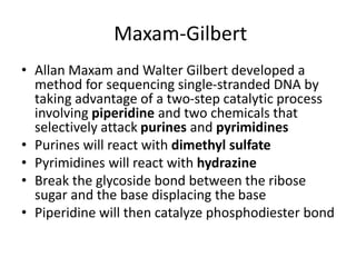
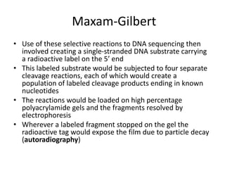
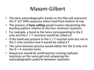
DNA Sequencing
Aman Ullah
B.Sc. Med. Lab. Technology
M. Phil. Microbiology
Certificate in Health Professional Education
Lecturer, Department of Medical Lab. Technology
Institute of Paramedical Sciences, Khyber Medical University, Peshawar, Pakistan
Introduction
• Prior to the mid-1970’s no method existed by
which DNA could be directly sequenced
• In the mid-1970’s two methods were
developed for directly sequencing DNA
1. Maxam-Gilbert chemical cleavage method
2. Sanger chain-termination method
Maxam-Gilbert
• Allan Maxam and Walter Gilbert developed a
method for sequencing single-stranded DNA by
taking advantage of a two-step catalytic process
involving piperidine and two chemicals that
selectively attack purines and pyrimidines
• Purines will react with dimethyl sulfate
• Pyrimidines will react with hydrazine
• Break the glycoside bond between the ribose
sugar and the base displacing the base
• Piperidine will then catalyze phosphodiester bond
Maxam-Gilbert
• Dimethyl sulfate and piperidine alone will
selectively cleave guanine nucleotides but
dimethyl sulfate and piperidine in formic acid
will cleave both guanine andadenine
nucleotides
• Similarly, hydrazine and piperidine will cleave
both thymine and cytosine nucleotides
whereas hydrazine and piperidine in 1.5M
NaCl will only cleave cytosine nucleotides
Maxam-Gilbert
• Use of these selective reactions to DNA sequencing then
involved creating a single-stranded DNA substrate carrying
a radioactive label on the 5’ end
• This labeled substrate would be subjected to four separate
cleavage reactions, each of which would create a
population of labeled cleavage products ending in known
nucleotides
• The reactions would be loaded on high percentage
polyacrylamide gels and the fragments resolved by
electrophoresis
• Wherever a labeled fragment stopped on the gel the
radioactive tag would expose the film due to particle decay
(autoradiography)
Maxam-Gilbert
• The dark autoradiographic bands on the film will represent
the 5’→3’ DNA sequence when read from bottom to top
• The process of base calling would involve interpreting the
banding pattern relative to the four chemical reactions
• For example, a band in the lanes corresponding to the C
only and the C + T reactions would be called a C
• If the band was present in the C + T reaction lane but not in
the C only reaction lane it would be called a T
• The same decision process would obtain for the G only and
the G + A reaction lanes
• Sequences would be confirmed by running replicate
reactions on the same gel and comparing the
autoradiographic patterns between replicates
Sanger method
• Fred Sanger developed an alternative method
• Sanger opted for a method involving a third form of the
ribose sugars
• Ribose has a hydroxyl group on both the 2’ and the 3’
carbons
• Deoxyribose has only the one hydroxyl group on the 3’
carbon
• This is not a concern for polynucleotide synthesis in vivo
since the coupling occurs through the 3’ carbon in both
RNA and DNA
• There is a third form of ribose in which the hydroxyl group
is missing from both the 2’ and the 3’ carbons
• This is dideoxyribose
Sanger method
• whenever a dideoxynucleotide was
incorporated into a polynucleotide, the chain
would irreversibly stop, or terminate
• Thus, the incorporation of specific
dideoxynucleotides in vitro would result in
selective chain termination
Sanger method
• Sanger proceeded to establish a protocol in
which four separate reactions, each
incorporating a different dideoxynucleotide
along with the four deoxynucleotides, would
produce a population of fragments all ending
in the same dideoxynucleotide in the presence
of a DNA polymerase if the ratio of the
dideoxynucleotide and the corresponding
deoxynucleotide was properly set.
Sanger method
• If the primer was radiolabeled instead of the
substrate, the resulting fragment population
would be labeled and could be resolved on
polyacrylamide gels
• Auto-radiography was the same but base
calling was easier
• The one new twist was that the sequence
fragments on the gel were the complement of
the actual template
Automated Fluorescence Sequencing
• The most dramatic advance in sequencing and the one that
carried DNA sequencing into a high throughput
environment was the introduction of automated
sequencing using fluorescence-labeled dideoxy-terminators
• In 1986, Leroy Hood and colleagues reported on a DNA
sequencing method in which the radioactive labels,
autoradiography, and manual base calling were all replaced
by fluorescent labels, laser induced fluorescence detection,
and computerized base calling
• In their method, the primer was labeled with one of four
different fluorescent dyes and each was placed in a
separate sequencing reaction with one of the four
dideoxynucleotides plus all four deoxynucleotides
Automated Fluorescence Sequencing
• Once the reactions were complete, the four
reactions were pooled and run together in one
lane of a polyacrylamide sequencing gel
• A four-color laser induced fluorescence detector
scanned the gel as the reaction fragments
migrated past
• The fluorescence signature of each fragment was
then sent to a computer where the software was
trained to perform base calling
• This method was commercialized in 1987 by
Applied Biosystems
Questions/Suggestions
khurramthalwi@hotamail.com

DNA Sequencing

  • 1.
    DNA Sequencing Aman Ullah B.Sc.Med. Lab. Technology M. Phil. Microbiology Certificate in Health Professional Education Lecturer, Department of Medical Lab. Technology Institute of Paramedical Sciences, Khyber Medical University, Peshawar, Pakistan
  • 2.
    Introduction • Prior tothe mid-1970’s no method existed by which DNA could be directly sequenced • In the mid-1970’s two methods were developed for directly sequencing DNA 1. Maxam-Gilbert chemical cleavage method 2. Sanger chain-termination method
  • 3.
    Maxam-Gilbert • Allan Maxamand Walter Gilbert developed a method for sequencing single-stranded DNA by taking advantage of a two-step catalytic process involving piperidine and two chemicals that selectively attack purines and pyrimidines • Purines will react with dimethyl sulfate • Pyrimidines will react with hydrazine • Break the glycoside bond between the ribose sugar and the base displacing the base • Piperidine will then catalyze phosphodiester bond
  • 4.
    Maxam-Gilbert • Dimethyl sulfateand piperidine alone will selectively cleave guanine nucleotides but dimethyl sulfate and piperidine in formic acid will cleave both guanine andadenine nucleotides • Similarly, hydrazine and piperidine will cleave both thymine and cytosine nucleotides whereas hydrazine and piperidine in 1.5M NaCl will only cleave cytosine nucleotides
  • 5.
    Maxam-Gilbert • Use ofthese selective reactions to DNA sequencing then involved creating a single-stranded DNA substrate carrying a radioactive label on the 5’ end • This labeled substrate would be subjected to four separate cleavage reactions, each of which would create a population of labeled cleavage products ending in known nucleotides • The reactions would be loaded on high percentage polyacrylamide gels and the fragments resolved by electrophoresis • Wherever a labeled fragment stopped on the gel the radioactive tag would expose the film due to particle decay (autoradiography)
  • 6.
    Maxam-Gilbert • The darkautoradiographic bands on the film will represent the 5’→3’ DNA sequence when read from bottom to top • The process of base calling would involve interpreting the banding pattern relative to the four chemical reactions • For example, a band in the lanes corresponding to the C only and the C + T reactions would be called a C • If the band was present in the C + T reaction lane but not in the C only reaction lane it would be called a T • The same decision process would obtain for the G only and the G + A reaction lanes • Sequences would be confirmed by running replicate reactions on the same gel and comparing the autoradiographic patterns between replicates
  • 9.
    Sanger method • FredSanger developed an alternative method • Sanger opted for a method involving a third form of the ribose sugars • Ribose has a hydroxyl group on both the 2’ and the 3’ carbons • Deoxyribose has only the one hydroxyl group on the 3’ carbon • This is not a concern for polynucleotide synthesis in vivo since the coupling occurs through the 3’ carbon in both RNA and DNA • There is a third form of ribose in which the hydroxyl group is missing from both the 2’ and the 3’ carbons • This is dideoxyribose
  • 11.
    Sanger method • whenevera dideoxynucleotide was incorporated into a polynucleotide, the chain would irreversibly stop, or terminate • Thus, the incorporation of specific dideoxynucleotides in vitro would result in selective chain termination
  • 12.
    Sanger method • Sangerproceeded to establish a protocol in which four separate reactions, each incorporating a different dideoxynucleotide along with the four deoxynucleotides, would produce a population of fragments all ending in the same dideoxynucleotide in the presence of a DNA polymerase if the ratio of the dideoxynucleotide and the corresponding deoxynucleotide was properly set.
  • 13.
    Sanger method • Ifthe primer was radiolabeled instead of the substrate, the resulting fragment population would be labeled and could be resolved on polyacrylamide gels • Auto-radiography was the same but base calling was easier • The one new twist was that the sequence fragments on the gel were the complement of the actual template
  • 15.
    Automated Fluorescence Sequencing •The most dramatic advance in sequencing and the one that carried DNA sequencing into a high throughput environment was the introduction of automated sequencing using fluorescence-labeled dideoxy-terminators • In 1986, Leroy Hood and colleagues reported on a DNA sequencing method in which the radioactive labels, autoradiography, and manual base calling were all replaced by fluorescent labels, laser induced fluorescence detection, and computerized base calling • In their method, the primer was labeled with one of four different fluorescent dyes and each was placed in a separate sequencing reaction with one of the four dideoxynucleotides plus all four deoxynucleotides
  • 16.
    Automated Fluorescence Sequencing •Once the reactions were complete, the four reactions were pooled and run together in one lane of a polyacrylamide sequencing gel • A four-color laser induced fluorescence detector scanned the gel as the reaction fragments migrated past • The fluorescence signature of each fragment was then sent to a computer where the software was trained to perform base calling • This method was commercialized in 1987 by Applied Biosystems
  • 17.