Electrophoretic
mobility shift Assay
Group no. 15
16604 Fatima Tahir
16630 Iqra akbar
16638 Hajra Illayas
Introduction
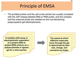
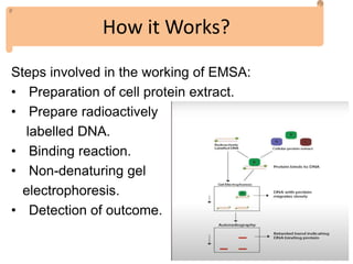
• The electrophoretic mobility shift assay is a
powerful technique for detecting specific-binding
of nucleic acid-protein complexes.
• Over the past 30 years, EMSA has been the “go
to assay” to investigate the qualitative interactions
between nucleic acids (DNA or RNA) and nucleic-
acid binding proteins.
• It is rapid and sensitive method to detect protein
complex with the nucleic acid.
What is EMSA?
It is also known as Gel shift assay or gel mobility shift assay,
band shift assay, or gel retardation assay.
It is technique to detect weather protein is attached with
DNA or RNA or not. As we know, there are many cellular
processes where a protein needs to interact with the DNA or
RNA like in transcription and translation.
Originally utilized broadly in the investigation of sequence-
specific DNA-binding proteins such as transcription factors,
EMSA has been additionally developed to explore DNA-
protein interactions, RNA- protein interactions.
EMSA is a sensitive method, using radioisotopes to label
nucleic acids and autoradiography, it is possible to use very
low concentrations and small sample volumes.
Principle of EMSA
• The purified protein and the cell crude extract are usually incubated
with the 32P isotope-labeled DNA or RNA probe, and the complex
and the unbound probe are isolated on the non-denaturing
polypropylene gel electrophoresis.
A mobility shift assay is
electrophoretic separation
of a protein-DNA or
protein-RNA mixture on a
polyacrylamide or agrose
gel for a short period.
The speed at which
different molecules
move through the gel
is determined by their
size, charge, and
sometime by shape.
Steps involved in the working of EMSA:
• Preparation of cell protein extract.
• Prepare radioactively
labelled DNA.
• Binding reaction.
• Non-denaturing gel
electrophoresis.
• Detection of outcome.
How it Works?
Working of EMSA
• The protein (P) binding to the unique site on a
DNA (D), will form a complex PD; in equilibrium
with the free components.
When there is a strong interaction between proteins and
DNA ka ≥ Kd and a distinct band is observed.
However, due to dissociation occurs during
electrophoresis, a faint smear a faint smear will also show
between two bands.
If single DNA molecule has multiple binding sites for an
individual protein, there will be multiple complexes formed,
and we observe many bands.
Protocol or steps we follow in EMSA
1- Making labeled DNA:
It has three ways:
1-
2-
• Biotinylation:
2-Preparation of Gel:
Ensure the gel plate, spacer and comb are clean.
Remove all fingerprints through glass plate by
methanol and dry all components before use.
Prepare polymerization mixture.
Pour the mixture into the glass plate and make
sure to avoid bubble formation. Insert the comb
immediately.
3- Pre-electrophoresis:
• Remove comb and bottom
spacer from gel and mount the
gel in the vertical
electrophoresis apparatus.
• Place glycerol and dye
solution in each well of the gel
and conduct pre-
electrophoresis.
• This is done to just to see of
the gel has formed properly,
rununig the gel one it does not
breakdown.
4- Sample preparation and equilibration:
 Prepare sample and equilibrate for 30 min at 20 + 1 ° C.
This can be carried out while the gel is undergoing pre-
electrophoresis.
5- Electrophoresis:
 Detection of electrophoretic bands on the basis of
radioactivity of samples.
 At the end, remove the gel and plate assembly from the
electrophoresis device and dry it.
6- Autoradiograph:
 To visualize gel bands.
 In a lab with dim light (for phosphorus screen) place film
or phosphorus in an exposure casset.
• Multiple protein binding:
• Super shift:
Control Study understudy and result
How and why titration is done?
• As we know that, we are going to have DNA
interacting with protein and for that to be
happen we need to have enough DNA and
protein in the solution.
It is used to study
conformational
changes in DNA . For
example a EMSA 2-D
variant with electron
microscopy is used for
the characterization of
conformation.
Analysis of cellular
extracts for the
presence of certain
DNA-binding proteins.
Bound and free DNA
are separated using
EMSA.
It proves useful
for stoichiometric
analysis. Number
of protein that
bind per DNA
fragment
It is also used too study wether transcription factor is attached to the DNA or not or if
transcription factor attached is our targeted or not and their activity.
Applications
Advantages of EMSA
It can be used with wide range of nucleic acid, sizes,
structure as well as wide range of proteins.
It is relatively simple method to perform. It is also used
for qualitative analysis.
It proves useful in detection of protein that binds to
DNA, and cause difficulty in migration of fragment on
gel.
Highly sensitive method and usually of Low cost.
No special equipment needed.
In this technique, it is possible to use both crude
protein extracts and purified recombinant proteins.
Drawbacks of EMSA:
EMSA doesn’t provide a straight forward measure of
the weights of the proteins as mobility is influenced by
several other methods.
Not an appropriate method for kinetic studies.
Dissociation is also one of the drawback of EMSA. It
occurs during electrophoresis thus prevents detection.
Interacting biomolecules must have different
electrophoretic mobilities.
Conclusion:
Electrophoretic mobility shift
assay (EMSA) is most widely
used method for the
detection of protein-DNA
interactions.
Used for various
purposes such as
quantifying
interactions
between DNA and
proteins
Works on the
observation that
protein-bounded
DNA migrate slowly
as compared to free
DNA
Electrophoretic mobility shift assay

Electrophoretic mobility shift assay

  • 1.
    Electrophoretic mobility shift Assay Groupno. 15 16604 Fatima Tahir 16630 Iqra akbar 16638 Hajra Illayas
  • 2.
    Introduction • The electrophoreticmobility shift assay is a powerful technique for detecting specific-binding of nucleic acid-protein complexes. • Over the past 30 years, EMSA has been the “go to assay” to investigate the qualitative interactions between nucleic acids (DNA or RNA) and nucleic- acid binding proteins. • It is rapid and sensitive method to detect protein complex with the nucleic acid.
  • 3.
    What is EMSA? Itis also known as Gel shift assay or gel mobility shift assay, band shift assay, or gel retardation assay. It is technique to detect weather protein is attached with DNA or RNA or not. As we know, there are many cellular processes where a protein needs to interact with the DNA or RNA like in transcription and translation. Originally utilized broadly in the investigation of sequence- specific DNA-binding proteins such as transcription factors, EMSA has been additionally developed to explore DNA- protein interactions, RNA- protein interactions. EMSA is a sensitive method, using radioisotopes to label nucleic acids and autoradiography, it is possible to use very low concentrations and small sample volumes.
  • 4.
    Principle of EMSA •The purified protein and the cell crude extract are usually incubated with the 32P isotope-labeled DNA or RNA probe, and the complex and the unbound probe are isolated on the non-denaturing polypropylene gel electrophoresis. A mobility shift assay is electrophoretic separation of a protein-DNA or protein-RNA mixture on a polyacrylamide or agrose gel for a short period. The speed at which different molecules move through the gel is determined by their size, charge, and sometime by shape.
  • 5.
    Steps involved inthe working of EMSA: • Preparation of cell protein extract. • Prepare radioactively labelled DNA. • Binding reaction. • Non-denaturing gel electrophoresis. • Detection of outcome. How it Works?
  • 6.
  • 7.
    • The protein(P) binding to the unique site on a DNA (D), will form a complex PD; in equilibrium with the free components. When there is a strong interaction between proteins and DNA ka ≥ Kd and a distinct band is observed. However, due to dissociation occurs during electrophoresis, a faint smear a faint smear will also show between two bands. If single DNA molecule has multiple binding sites for an individual protein, there will be multiple complexes formed, and we observe many bands.
  • 8.
    Protocol or stepswe follow in EMSA 1- Making labeled DNA: It has three ways: 1- 2-
  • 9.
    • Biotinylation: 2-Preparation ofGel: Ensure the gel plate, spacer and comb are clean. Remove all fingerprints through glass plate by methanol and dry all components before use. Prepare polymerization mixture. Pour the mixture into the glass plate and make sure to avoid bubble formation. Insert the comb immediately.
  • 10.
    3- Pre-electrophoresis: • Removecomb and bottom spacer from gel and mount the gel in the vertical electrophoresis apparatus. • Place glycerol and dye solution in each well of the gel and conduct pre- electrophoresis. • This is done to just to see of the gel has formed properly, rununig the gel one it does not breakdown.
  • 11.
    4- Sample preparationand equilibration:  Prepare sample and equilibrate for 30 min at 20 + 1 ° C. This can be carried out while the gel is undergoing pre- electrophoresis. 5- Electrophoresis:  Detection of electrophoretic bands on the basis of radioactivity of samples.  At the end, remove the gel and plate assembly from the electrophoresis device and dry it. 6- Autoradiograph:  To visualize gel bands.  In a lab with dim light (for phosphorus screen) place film or phosphorus in an exposure casset.
  • 12.
    • Multiple proteinbinding: • Super shift:
  • 13.
  • 14.
    How and whytitration is done? • As we know that, we are going to have DNA interacting with protein and for that to be happen we need to have enough DNA and protein in the solution.
  • 15.
    It is usedto study conformational changes in DNA . For example a EMSA 2-D variant with electron microscopy is used for the characterization of conformation. Analysis of cellular extracts for the presence of certain DNA-binding proteins. Bound and free DNA are separated using EMSA. It proves useful for stoichiometric analysis. Number of protein that bind per DNA fragment It is also used too study wether transcription factor is attached to the DNA or not or if transcription factor attached is our targeted or not and their activity. Applications
  • 16.
    Advantages of EMSA Itcan be used with wide range of nucleic acid, sizes, structure as well as wide range of proteins. It is relatively simple method to perform. It is also used for qualitative analysis. It proves useful in detection of protein that binds to DNA, and cause difficulty in migration of fragment on gel. Highly sensitive method and usually of Low cost. No special equipment needed. In this technique, it is possible to use both crude protein extracts and purified recombinant proteins.
  • 17.
    Drawbacks of EMSA: EMSAdoesn’t provide a straight forward measure of the weights of the proteins as mobility is influenced by several other methods. Not an appropriate method for kinetic studies. Dissociation is also one of the drawback of EMSA. It occurs during electrophoresis thus prevents detection. Interacting biomolecules must have different electrophoretic mobilities.
  • 18.
    Conclusion: Electrophoretic mobility shift assay(EMSA) is most widely used method for the detection of protein-DNA interactions. Used for various purposes such as quantifying interactions between DNA and proteins Works on the observation that protein-bounded DNA migrate slowly as compared to free DNA