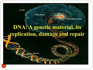
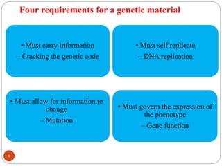
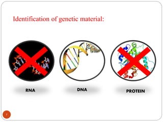
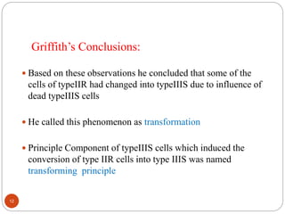
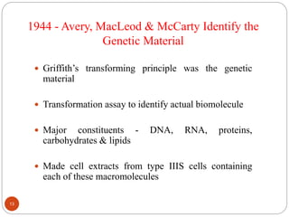
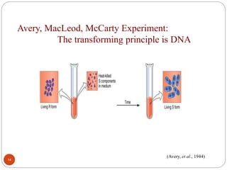
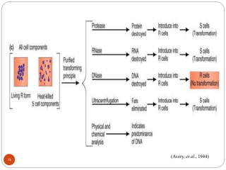
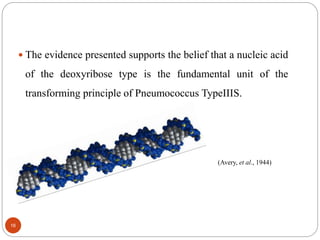
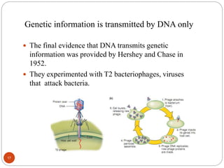
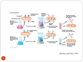
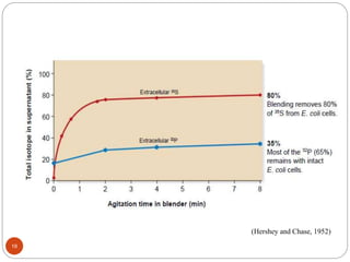
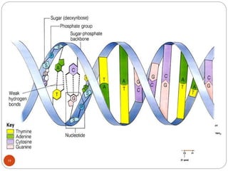
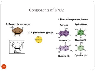
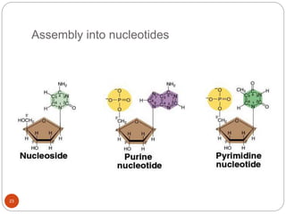
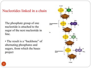
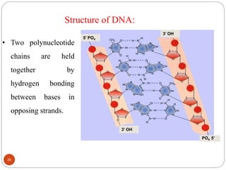
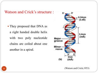
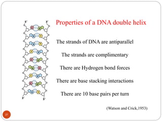
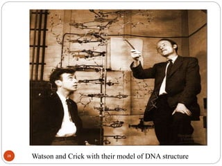
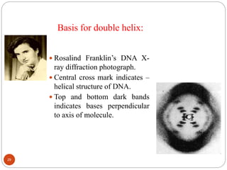
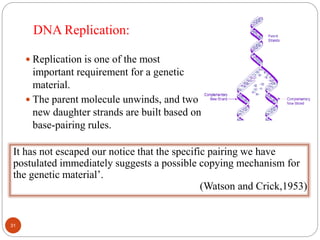
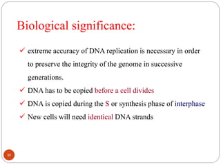
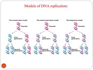
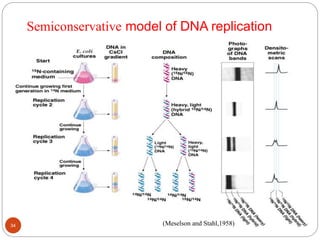
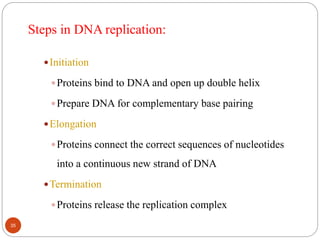
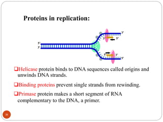
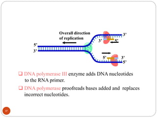
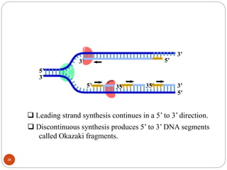
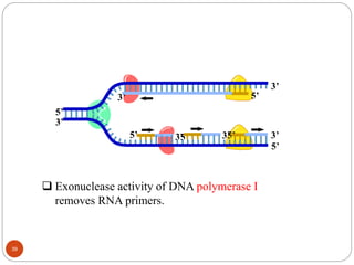
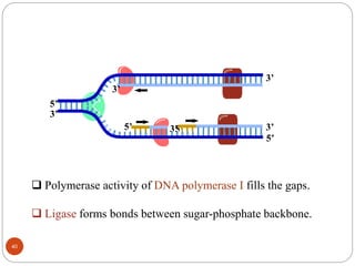
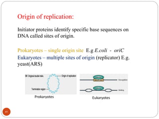
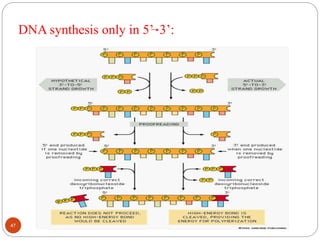
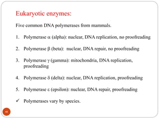
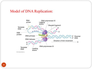
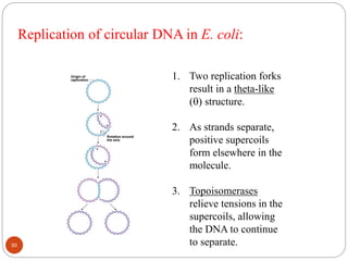
The document provides information on DNA, including its identification as the genetic material, its structure and replication. Some key points:
- Experiments in the 1920s-1950s identified DNA as the genetic material, including Griffith's work on bacterial transformation and the experiments of Avery, MacLeod and McCarty and Hershey and Chase.
- DNA is made up of nucleotides containing phosphate groups, sugars and nitrogenous bases. Watson and Crick discovered its double helix structure in 1953, with two anti-parallel strands held together by hydrogen bonds between complementary bases.
- DNA replication is semi-conservative and involves unwinding of the DNA strands, synthesis of new complementary strands, and production of identical double
Transcription in eukaryotes: A brief view
Transcription is the process by which single stranded RNA is synthesized by double stranded DNA. Transcription in eukaryotes and prokaryotes has many similarities while at the same time both showing their individual characteristics due to the differences in organization. RNA Polymerase (RNAP or RNA Pol) is different in prokaryotes and eukaryotes. Coupled transcription is seen in prokaryotes but not in Eukaryotes. In eukaryotes the pre-RNA should be spliced first to be translated.
In Eukaryotic transcription, synthesis of RNA occurs in the 3’→5’ direction. The 3’ end is more reactive due to the hydroxide group. 5’ end containing phosphate groups meanwhile, is not very reactive when it comes to adding new nucleotides. In Eukaryotes, the whole genome is not transcribed at once. Only a part of the genome is transcribed which also acts as the first, principle stage of genetic regulation.
Eukaryotes have five nuclear polymerases:
• RNA Polymerase I: This produces rRNA (23S, 5.8S, and 18S) which are the major components in a ribosome. This also produces pre-rRNA in yeasts.
• RNA Polymerase II: Helps in the production of mRNA (messenger RNA), snRNA (small, nuclear RNA), miRNA. This is the most studied type and requires several transcription factors for its binding
• RNA Polymerase III: This synthesizes tRNA (transfer RNA), 5S rRNA and other small RNAs required in the cytosol and nucleus.
• RNA Polymerase IV: Synthesizes siRNA (small interfering RNA) in plants.
• RNA Polymerase V: This is the least studied polymerase and synthesizes siRNA-directed heterochromatin in plants.
Eukaryotic transcription can be broadly divided into 4 stages:
• Pre-Initiation
• Initiation
• Elongation
• Termination
Transcription is an elaborate process which cells use to copy the genetic information stored in DNA into RNA. This pre-RNA is modified into mRNA before being transcribed to proteins. Transcription is the first step to utilizing the genetic information in a cell. Both Eukaryotes and Prokaryotes employ this process with the basic phases remaining the same. However eukaryotic transcription is more complex indicating the changes transcription has undergone towards perfection during evolution.
This power point presentation explains double helical structure of DNA as proposed by Watson and Crick (1953).Attempts have also been made to high light the valuable contributions made by Rosalind Franklin and Wilkins. Brief details of different types of DNA have also been included.
wooble hypothesis is the important topic for the molecular biology . this will help u to learn about new type of base pairing method and a new base pair that was discovered. this method is also opposite of watson crick pairing
Prokaryotic and eukaryotic dna replication with their clinical applicationsrohini sane
A comprehensive presentation on Prokaryotic and Eukaryotic DNA Replication with their clinical applications for MBBS , BDS, B Pharm & Biotechnology students to facilitate self- study.
This presentation is about the transcription machinery that is required for the transcription in eukaryotes. The comparison between the transcription factors involved in prokaryotes and eukaryotes. The initiation of transcription and how it helps in producing a mRNA.
Dna methylation ppt
definition of Dna methylation ppt
discovery of Dna methylation ppt
types of Dna methylation ppt
history of Dna methylation ppt
process of Dna methylation ppt
mechanism of Dna methylation ppt
methylation in cancer
cytosine methylation
genomic imprinting
Replication (prokaryotes and eukaryotes) FN 312.pptsultanasadia912
The traditional monoclonal antibody (mAb) production process usually starts with generation of mAb-producing cells (i.e. hybridomas) by fusing myeloma cells with desired antibody-producing splenocytes (e.g. B cells). These B cells are typically sourced from animals, usually mice. After cell fusion, large numbers of clones are screened and selected on the basis of antigen specificity and immunoglobulin class. Once candidate hybridoma cell lines are identified, each "hit" is confirmed, validated, and characterized using a variety of downstream functional assays. Upon completion, the clones are scaled up where additional downstream bioprocesses occur.
DNA is a double helical structure that transfers the genetic information from one generation to another. it consists of two strands with the four nucleotide basis .The four nucleotides contains adenine, cytosine, guanine, thymine .These four nuclic basis are such arranged and coiled with the help of hydrogen bonds and forms the helical structure of DNA. In RNA the thymine is replaced with uracil. Here you will learn the replication ,transcription and translation process in DNA.
DNA is made of two linked strands that wind around each other to resemble a twisted ladder — a shape known as a double helix. Each strand has a backbone made of alternating sugar (deoxyribose) and phosphate groups. Attached to each sugar is one of four bases: adenine (A), cytosine (C), guanine (G) or thymine (T).
Introduction:
RNA interference (RNAi) or Post-Transcriptional Gene Silencing (PTGS) is an important biological process for modulating eukaryotic gene expression.
It is highly conserved process of posttranscriptional gene silencing by which double stranded RNA (dsRNA) causes sequence-specific degradation of mRNA sequences.
dsRNA-induced gene silencing (RNAi) is reported in a wide range of eukaryotes ranging from worms, insects, mammals and plants.
This process mediates resistance to both endogenous parasitic and exogenous pathogenic nucleic acids, and regulates the expression of protein-coding genes.
What are small ncRNAs?
micro RNA (miRNA)
short interfering RNA (siRNA)
Properties of small non-coding RNA:
Involved in silencing mRNA transcripts.
Called “small” because they are usually only about 21-24 nucleotides long.
Synthesized by first cutting up longer precursor sequences (like the 61nt one that Lee discovered).
Silence an mRNA by base pairing with some sequence on the mRNA.
Discovery of siRNA?
The first small RNA:
In 1993 Rosalind Lee (Victor Ambros lab) was studying a non- coding gene in C. elegans, lin-4, that was involved in silencing of another gene, lin-14, at the appropriate time in the
development of the worm C. elegans.
Two small transcripts of lin-4 (22nt and 61nt) were found to be complementary to a sequence in the 3' UTR of lin-14.
Because lin-4 encoded no protein, she deduced that it must be these transcripts that are causing the silencing by RNA-RNA interactions.
Types of RNAi ( non coding RNA)
MiRNA
Length (23-25 nt)
Trans acting
Binds with target MRNA in mismatch
Translation inhibition
Si RNA
Length 21 nt.
Cis acting
Bind with target Mrna in perfect complementary sequence
Piwi-RNA
Length ; 25 to 36 nt.
Expressed in Germ Cells
Regulates trnasposomes activity
MECHANISM OF RNAI:
First the double-stranded RNA teams up with a protein complex named Dicer, which cuts the long RNA into short pieces.
Then another protein complex called RISC (RNA-induced silencing complex) discards one of the two RNA strands.
The RISC-docked, single-stranded RNA then pairs with the homologous mRNA and destroys it.
THE RISC COMPLEX:
RISC is large(>500kD) RNA multi- protein Binding complex which triggers MRNA degradation in response to MRNA
Unwinding of double stranded Si RNA by ATP independent Helicase
Active component of RISC is Ago proteins( ENDONUCLEASE) which cleave target MRNA.
DICER: endonuclease (RNase Family III)
Argonaute: Central Component of the RNA-Induced Silencing Complex (RISC)
One strand of the dsRNA produced by Dicer is retained in the RISC complex in association with Argonaute
ARGONAUTE PROTEIN :
1.PAZ(PIWI/Argonaute/ Zwille)- Recognition of target MRNA
2.PIWI (p-element induced wimpy Testis)- breaks Phosphodiester bond of mRNA.)RNAse H activity.
MiRNA:
The Double-stranded RNAs are naturally produced in eukaryotic cells during development, and they have a key role in regulating gene expression .
Richard's entangled aventures in wonderlandRichard Gill
Since the loophole-free Bell experiments of 2020 and the Nobel prizes in physics of 2022, critics of Bell's work have retreated to the fortress of super-determinism. Now, super-determinism is a derogatory word - it just means "determinism". Palmer, Hance and Hossenfelder argue that quantum mechanics and determinism are not incompatible, using a sophisticated mathematical construction based on a subtle thinning of allowed states and measurements in quantum mechanics, such that what is left appears to make Bell's argument fail, without altering the empirical predictions of quantum mechanics. I think however that it is a smoke screen, and the slogan "lost in math" comes to my mind. I will discuss some other recent disproofs of Bell's theorem using the language of causality based on causal graphs. Causal thinking is also central to law and justice. I will mention surprising connections to my work on serial killer nurse cases, in particular the Dutch case of Lucia de Berk and the current UK case of Lucy Letby.
Observation of Io’s Resurfacing via Plume Deposition Using Ground-based Adapt...Sérgio Sacani
Since volcanic activity was first discovered on Io from Voyager images in 1979, changes
on Io’s surface have been monitored from both spacecraft and ground-based telescopes.
Here, we present the highest spatial resolution images of Io ever obtained from a groundbased telescope. These images, acquired by the SHARK-VIS instrument on the Large
Binocular Telescope, show evidence of a major resurfacing event on Io’s trailing hemisphere. When compared to the most recent spacecraft images, the SHARK-VIS images
show that a plume deposit from a powerful eruption at Pillan Patera has covered part
of the long-lived Pele plume deposit. Although this type of resurfacing event may be common on Io, few have been detected due to the rarity of spacecraft visits and the previously low spatial resolution available from Earth-based telescopes. The SHARK-VIS instrument ushers in a new era of high resolution imaging of Io’s surface using adaptive
optics at visible wavelengths.
This pdf is about the Schizophrenia.
For more details visit on YouTube; @SELF-EXPLANATORY;
https://www.youtube.com/channel/UCAiarMZDNhe1A3Rnpr_WkzA/videos
Thanks...!
Cancer cell metabolism: special Reference to Lactate PathwayAADYARAJPANDEY1
Normal Cell Metabolism:
Cellular respiration describes the series of steps that cells use to break down sugar and other chemicals to get the energy we need to function.
Energy is stored in the bonds of glucose and when glucose is broken down, much of that energy is released.
Cell utilize energy in the form of ATP.
The first step of respiration is called glycolysis. In a series of steps, glycolysis breaks glucose into two smaller molecules - a chemical called pyruvate. A small amount of ATP is formed during this process.
Most healthy cells continue the breakdown in a second process, called the Kreb's cycle. The Kreb's cycle allows cells to “burn” the pyruvates made in glycolysis to get more ATP.
The last step in the breakdown of glucose is called oxidative phosphorylation (Ox-Phos).
It takes place in specialized cell structures called mitochondria. This process produces a large amount of ATP. Importantly, cells need oxygen to complete oxidative phosphorylation.
If a cell completes only glycolysis, only 2 molecules of ATP are made per glucose. However, if the cell completes the entire respiration process (glycolysis - Kreb's - oxidative phosphorylation), about 36 molecules of ATP are created, giving it much more energy to use.
IN CANCER CELL:
Unlike healthy cells that "burn" the entire molecule of sugar to capture a large amount of energy as ATP, cancer cells are wasteful.
Cancer cells only partially break down sugar molecules. They overuse the first step of respiration, glycolysis. They frequently do not complete the second step, oxidative phosphorylation.
This results in only 2 molecules of ATP per each glucose molecule instead of the 36 or so ATPs healthy cells gain. As a result, cancer cells need to use a lot more sugar molecules to get enough energy to survive.
introduction to WARBERG PHENOMENA:
WARBURG EFFECT Usually, cancer cells are highly glycolytic (glucose addiction) and take up more glucose than do normal cells from outside.
Otto Heinrich Warburg (; 8 October 1883 – 1 August 1970) In 1931 was awarded the Nobel Prize in Physiology for his "discovery of the nature and mode of action of the respiratory enzyme.
WARNBURG EFFECT : cancer cells under aerobic (well-oxygenated) conditions to metabolize glucose to lactate (aerobic glycolysis) is known as the Warburg effect. Warburg made the observation that tumor slices consume glucose and secrete lactate at a higher rate than normal tissues.
Multi-source connectivity as the driver of solar wind variability in the heli...Sérgio Sacani
The ambient solar wind that flls the heliosphere originates from multiple
sources in the solar corona and is highly structured. It is often described
as high-speed, relatively homogeneous, plasma streams from coronal
holes and slow-speed, highly variable, streams whose source regions are
under debate. A key goal of ESA/NASA’s Solar Orbiter mission is to identify
solar wind sources and understand what drives the complexity seen in the
heliosphere. By combining magnetic feld modelling and spectroscopic
techniques with high-resolution observations and measurements, we show
that the solar wind variability detected in situ by Solar Orbiter in March
2022 is driven by spatio-temporal changes in the magnetic connectivity to
multiple sources in the solar atmosphere. The magnetic feld footpoints
connected to the spacecraft moved from the boundaries of a coronal hole
to one active region (12961) and then across to another region (12957). This
is refected in the in situ measurements, which show the transition from fast
to highly Alfvénic then to slow solar wind that is disrupted by the arrival of
a coronal mass ejection. Our results describe solar wind variability at 0.5 au
but are applicable to near-Earth observatories.
4. Introduction
The progeny of organism develops characters similar to that
organism
The resemblance of offspring to their parents depends on the
precise transmission of principle component from one generation
to the next
That component is-
The Genetic Material
4
6. Four requirements for a genetic material
6
• Must carry information
– Cracking the genetic code
• Must self replicate
– DNA replication
• Must allow for information to
change
– Mutation
• Must govern the expression of
the phenotype
– Gene function
8. The process of identification of genetic material began in 1928
with experiments of Griffith and concluded in 1952 with the
studies of Hershey and Chase.
Between these two experiments other three scientists, Avery,
Macloed and McCarty were did an experiment to identify the
genetic material.
8
9. Discovery of Transformation in Bacteria:
In 1928, Frederick Griffith discovered bacterial transformation.
He worked on Streptococcus pneumonieae (Pneumococcus)
Pneumococci have various strains which can be classified by-
1. The presence or absence of a polysaccharide capsule
2. The molecular composition of the capsule
When grown on blood agar medium, pneumococci with capsules
are virulent and form large, smooth colonies and designated as
typeIII S
9
10. S pneumococci mutate to an avirulent form that has no
capsules.
When grown on blood agar medium, these noncapsulated
pnuemococci form small, rough-surfaced colonies and
designated as typeII R
Based on the molecular composition of the capsule, these
pneumococci cells are type I, II, III, and so forth.
10
12. Based on these observations he concluded that some of the
cells of typeIIR had changed into typeIIIS due to influence of
dead typeIIIS cells
He called this phenomenon as transformation
Principle Component of typeIIIS cells which induced the
conversion of type IIR cells into type IIIS was named
transforming principle
Griffith’s Conclusions:
12
13. Griffith’s transforming principle was the genetic
material
Transformation assay to identify actual biomolecule
Major constituents - DNA, RNA, proteins,
carbohydrates & lipids
Made cell extracts from type IIIS cells containing
each of these macromolecules
1944 - Avery, MacLeod & McCarty Identify the
Genetic Material
13
14. Avery, MacLeod, McCarty Experiment:
The transforming principle is DNA
14
(Avery, et al., 1944)
16. The evidence presented supports the belief that a nucleic acid
of the deoxyribose type is the fundamental unit of the
transforming principle of Pneumococcus TypeIIIS.
(Avery, et al., 1944)
16
17. Genetic information is transmitted by DNA only
The final evidence that DNA transmits genetic
information was provided by Hershey and Chase in
1952.
They experimented with T2 bacteriophages, viruses
that attack bacteria.
17
20. • The sulphur containing protein of resting phage particles is
confined to a protective coat that is responsible for the adsorption
to bacteria, and functions as an instrument for injection of the
phage DNA into the cell. This protein probably has no role in
growth of intracellular phage. The DNA has some function.
Their conclusion:
(Hershey and Chase, 1952)
20
21. What is DNA?
• nitrogen base and sugar make a nucleoside.
• Phosphate group and a nucleoside make a
nucleotide.
•DNA is deoxyribo nucleic acid. A German
chemist,Friedrich Miescher, discovered
DNA in 1869.
19
•DNA contains three main components
(1) Phosphate (PO4) groups;
(2) Five-carbon sugars; and
(3) Nitrogen-containing bases called
purines and pyrimidines.
24. Nucleotides linked in a chain
The phosphate group of one
nucleotide is attached to the
sugar of the next nucleotide in
line.
• The result is a “backbone” of
alternating phosphates and
sugars, from which the bases
project
24
25. 5’ PO4
PO4 5’
3’ OH
3’ OH
Structure of DNA:
• Two polynucleotide
chains are held
together by
hydrogen bonding
between bases in
opposing strands.
25
26. Watson and Crick’s structure :
They proposed that DNA as
a right handed double helix
with two poly nucleotide
chains are coiled about one
another in a spiral.
(Watson and Crick,1953)26
27. The strands of DNA are antiparallel
The strands are complimentary
There are Hydrogen bond forces
There are base stacking interactions
There are 10 base pairs per turn
Properties of a DNA double helix
(Watson and Crick,1953)
27
28. 28 Watson and Crick with their model of DNA structure
29. Basis for double helix:
Rosalind Franklin’s DNA X-
ray diffraction photograph.
Central cross mark indicates –
helical structure of DNA.
Top and bottom dark bands
indicates bases perpendicular
to axis of molecule.
29
30. Chargaff’s base pairing rule:
Percent of adenine = percent of thymine (A=T)
Percent of cytosine = percent of guanine (C=G)
A+G = T+C (or purines = pyrimidines)
(Chargaff et al.,1950)
30
31. DNA Replication:
Replication is one of the most
important requirement for a genetic
material.
The parent molecule unwinds, and two
new daughter strands are built based on
base-pairing rules.
It has not escaped our notice that the specific pairing we have
postulated immediately suggests a possible copying mechanism for
the genetic material’.
(Watson and Crick,1953)
31
32. extreme accuracy of DNA replication is necessary in order
to preserve the integrity of the genome in successive
generations.
DNA has to be copied before a cell divides
DNA is copied during the S or synthesis phase of interphase
New cells will need identical DNA strands
Biological significance:
32
35. Steps in DNA replication:
Initiation
Proteins bind to DNA and open up double helix
Prepare DNA for complementary base pairing
Elongation
Proteins connect the correct sequences of nucleotides
into a continuous new strand of DNA
Termination
Proteins release the replication complex
35
36. Binding proteins prevent single strands from rewinding.
Helicase protein binds to DNA sequences called origins and
unwinds DNA strands.
5’
3’
5’
3’
Primase protein makes a short segment of RNA
complementary to the DNA, a primer.
3’5’
5’3’
Proteins in replication:
36
40. Polymerase activity of DNA polymerase I fills the gaps.
Ligase forms bonds between sugar-phosphate backbone.
3’
5’
3’
5’ 3’
5’
3’
3’
5’
40
41. Origin of replication:
Initiator proteins identify specific base sequences on
DNA called sites of origin.
Prokaryotes – single origin site E.g E.coli - oriC
Eukaryotes – multiple sites of origin (replicator) E.g.
yeast(ARS)
Prokaryotes Eukaryotes
41
42. Most eukaryotes except for budding yeast have ill-defined
origins of replication that rely on epigenetic mechanisms for
molecular recognition by initiator proteins.
Replication is initiated at multiple origins along the DNA
using a conserved mechanism that consists of four steps:
origin recognition, assembly of a prereplicative initiation
complex, followed by activation of the helicase and loading of
the replisome.
(Sclafani and Holzen,2007)
42
48. Eukaryotic enzymes:
Five common DNA polymerases from mammals.
1. Polymerase (alpha): nuclear, DNA replication, no proofreading
2. Polymerase (beta): nuclear, DNA repair, no proofreading
3. Polymerase (gamma): mitochondria, DNA replication,
proofreading
4. Polymerase (delta): nuclear, DNA replication, proofreading
5. Polymerase (epsilon): nuclear, DNA repair, proofreading
Polymerases vary by species.
48
50. Replication of circular DNA in E. coli:
1. Two replication forks
result in a theta-like
() structure.
2. As strands separate,
positive supercoils
form elsewhere in the
molecule.
3. Topoisomerases
relieve tensions in the
supercoils, allowing
the DNA to continue
to separate.50
51. 1. Common in several bacteriophages
including .
2. Begins with a nick at the origin of
replication.
3. 5’ end of the molecule is displaced and
acts as primer for DNA synthesis.
4. Can result in a DNA molecule many
multiples of the genome length
5. During viral assembly the DNA is cut
into individual viral chromosomes.51
Rolling circle model of DNA Replication:
52. End-replication problem:
Every time a linear chromosome replicates, the laggaing strand at each end
gets shorter by about 150 nucleotides. Because there is a minimum length
of DNA needed for initiation of an Okazaki fragment.
DNA polymerase/ligase cannot fill gap at end of chromosome after RNA
primer is removed. If this gap is not filled, chromosomes would become
shorter each round of replication.
Eukaryotes have tandemly repeated sequences at the ends of their
chromosomes.
Telomerase binds to the terminal telomere repeat and catalyzes the
addition of of new repeats.
Compensates by lengthening the chromosome.
52
53. DNA Damage and Repair:
DNA polymerase do great job during DNA replication by
proof reading the new DNA strand.
But its not enough to maintain the 100% fidelity in
replication.
Several kinds of damage occurs by endogenous and
exogenous agents.
DNA has its own mechanisms to repair this damages and
maintain the accuracy of copying mechanism.
53
54. 54
Natural polymerase error
Endogenous DNA damage
oxidative damage
depurination
Exogenous DNA damage
radiation
chemical adducts
“Error-prone” DNA repair
Sources of
damage
55. DNA Damage Response(DDR):
To respond to these threats, eukaryotes have evolved the
DNA Damage Response (DDR).
The DDR is a complex signal transduction pathway that has
the ability to sense DNA damage and transduce this
information to the cell to influence cellular responses to
DNA damage.
(Ciccia and Elledge, 2010)
55
56. “Mutation is rare because of repair”
Over 200 human genes known to be involved in DNA repair
Major DNA repair pathways:
1. Base excision repair (BER)
2. DNA Mismatch repair (MMR)
3. Nucleotide excision repair (NER)
4. DNA strand break repair pathways:
Single strand break repair (SSBR)
Double-strand break repair pathways (DSBR)
Homologous Recombination (HR)
Nonhomologous end joining (NHEJ)
56
57. Direct reversal of damage - Photoreactivation (bacteria, yeast,
some vertebrates - not humans) Two thymines connected together
by UV light.
Excision Repair - removal of defective DNA. There are three
distinct types
1) base-excision
2) nucleotide-excision
3) mismatch repair
57
58. Base-excision repair(BER):
Presence of the Uracil in DNA is a great example of this type
Special enzymes replace just the defective base
snip out the defective base
cut the DNA strand
Add fresh nucleotide
Ligate gap
N
N
NH2
O
O
H2
C
O
O
N
H
N
O
O
O
H2
C
O
O
deoxycytosine deoxyuracil
1’
2’3’
4’
5’
12
3
4
5
6
CH3
thymine
glycosidic bond
58
59. Nucleotide-excision repair(NER):
Same as previous except that-
It removes entire dmaged nucleotide
Remove larger segments of DNA
Example:Xeroderma pigmentosum
• Extreme sensitivity to sunlight
• Predisposition to skin cancer
59
60. Mismatch repair (MMR):
Despite extraordinary fidelity of DNA synthesis, errors do
persist
Such errors can be detected and repaired by the post-
replication mismatch repair system
Special enzymes scan the DNA for bulky alterations in the
DNA double helix
These are normally caused by mismatched bases
A G
A C
C T
These are excised and the DNA repaired
60
61. MMR also processes mispairs that result from heteroduplex DNA
formed during genetic recombination: act to exclude
“homeologous” recombination.
Repair involving two or more close sites in same heteroduplex
occur much more often on the same strand than the opposite
strands.
Analysis of the pattern of repair suggest that repair tracks initiates
at mismatches and propagate preferentially in 5’ to 3’ direction.
(Wagner and Meselson, 1976)
61
62. The problem of strand discrimination:
MMR can only aid replication fidelity if repair is targeted to
newly synthesized strand
The cell has a mechanism of identifying new strand synthesis by
leaving nicks that DNA. There are enzymes which scan these
new regions looking for errors.
62
63. Other forms of DNA damage:
Depurination - the base is simply ripped out of the DNA molecule
leaving a gap.
Deamination - An amino group of Cytosine is removed and the
base becomes Uracil.
63
64. Basic mechanism is the
same for all three types
1) Remove damaged
region
2) Resynthesis DNA
3) Ligate
64