SlideShare a Scribd company logo
• In the mid-1800s; Gregor Mendel; studied the inheritance of various traits in pea
plants.
• In 1920s; P. A. Levene; elucidated basic chemical composition of nucleic acids.
• Fred Griffith; 1928; transfer of virulence in the pathogen Streptococcus pneumoniae
(pneumococcus).
• Between 1941 and 1944; Archer Martin and Richard Synge; invention of paper
chromatography.
• By 1948; Erwin Chargaff; had begun using paper chromatography to analyse the base
composition of DNA from a number of species.
• In 1951; Rosalind Franklin arrived at King’s College, London, and joined Maurice Wilkins
in his efforts to prepare highly oriented DNA fibers and study them by X-ray
crystallography. By the winter of 1952–1953, Franklin had obtained an excellent X-ray
diffraction photograph of DNA.
• 1953; Watson and Crick propose DNA double helix.
• 1962; Watson, Crick, and Wilkins received the Nobel Prize in 1962 for their discoveries.
Historical Highlights
The Search for the Genetic Material
1. Some substance must be responsible for passage of traits from
parents to offspring. For a substance to do this it must be:
a. Stable enough to store information for long periods.
b. Able to replicate accurately.
c. Capable of change to allow evolution.
DNA as Genetic Material
In the early 1900s, chromosomes were shown to be the
carriers of hereditary information.
DNA and Protein
Most scientists initially believed that protein must be the
genetic material.
• Frederick Griffith’s 1928 experiment with Streptococcus
pneumoniae bacteria in mice showed that something passed from dead
bacteria into nearby living ones, allowing them to change their cell
surface.
• He called this agent the transforming principle
Transformation: Griffith’s Experiments
Transformation
and
Recombination
Avery’s Transformation Experiment
DNA as Genetic Material: The Avery Experiment
1. In 1944, Avery, MacLeod and McCarty published results of a study that
identified the transforming principle from S. pneumoniae.
2. Only the nucleic acid fraction was capable of transforming the bacteria.
3. Critics noted that the nucleic acid fraction was contaminated with
proteins.
Oswald Avery with his colleagues set
out to discover which constituent in
the heat-killed virulent pneumococci
was responsible for Griffith’s
transformation.
The Hershey-Chase Bacteriophage Experiment
DNA as Genetic Material: The Hershey-Chase Experiment
1. More evidence for DNA as the genetic material came in 1953 with Alfred
Hershey and Martha Chase’s work on E. coli infected with bacteriophage
T2.
2. In one part of the experiment, T2 proteins were labeled with 35S, and in
the other part, T2 DNA was labeled with 32P. Then each group of labeled
viruses was mixed separately with the E. coli host.
3. The 35S-labeled protein was found outside the infected cells, while the
32P-labeled DNA was inside the E. coli.
The Hershey-Chase Experiment
(a) When E. coli was infected with a T2 phage containing 35S protein, most of the
radioactivity remained outside the host cell. (b) When a T2 phage containing 32P DNA was
mixed with the host bacterium, the radioactive DNA was injected into the cell and phages
were produced. Thus DNA was carrying the virus’s genetic information.
The Hershey-Chase Waring Blender
Martha Chase and Alfred Hershey, 1953
The Composition of Nucleic Acids
The Composition and Structure of DNA and RNA
• DNA and RNA are polymers composed of monomers called nucleotides.
• Each nucleotide has three parts:
a. A pentose (5-carbon) sugar.
b. A nitrogenous base.
c. A phosphate group.
• The pentose sugar in RNA is ribose, and in DNA it’s deoxyribose. The only
difference is at the 2’ position, where RNA has a hydroxyl (OH) group, while DNA
has only a hydrogen.
Structures of deoxyribose and ribose in DNA and RNA
There are two classes of nitrogenous bases:
a. Purines (double-ring, nine-membered structures) include adenine (A) and
guanine (G).
b. Pyrimidines (one-ring, six-membered structures) include cytosine (C),
thymine (T) in DNA and uracil (U) in RNA.
Structures of the nitrogenous bases in DNA and RNA
• The structure of nucleotides has these features:
a. The base is always attached by a covalent bond between the 1′ carbon of the
pentose sugar and a nitrogen in the base (specifically, the nine nitrogen in
purines and the one nitrogen in pyrimidines).
b. The sugar-base combination is a nucleoside. When a phosphate is added
(always to the 5′ carbon of the pentose sugar), it becomes a nucleoside
phosphate, or simply nucleotide.
c. Nucleotide examples are shown in Figure 2.11, and naming conventions are
given in Table 2.1.
• Polynucleotides of both DNA and RNA are formed by stable covalent bonds
(phosphodiester linkages) between the phosphate group on the 5′ carbon of one
nucleotide, and the 3′ hydroxyl on another nucleotide. This creates the
“backbone” of a nucleic acid molecule.
• The asymmetry of phosphodiester bonds creates 3′-5′ polarity within the nucleic
acid chain.
Fig. 2.11 Chemical structures of DNA and RNA
The Discovery of the DNA Double Helix
• James Watson and Francis Crick published the famous double-helix structure in 1953.
• When they began their work, it was known that DNA is composed of nucleotides, but
how the nucleotides are assembled into nucleic acid was unknown.
• Two additional sources of data assisted Watson and Crick with their model:
a. Erwin Chargaff’s ratios obtained for DNA derived from a variety of sources
showed that the amount of purine always equals the amount of pyrimidine, and
further, that the amount of G equals C, and the amount of A equals T.
b. Rosalind Franklin’s X ray diffraction images of DNA showed a helical structure with
regularities at 0.34 nm and 3.4 nm along the axis of the molecule (Figure 8.9).
Molecular structure of DNA
Watson and Crick’s three-dimensional
model has these main features:
a. It is two polynucleotide chains
wound around each other in a
right-handed helix.
b. The two chains are antiparallel.
c. The sugar-phosphate backbones are
on the outside of the helix, and the
bases are on the inside, stacked
perpendicularly to the long axis like
the steps of a spiral staircase.
d. The bases of the two strands are held
together by hydrogen bonds between
complementary bases (two for A-T pairs
and three for G-C pairs). Individual H-
bonds are relatively weak and so the
strands can be separated (by heating, for
example). Complementary base pairing
means that the sequence of one strand
dictates the sequence of the other strand.
e. The base pairs are 0.34 nm apart, and one full
turn of the DNA helix takes 3.4 nm, so there
are 10 bp in a complete turn. The diameter of a
dsDNA helix is 2 nm.
f. Because of the way the bases H-bond with
each other, the opposite sugar-phosphate
backbones are not equally spaced, resulting in
a major and minor groove. This feature of
DNA structure is important for protein binding.
• The 1962 Nobel Prize in Physiology or Medicine
was awarded to Francis Crick, James Watson and
Maurice Wilkins (the head of the lab in which
Franklin worked). Franklin had already died, and
so was not eligible.
Assignment on –
Different DNA Structures
The Genetic Code
• The final step in the expression of genes that encode proteins is translation.
• The mRNA nucleotide sequence is translated into the amino acid sequence of a
polypeptide chain.
• Protein synthesis is called translation because it is a decoding process.
• The information encoded in the language of nucleic acids must be rewritten in the
language of proteins.
• There is code degeneracy. i.e., there are up to 6 different codons for a given amino acid.
• Only 61 codons, the sense codons, direct amino acid incorporation into protein.
• The remaining 3 codons (UGA, UAG, and UAA) are involved in the termination of
translation and are called stop or nonsense codons.
• The 5′ nucleotide in the anticodon can vary, but generally, if the nucleotides in the second
and third anticodon positions complement the first two bases of the mRNA codon, an
aminoacyl-tRNA with the proper amino acid will bind to the mRNA-ribosome complex.
• This pattern is evident on inspection of changes in the amino acid specified with variation
in the third position (see table).
• This loose base pairing is known as wobble and relieves cells of the need to synthesize so
many tRNAs.
• Wobble also decreases the effects of DNA mutations.
• Genetic codons Discovered by Marshall Nirenberg ,Heinrich
Matthaei,philip,Leder and Har Govind Khorana in 1968.
• Nirenberg and Khorana the Nobel prize.
History and definition of Genetic Code
• Genetic code is a triplet of neiutides that coded that coded the
specific amino acid.
• Ex. AUG-met
• UUU-Phe
The amino acids encoded
by all 64 possible codons
were determined.
The genetic code required determining how 4 nucleotides (A, T, G, C) could
encode more than 20 amino acids.
Francis Crick and Sydney Brenner determined that the DNA is read in sets of 3
nucleotides for each amino acid.
Each codon consists of three bases (triplet). There are 64 codons.
They are all written in the 5' to 3' direction.
61 codons code for amino acids. The other three (UAA, UGA, UAG) are stop
codons (or nonsense codons) that terminate translation.
There is one start codon (initiation codon), AUG, coding for methionine. Protein
synthesis begins with methionine (Met) in eukaryotes, and formylmethionine
(fmet) in prokaryotes.
The code is unambiguous.
The Genetic Code
• The code is degenerate. More than one codon can specify a single
amino acid.
• All amino acids, except Met and tryptophan (Trp), have more than one
codon.
• For those amino acids having more than one codon, the first two bases
in the codon are usually the same. The base in the third position often
varies.
• The code is almost universal (the same in all organisms). Some minor
exceptions to this occur in mitochondria and some organisms.
• The code is commaless (contiguous).
• There are no spacers or "commas" between codons on an mRNA.
• Neighboring codons on a message are non-overlapping.
reading frame: the series of nucleotides read in sets of 3 (codon)
• only 1 reading frame is correct for encoding the correct sequence UU of amino
acids
UUC GAG
UUU
AUG
UCG AAU
U UCU CGA UGU UUG AGA
UU
AU
CUC GAU GUU UGA GAA U
Reading frame 1
Reading frame 2
Reading frame 3
Genetic Code
stop codons: 3 codons (UUA, UGA, UAG) in the genetic code used to
terminate translation.
start codon: the codon (AUG) used to signify the start of translation.
31
Types of codons
Unambiguous
 Each codon specifies a particular amino acid, the codon ACG codes for the
amino acid threonine, and only threonine.
Non overlapping
 This means that successive triplets are read in order. Each nucleotide is part of
only one triplet codon.
Commanelss which mean there is no punctuation witbeen two
codans
32
Characteristics of the Genetic Code
DNA Codon RNA Codon
• The first two bases of the codon make normal (canonical) H-bond pairs
with the 2nd and 3rd bases of the anticodon
• At the remaining position, less stringent rules apply and non-canonical
pairing may occur
• The rules: first base U can recognize A or G, first base G can recognize U
or C, and first base I can recognize U, C or A (I comes from deamination
of A)
• Advantage of wobble: dissociation of tRNA from mRNA is faster and
protein synthesis too
The Wobble Hypothesis
• It is a triplet code.
• – Each three-nucleotide codon in the mRNA specifies one amino acid
• • It is comma free.
• – mRNA is read three bases at a time without skipping any bases.
• • It is non-overlapping/non-ambiguous.
• • It is almost universal.
• – In nearly all organisms, most codons have the same amino acid meaning.
• – Of 20 amino acids, 18 are encoded by 2 or more codons.
• • The code has start and stop signals.
• – AUG is the usual start signal and defines the open reading frame.
Points to remember:
• • Stop signals are codons with no corresponding tRNA
• – the nonsense or chain-terminating codons.
• – generally three stop codons: UAG, UAA, and UGA.
• More than one codon can code for each amino acid.
• Each amino acid can be coded for by more than one codon
UNIT 2
Organization and function of genetic material
• Bacterial
• Viral
The Organization of DNA in Chromosomes
1. Cellular DNA is organized into chromosomes. A genome is the
chromosome or set of chromosomes that contains all the DNA of an
organism.
2. In prokaryotes the genome is usually a single circular chromsome.
In eukaryotes, the genome is one complete haploid set of nuclear
chromosomes; mitochondrial and chloroplast DNA are not included.
Viral Chromosomes
1. A virus is nucleic acid surrounded by a protein coat. The nucleic acid
may be dsDNA, ssDNA, dsRNA or ssRNA, and it may be linear or
circular, a single molecule or several segments.
2. Bacteriophages are viruses that infect bacteria. Three different types
that infect E. coli are good examples of the variety of chromosome
structure found in viruses.
T-even phage
3. The T-even phages (T2, T4 and T6) have similar structures; all have
dsDNA genomes composed of a one linear DNA molecule surrounded
by a protein coat.
ΦX174 phage
4. ΦX174 is a small, simple virus with one short ssDNA chromosome. In 1959,
Robert Sinsheimer found that the DNA of ΦX174 has a base composition
that does not fit the complementary base-pair-rules.  single strand DNA
rather than dsDNA
λ phage
5. Bacteriophage λ is somewhat like the T-
even phages in structure. However, its
chromosome changes form. A linear
molecule of dsDNA is packaged inside the
protein head, but after the virus infects its
host the chromosome becomes circular due
to base-pairing of complementary 12-base
single-stranded regions at the ends of the
linear molecule (Fig. 2.18)
Fig. 2.18  chromosome structure varies at stages of lytic infection of E. coli
Prokaryotic Chromosomes
1. The typical prokaryotic genome is one circular dsDNA chromosome, but
some prokaryotes are more exotic, with a main chromosome and one or
more smaller ones. When a minor chromosome is dispensable to the life
of the cell, it is called a plasmid. Some examples:
a. Borrelia burgdorferi (Lyme disease in humans) has a 0.91-Mb linear
chromosome, plus an additional 0.53-Mb of DNA in 17 different linear
and circular molecules.
b. Agrobacterium tumefaciens (crown gall disease of plants) has a 3.0-Mb
circular chromosome and a 2.1-Mb linear one.
2. Archaebacteria also vary in chromosomal organization, but only circular
forms have been found. Examples:
a. Methanococcus jannaschii has three chromosomes of 1.66-Mb, 58-kb
and 16-kb.
b. Archaeoglobus fulgidus has one 2.2-Mb circular chromosome.
3. Both Eubacteria and Archaebacteria lack a membrane-bounded nucleus,
hence their classification as prokaryotes. Their DNA is densely arranged in
a cytoplasmic region called the nucleoid.
4. In an experiment where E. coli is gently lysed, it releases one 4.6-Mb
circular chromosome, highly supercoiled (Figures 2.20). A 4.6-Mb double
helix is about 1mm in length, about 103 times longer than an E. coli cell.
DNA supercoiling helps it fit into the cell.
Fig. 2.20 Illustration of DNA supercoiling
5. A molecule of B-DNA, with 10bp/turn of the helix, is in relaxed conformation. If turns
of the helix are removed and the molecule circularized, the DNA will form
superhelical turns to compensate for the added tension.
6. Either overwinding or underwinding DNA will create a structure where 10bp/turn of
the helix is not the most energetically favored conformation, and supercoils will be
induced. Both positive and negative supercoils will condense the DNA.
7. All organisms contain topoisomerase enzymes to supercoil their DNA.
8. Prokaryotes also organize their DNA into looped domains, with the ends of the
domains held so that each is supercoiled independently (Figure 2.22).
9. The compaction factor for looped domains is about 10-fold. In E. coli there are
about 100 domains of about 40kb each.
Fig. 2.22 Model for the structure of a bacterial chromosome
UNIT 3
Brief account of plasmids-
• Structure
• Types
• In addition to the genetic material present in the nucleoid, many procaryotes (and
some yeasts and other fungi) contain extrachromosomal DNA molecules called
plasmids.
• In some cases, numerous different plasmids within a single species have been
identified. (B. burgdorferi, carries 12 linear and 9 circular plasmids)
• Plasmids-
small
double-stranded DNA molecules
can exist independently of the chromosome
replicate autonomously
mostly circular
• Linear plasmids possess special structures or sequences at their ends to prevent their
degradation and to permit their replication.
• Plasmids have relatively few genes, generally less than 30. Their genetic information
is not essential to the host, and cells that lack them usually function normally.
• However, many plasmids carry genes that confer a selective advantage to their hosts
in certain environments.
• Episomes: Plasmids which are able to integrate into the chromosome and are thus
replicated with the chromosome.
• Plasmids are not always equally apportioned into daughter cells and sometimes are
lost. The loss of a plasmid is called curing.
• Curing can be induced by acridine mutagens, UV and ionizing radiation, thymine
starvation, antibiotics, and growth above optimal temperatures.
• Conjugative plasmids have genes for the construction of hair-like structures called
pili and can transfer copies of themselves to other bacteria during conjugation.
• F plasmid (F factor or fertility factor) of E. coli, was the first conjugative factor to be
described; contains genes that direct the formation of sex pili that attach an F+ cell
(a cell containing an F plasmid) to an F- cell (a cell lacking an F plasmid).
• F factor is also an episome.
• R plasmids (Resistance factors or R factors) give antibiotic resistance on the cells that
contain them.
• R factors typically have genes that code for enzymes capable of destroying or
modifying antibiotics.
• Bacteriocin-encoding plasmids may give the bacteria that harbor them a competitive
advantage in the microbial world. Bacteriocins are bacterial proteins that destroy
other bacteria.
• Col plasmids contain genes for the synthesis of bacteriocins known as colicins, which
are directed against E. coli. Example; cloacins kill Enterobacter species.
• Virulence plasmids encode factors that make their hosts more pathogenic. For
example, enterotoxigenic strains of E. coli cause traveler’s diarrhea because they
contain a plasmid that codes for an enterotoxin.
• Metabolic plasmids carry genes for enzymes that degrade substances such as
aromatic compounds (toluene), pesticides (2,4-dichlorophenoxyacetic acid), and
sugars (lactose).
• Metabolic plasmids even carry the genes required for some strains of Rhizobium to
induce legume nodulation and carry out nitrogen fixation.
UNIT 4
Replication of DNA-
• Rolling circle model
Replication of RNA-
• Reverse transcriptase
A process of producing two identical copies from one original DNA molecule
Three basic steps:
i) Initiation
ii) Elongation
iii) Termination
i) Initiation:
During initiation, some enzymes like helicase, cut the hydrogen bonds or produce
the nick in the strands of DNA to make it available for the enzymes to carry out further
process of replication. Single strand binding proteins restrict the rebinding of hydrogen
bonds. Enzymes & proteins reach to the nick; make the complex and replication starts.
DNA Replication a general account
Real difference is present at the step of elongation.
ii) Elongation:
Polymerase proceeds on the DNA and formation or elongation starts.
In Eukaryotes, the DNA is much larger so the replication forms a linear structure often.
But in some Prokaryotes, specially in bacteria who possess the plasmid, a unique type of
replication occurs known as “Rolling circle Replication”.
• Replication in eukaryotes is bidirectional, this type is unidirectional (i.e. in prokaryotes)
• Ideal example of this type is the circular plasmid of bacteria, as it happens only in
circular genomes.
Initiation
• Initiates by the production of nick on one of the two strands producing free 3́-OH and 5́
phosphate ends, by the action of:
• Helicase
• Topoisomerases
• Single stranded binding proteins (SSBPs).
Helicase
Bacterial Plasmid
SSBPs
Rolling Circle Model of DNA Replication
Elongation
• For Elongation, the DNA polymerase III binds to the 3́-OH group of broken strand, using
the unbroken strand as a template.
• The polymerase will start to move in a circle for elongation, due to which it is named as
Rolling circle model. As the elongation proceeds, the 5́end will be displaced and will
grow out like a waving thread.
DNA Polymerase III
Broken Strand
Origin Point
Termination
• At the point of termination, the linear DNA molecule is cleaved from the circle,
resulting in a double stranded circular DNA molecule and a single-stranded linear DNA
molecule.
• The linear single stranded molecule is circularized by the action of ligase and then
replication to double stranded circular plasmid molecule.
Origin Point
Example of Rolling Circle Model: The conjugation between F+ and F- Bacteria
Simplified Diagram of Rolling Circle Model of
DNA Replication

More Related Content

What's hot

Models of mechanism of dna helicase action
Models of mechanism of dna helicase actionModels of mechanism of dna helicase action
Models of mechanism of dna helicase action
Mital Chandegara
 
Milestones in genetics and molecular biology
Milestones in genetics and molecular biologyMilestones in genetics and molecular biology
Milestones in genetics and molecular biology
LoveleenKaur76
 
DNA Analysis
DNA Analysis DNA Analysis
DNA Analysis
Yosok Pun
 
Dna sequencing
Dna sequencingDna sequencing
Dna sequencing
Rachana Tiwari
 
RAPD
RAPDRAPD
Real time pcr
Real time pcrReal time pcr
The brief history of molecular biology
The brief history of molecular biologyThe brief history of molecular biology
The brief history of molecular biology
Zohaib HUSSAIN
 
Genetic recombination in bacteria
Genetic recombination in bacteriaGenetic recombination in bacteria
Genetic recombination in bacteria
DhruvGohel5
 
Organization of DNA
Organization of DNAOrganization of DNA
Organization of DNA
Karthikeyan Pethusamy
 
Bacterial Conjugation
Bacterial ConjugationBacterial Conjugation
Bacterial Conjugation
Richa Banthia
 
Pcr
PcrPcr
TRANSPOSABLE ELEMENTS
TRANSPOSABLE   ELEMENTSTRANSPOSABLE   ELEMENTS
TRANSPOSABLE ELEMENTS
seetugulia
 
Transcription in prokaryotes
Transcription in prokaryotesTranscription in prokaryotes
Transcription in prokaryotes
Kaayathri Devi
 
DNA organization in Eukaryotic cells
DNA organization in Eukaryotic cellsDNA organization in Eukaryotic cells
DNA organization in Eukaryotic cells
Subhradeep sarkar
 
Kenyatta university barr bodies
Kenyatta university barr bodiesKenyatta university barr bodies
Kenyatta university barr bodies
Lando Elvis
 
conjugation
conjugationconjugation
Chromosome
ChromosomeChromosome
Chromosome
arti yadav
 
Genome organization in prokaryotes
Genome organization in prokaryotesGenome organization in prokaryotes
Genome organization in prokaryotes
Sangeeta Das
 

What's hot (20)

Models of mechanism of dna helicase action
Models of mechanism of dna helicase actionModels of mechanism of dna helicase action
Models of mechanism of dna helicase action
 
Milestones in genetics and molecular biology
Milestones in genetics and molecular biologyMilestones in genetics and molecular biology
Milestones in genetics and molecular biology
 
DNA Analysis
DNA Analysis DNA Analysis
DNA Analysis
 
Dna sequencing
Dna sequencingDna sequencing
Dna sequencing
 
RAPD
RAPDRAPD
RAPD
 
Real time pcr
Real time pcrReal time pcr
Real time pcr
 
The brief history of molecular biology
The brief history of molecular biologyThe brief history of molecular biology
The brief history of molecular biology
 
Genetic recombination in bacteria
Genetic recombination in bacteriaGenetic recombination in bacteria
Genetic recombination in bacteria
 
Organization of DNA
Organization of DNAOrganization of DNA
Organization of DNA
 
Bacterial Conjugation
Bacterial ConjugationBacterial Conjugation
Bacterial Conjugation
 
Pcr
PcrPcr
Pcr
 
TRANSPOSABLE ELEMENTS
TRANSPOSABLE   ELEMENTSTRANSPOSABLE   ELEMENTS
TRANSPOSABLE ELEMENTS
 
Transcription in prokaryotes
Transcription in prokaryotesTranscription in prokaryotes
Transcription in prokaryotes
 
DNA organization in Eukaryotic cells
DNA organization in Eukaryotic cellsDNA organization in Eukaryotic cells
DNA organization in Eukaryotic cells
 
Microbial genetics lectures 4, 5, and 6
Microbial genetics lectures 4, 5, and 6Microbial genetics lectures 4, 5, and 6
Microbial genetics lectures 4, 5, and 6
 
Bacterial genetics
Bacterial geneticsBacterial genetics
Bacterial genetics
 
Kenyatta university barr bodies
Kenyatta university barr bodiesKenyatta university barr bodies
Kenyatta university barr bodies
 
conjugation
conjugationconjugation
conjugation
 
Chromosome
ChromosomeChromosome
Chromosome
 
Genome organization in prokaryotes
Genome organization in prokaryotesGenome organization in prokaryotes
Genome organization in prokaryotes
 

Similar to Principles of microbial genetics

genetic material in organization, Central dogma,transcription in prokaryotes ...
genetic material in organization, Central dogma,transcription in prokaryotes ...genetic material in organization, Central dogma,transcription in prokaryotes ...
genetic material in organization, Central dogma,transcription in prokaryotes ...
Patelrushi11
 
2 (1).pptx
2 (1).pptx2 (1).pptx
2 (1).pptx
atabdlih
 
Std 12 chapter 6
Std 12 chapter 6Std 12 chapter 6
research done to prove DNA a genetic material
research done to prove DNA a genetic materialresearch done to prove DNA a genetic material
research done to prove DNA a genetic material
Partha Sarathi
 
Genetics dentistry part 2 2017
Genetics dentistry part  2 2017Genetics dentistry part  2 2017
Genetics dentistry part 2 2017
Lama K Banna
 
1. modern genetics (2010)
1. modern genetics (2010)1. modern genetics (2010)
1. modern genetics (2010)Bob Smullen
 
DNA Structure
DNA StructureDNA Structure
DNA Structure
HariswamiDas
 
BIOLOGY FORM 5 CHAPTER 5 - 5.3 A (DNA)
BIOLOGY FORM 5 CHAPTER 5 - 5.3 A (DNA)BIOLOGY FORM 5 CHAPTER 5 - 5.3 A (DNA)
BIOLOGY FORM 5 CHAPTER 5 - 5.3 A (DNA)
Nirmala Josephine
 
The molecular basis of inheritance
The molecular basis of inheritanceThe molecular basis of inheritance
The molecular basis of inheritance
tanzeem khan
 
nucleic acid and its chemistry .pptx
nucleic acid and its chemistry .pptxnucleic acid and its chemistry .pptx
nucleic acid and its chemistry .pptx
irfan350885
 
Genetics : Molecular basis of Inheritance
Genetics : Molecular basis of InheritanceGenetics : Molecular basis of Inheritance
Genetics : Molecular basis of Inheritance
Eneutron
 
Dna and replication
Dna and replication Dna and replication
Dna and replication
university of karachi
 
Genetic materials.ppt
Genetic materials.pptGenetic materials.ppt
Genetic materials.ppt
midolyon1990gmailcom
 
Genetic materials .ppt
Genetic materials                   .pptGenetic materials                   .ppt
Genetic materials .ppt
HanySaid33
 
Mol bio
Mol bio Mol bio
Mol bio
MSCW Mysore
 
CH- 6 MOLECULAR BASIS OF INHERITANCE (1).pdf
CH- 6 MOLECULAR BASIS OF INHERITANCE (1).pdfCH- 6 MOLECULAR BASIS OF INHERITANCE (1).pdf
CH- 6 MOLECULAR BASIS OF INHERITANCE (1).pdf
SunitaKumar24
 
Structure and forms of dna&rna
Structure and forms of dna&rnaStructure and forms of dna&rna
Structure and forms of dna&rnaRachana Eshwaran
 
FISH GENETICS AND BREEDING NOgdgTES.docx
FISH GENETICS AND BREEDING NOgdgTES.docxFISH GENETICS AND BREEDING NOgdgTES.docx
FISH GENETICS AND BREEDING NOgdgTES.docx
derhojohn
 
Dna structure and analysis
Dna structure and analysisDna structure and analysis
Dna structure and analysis
Numan Sharif
 
Molecular basis of inheritance
Molecular basis of inheritanceMolecular basis of inheritance
Molecular basis of inheritance
Shashi Kumar Shekhar
 

Similar to Principles of microbial genetics (20)

genetic material in organization, Central dogma,transcription in prokaryotes ...
genetic material in organization, Central dogma,transcription in prokaryotes ...genetic material in organization, Central dogma,transcription in prokaryotes ...
genetic material in organization, Central dogma,transcription in prokaryotes ...
 
2 (1).pptx
2 (1).pptx2 (1).pptx
2 (1).pptx
 
Std 12 chapter 6
Std 12 chapter 6Std 12 chapter 6
Std 12 chapter 6
 
research done to prove DNA a genetic material
research done to prove DNA a genetic materialresearch done to prove DNA a genetic material
research done to prove DNA a genetic material
 
Genetics dentistry part 2 2017
Genetics dentistry part  2 2017Genetics dentistry part  2 2017
Genetics dentistry part 2 2017
 
1. modern genetics (2010)
1. modern genetics (2010)1. modern genetics (2010)
1. modern genetics (2010)
 
DNA Structure
DNA StructureDNA Structure
DNA Structure
 
BIOLOGY FORM 5 CHAPTER 5 - 5.3 A (DNA)
BIOLOGY FORM 5 CHAPTER 5 - 5.3 A (DNA)BIOLOGY FORM 5 CHAPTER 5 - 5.3 A (DNA)
BIOLOGY FORM 5 CHAPTER 5 - 5.3 A (DNA)
 
The molecular basis of inheritance
The molecular basis of inheritanceThe molecular basis of inheritance
The molecular basis of inheritance
 
nucleic acid and its chemistry .pptx
nucleic acid and its chemistry .pptxnucleic acid and its chemistry .pptx
nucleic acid and its chemistry .pptx
 
Genetics : Molecular basis of Inheritance
Genetics : Molecular basis of InheritanceGenetics : Molecular basis of Inheritance
Genetics : Molecular basis of Inheritance
 
Dna and replication
Dna and replication Dna and replication
Dna and replication
 
Genetic materials.ppt
Genetic materials.pptGenetic materials.ppt
Genetic materials.ppt
 
Genetic materials .ppt
Genetic materials                   .pptGenetic materials                   .ppt
Genetic materials .ppt
 
Mol bio
Mol bio Mol bio
Mol bio
 
CH- 6 MOLECULAR BASIS OF INHERITANCE (1).pdf
CH- 6 MOLECULAR BASIS OF INHERITANCE (1).pdfCH- 6 MOLECULAR BASIS OF INHERITANCE (1).pdf
CH- 6 MOLECULAR BASIS OF INHERITANCE (1).pdf
 
Structure and forms of dna&rna
Structure and forms of dna&rnaStructure and forms of dna&rna
Structure and forms of dna&rna
 
FISH GENETICS AND BREEDING NOgdgTES.docx
FISH GENETICS AND BREEDING NOgdgTES.docxFISH GENETICS AND BREEDING NOgdgTES.docx
FISH GENETICS AND BREEDING NOgdgTES.docx
 
Dna structure and analysis
Dna structure and analysisDna structure and analysis
Dna structure and analysis
 
Molecular basis of inheritance
Molecular basis of inheritanceMolecular basis of inheritance
Molecular basis of inheritance
 

More from Rinaldo John

Virtual machine
Virtual machineVirtual machine
Virtual machine
Rinaldo John
 
Handling fermenters and troubleshooting
Handling fermenters and troubleshootingHandling fermenters and troubleshooting
Handling fermenters and troubleshooting
Rinaldo John
 
Fermenter designs and setup
Fermenter designs and setupFermenter designs and setup
Fermenter designs and setup
Rinaldo John
 
Bioprocessing and its significance
Bioprocessing and its significanceBioprocessing and its significance
Bioprocessing and its significance
Rinaldo John
 
Luke 18:18-30
Luke 18:18-30Luke 18:18-30
Luke 18:18-30
Rinaldo John
 
Lost in communication
Lost in communicationLost in communication
Lost in communication
Rinaldo John
 
Role of Environmental Biotechnology
Role of Environmental BiotechnologyRole of Environmental Biotechnology
Role of Environmental Biotechnology
Rinaldo John
 
Molecular analysis of Microbial Community
Molecular analysis of Microbial CommunityMolecular analysis of Microbial Community
Molecular analysis of Microbial Community
Rinaldo John
 
Social entrepreneurship
Social entrepreneurshipSocial entrepreneurship
Social entrepreneurship
Rinaldo John
 
Genetic counselling
Genetic counsellingGenetic counselling
Genetic counselling
Rinaldo John
 
Journal Club Presentation PPT Format
Journal Club Presentation PPT FormatJournal Club Presentation PPT Format
Journal Club Presentation PPT Format
Rinaldo John
 
Microbial loop
Microbial loopMicrobial loop
Microbial loop
Rinaldo John
 
Distribution of microbes in aquatic environment
Distribution of microbes in aquatic environmentDistribution of microbes in aquatic environment
Distribution of microbes in aquatic environment
Rinaldo John
 
Microbial contamination
Microbial contamination Microbial contamination
Microbial contamination
Rinaldo John
 
Antibiotics and Synthetic Antimicrobial agents
Antibiotics and Synthetic Antimicrobial agentsAntibiotics and Synthetic Antimicrobial agents
Antibiotics and Synthetic Antimicrobial agents
Rinaldo John
 
Antibiotics part 4
Antibiotics part 4Antibiotics part 4
Antibiotics part 4
Rinaldo John
 
Antibiotics part 3
Antibiotics part 3Antibiotics part 3
Antibiotics part 3
Rinaldo John
 
Antibiotics part 2
Antibiotics part 2Antibiotics part 2
Antibiotics part 2
Rinaldo John
 
Proverbs 2
Proverbs 2Proverbs 2
Proverbs 2
Rinaldo John
 
Soil as microbial habitat
Soil as microbial habitatSoil as microbial habitat
Soil as microbial habitat
Rinaldo John
 

More from Rinaldo John (20)

Virtual machine
Virtual machineVirtual machine
Virtual machine
 
Handling fermenters and troubleshooting
Handling fermenters and troubleshootingHandling fermenters and troubleshooting
Handling fermenters and troubleshooting
 
Fermenter designs and setup
Fermenter designs and setupFermenter designs and setup
Fermenter designs and setup
 
Bioprocessing and its significance
Bioprocessing and its significanceBioprocessing and its significance
Bioprocessing and its significance
 
Luke 18:18-30
Luke 18:18-30Luke 18:18-30
Luke 18:18-30
 
Lost in communication
Lost in communicationLost in communication
Lost in communication
 
Role of Environmental Biotechnology
Role of Environmental BiotechnologyRole of Environmental Biotechnology
Role of Environmental Biotechnology
 
Molecular analysis of Microbial Community
Molecular analysis of Microbial CommunityMolecular analysis of Microbial Community
Molecular analysis of Microbial Community
 
Social entrepreneurship
Social entrepreneurshipSocial entrepreneurship
Social entrepreneurship
 
Genetic counselling
Genetic counsellingGenetic counselling
Genetic counselling
 
Journal Club Presentation PPT Format
Journal Club Presentation PPT FormatJournal Club Presentation PPT Format
Journal Club Presentation PPT Format
 
Microbial loop
Microbial loopMicrobial loop
Microbial loop
 
Distribution of microbes in aquatic environment
Distribution of microbes in aquatic environmentDistribution of microbes in aquatic environment
Distribution of microbes in aquatic environment
 
Microbial contamination
Microbial contamination Microbial contamination
Microbial contamination
 
Antibiotics and Synthetic Antimicrobial agents
Antibiotics and Synthetic Antimicrobial agentsAntibiotics and Synthetic Antimicrobial agents
Antibiotics and Synthetic Antimicrobial agents
 
Antibiotics part 4
Antibiotics part 4Antibiotics part 4
Antibiotics part 4
 
Antibiotics part 3
Antibiotics part 3Antibiotics part 3
Antibiotics part 3
 
Antibiotics part 2
Antibiotics part 2Antibiotics part 2
Antibiotics part 2
 
Proverbs 2
Proverbs 2Proverbs 2
Proverbs 2
 
Soil as microbial habitat
Soil as microbial habitatSoil as microbial habitat
Soil as microbial habitat
 

Recently uploaded

GBSN- Microbiology (Lab 3) Gram Staining
GBSN- Microbiology (Lab 3) Gram StainingGBSN- Microbiology (Lab 3) Gram Staining
GBSN- Microbiology (Lab 3) Gram Staining
Areesha Ahmad
 
NuGOweek 2024 Ghent - programme - final version
NuGOweek 2024 Ghent - programme - final versionNuGOweek 2024 Ghent - programme - final version
NuGOweek 2024 Ghent - programme - final version
pablovgd
 
Citrus Greening Disease and its Management
Citrus Greening Disease and its ManagementCitrus Greening Disease and its Management
Citrus Greening Disease and its Management
subedisuryaofficial
 
Astronomy Update- Curiosity’s exploration of Mars _ Local Briefs _ leadertele...
Astronomy Update- Curiosity’s exploration of Mars _ Local Briefs _ leadertele...Astronomy Update- Curiosity’s exploration of Mars _ Local Briefs _ leadertele...
Astronomy Update- Curiosity’s exploration of Mars _ Local Briefs _ leadertele...
NathanBaughman3
 
Hemostasis_importance& clinical significance.pptx
Hemostasis_importance& clinical significance.pptxHemostasis_importance& clinical significance.pptx
Hemostasis_importance& clinical significance.pptx
muralinath2
 
platelets_clotting_biogenesis.clot retractionpptx
platelets_clotting_biogenesis.clot retractionpptxplatelets_clotting_biogenesis.clot retractionpptx
platelets_clotting_biogenesis.clot retractionpptx
muralinath2
 
In silico drugs analogue design: novobiocin analogues.pptx
In silico drugs analogue design: novobiocin analogues.pptxIn silico drugs analogue design: novobiocin analogues.pptx
In silico drugs analogue design: novobiocin analogues.pptx
AlaminAfendy1
 
SCHIZOPHRENIA Disorder/ Brain Disorder.pdf
SCHIZOPHRENIA Disorder/ Brain Disorder.pdfSCHIZOPHRENIA Disorder/ Brain Disorder.pdf
SCHIZOPHRENIA Disorder/ Brain Disorder.pdf
SELF-EXPLANATORY
 
Leaf Initiation, Growth and Differentiation.pdf
Leaf Initiation, Growth and Differentiation.pdfLeaf Initiation, Growth and Differentiation.pdf
Leaf Initiation, Growth and Differentiation.pdf
RenuJangid3
 
4. An Overview of Sugarcane White Leaf Disease in Vietnam.pdf
4. An Overview of Sugarcane White Leaf Disease in Vietnam.pdf4. An Overview of Sugarcane White Leaf Disease in Vietnam.pdf
4. An Overview of Sugarcane White Leaf Disease in Vietnam.pdf
ssuserbfdca9
 
Deep Behavioral Phenotyping in Systems Neuroscience for Functional Atlasing a...
Deep Behavioral Phenotyping in Systems Neuroscience for Functional Atlasing a...Deep Behavioral Phenotyping in Systems Neuroscience for Functional Atlasing a...
Deep Behavioral Phenotyping in Systems Neuroscience for Functional Atlasing a...
Ana Luísa Pinho
 
platelets- lifespan -Clot retraction-disorders.pptx
platelets- lifespan -Clot retraction-disorders.pptxplatelets- lifespan -Clot retraction-disorders.pptx
platelets- lifespan -Clot retraction-disorders.pptx
muralinath2
 
Multi-source connectivity as the driver of solar wind variability in the heli...
Multi-source connectivity as the driver of solar wind variability in the heli...Multi-source connectivity as the driver of solar wind variability in the heli...
Multi-source connectivity as the driver of solar wind variability in the heli...
Sérgio Sacani
 
RNA INTERFERENCE: UNRAVELING GENETIC SILENCING
RNA INTERFERENCE: UNRAVELING GENETIC SILENCINGRNA INTERFERENCE: UNRAVELING GENETIC SILENCING
RNA INTERFERENCE: UNRAVELING GENETIC SILENCING
AADYARAJPANDEY1
 
PRESENTATION ABOUT PRINCIPLE OF COSMATIC EVALUATION
PRESENTATION ABOUT PRINCIPLE OF COSMATIC EVALUATIONPRESENTATION ABOUT PRINCIPLE OF COSMATIC EVALUATION
PRESENTATION ABOUT PRINCIPLE OF COSMATIC EVALUATION
ChetanK57
 
Comparative structure of adrenal gland in vertebrates
Comparative structure of adrenal gland in vertebratesComparative structure of adrenal gland in vertebrates
Comparative structure of adrenal gland in vertebrates
sachin783648
 
THE IMPORTANCE OF MARTIAN ATMOSPHERE SAMPLE RETURN.
THE IMPORTANCE OF MARTIAN ATMOSPHERE SAMPLE RETURN.THE IMPORTANCE OF MARTIAN ATMOSPHERE SAMPLE RETURN.
THE IMPORTANCE OF MARTIAN ATMOSPHERE SAMPLE RETURN.
Sérgio Sacani
 
Seminar of U.V. Spectroscopy by SAMIR PANDA
 Seminar of U.V. Spectroscopy by SAMIR PANDA Seminar of U.V. Spectroscopy by SAMIR PANDA
Seminar of U.V. Spectroscopy by SAMIR PANDA
SAMIR PANDA
 
erythropoiesis-I_mechanism& clinical significance.pptx
erythropoiesis-I_mechanism& clinical significance.pptxerythropoiesis-I_mechanism& clinical significance.pptx
erythropoiesis-I_mechanism& clinical significance.pptx
muralinath2
 
general properties of oerganologametal.ppt
general properties of oerganologametal.pptgeneral properties of oerganologametal.ppt
general properties of oerganologametal.ppt
IqrimaNabilatulhusni
 

Recently uploaded (20)

GBSN- Microbiology (Lab 3) Gram Staining
GBSN- Microbiology (Lab 3) Gram StainingGBSN- Microbiology (Lab 3) Gram Staining
GBSN- Microbiology (Lab 3) Gram Staining
 
NuGOweek 2024 Ghent - programme - final version
NuGOweek 2024 Ghent - programme - final versionNuGOweek 2024 Ghent - programme - final version
NuGOweek 2024 Ghent - programme - final version
 
Citrus Greening Disease and its Management
Citrus Greening Disease and its ManagementCitrus Greening Disease and its Management
Citrus Greening Disease and its Management
 
Astronomy Update- Curiosity’s exploration of Mars _ Local Briefs _ leadertele...
Astronomy Update- Curiosity’s exploration of Mars _ Local Briefs _ leadertele...Astronomy Update- Curiosity’s exploration of Mars _ Local Briefs _ leadertele...
Astronomy Update- Curiosity’s exploration of Mars _ Local Briefs _ leadertele...
 
Hemostasis_importance& clinical significance.pptx
Hemostasis_importance& clinical significance.pptxHemostasis_importance& clinical significance.pptx
Hemostasis_importance& clinical significance.pptx
 
platelets_clotting_biogenesis.clot retractionpptx
platelets_clotting_biogenesis.clot retractionpptxplatelets_clotting_biogenesis.clot retractionpptx
platelets_clotting_biogenesis.clot retractionpptx
 
In silico drugs analogue design: novobiocin analogues.pptx
In silico drugs analogue design: novobiocin analogues.pptxIn silico drugs analogue design: novobiocin analogues.pptx
In silico drugs analogue design: novobiocin analogues.pptx
 
SCHIZOPHRENIA Disorder/ Brain Disorder.pdf
SCHIZOPHRENIA Disorder/ Brain Disorder.pdfSCHIZOPHRENIA Disorder/ Brain Disorder.pdf
SCHIZOPHRENIA Disorder/ Brain Disorder.pdf
 
Leaf Initiation, Growth and Differentiation.pdf
Leaf Initiation, Growth and Differentiation.pdfLeaf Initiation, Growth and Differentiation.pdf
Leaf Initiation, Growth and Differentiation.pdf
 
4. An Overview of Sugarcane White Leaf Disease in Vietnam.pdf
4. An Overview of Sugarcane White Leaf Disease in Vietnam.pdf4. An Overview of Sugarcane White Leaf Disease in Vietnam.pdf
4. An Overview of Sugarcane White Leaf Disease in Vietnam.pdf
 
Deep Behavioral Phenotyping in Systems Neuroscience for Functional Atlasing a...
Deep Behavioral Phenotyping in Systems Neuroscience for Functional Atlasing a...Deep Behavioral Phenotyping in Systems Neuroscience for Functional Atlasing a...
Deep Behavioral Phenotyping in Systems Neuroscience for Functional Atlasing a...
 
platelets- lifespan -Clot retraction-disorders.pptx
platelets- lifespan -Clot retraction-disorders.pptxplatelets- lifespan -Clot retraction-disorders.pptx
platelets- lifespan -Clot retraction-disorders.pptx
 
Multi-source connectivity as the driver of solar wind variability in the heli...
Multi-source connectivity as the driver of solar wind variability in the heli...Multi-source connectivity as the driver of solar wind variability in the heli...
Multi-source connectivity as the driver of solar wind variability in the heli...
 
RNA INTERFERENCE: UNRAVELING GENETIC SILENCING
RNA INTERFERENCE: UNRAVELING GENETIC SILENCINGRNA INTERFERENCE: UNRAVELING GENETIC SILENCING
RNA INTERFERENCE: UNRAVELING GENETIC SILENCING
 
PRESENTATION ABOUT PRINCIPLE OF COSMATIC EVALUATION
PRESENTATION ABOUT PRINCIPLE OF COSMATIC EVALUATIONPRESENTATION ABOUT PRINCIPLE OF COSMATIC EVALUATION
PRESENTATION ABOUT PRINCIPLE OF COSMATIC EVALUATION
 
Comparative structure of adrenal gland in vertebrates
Comparative structure of adrenal gland in vertebratesComparative structure of adrenal gland in vertebrates
Comparative structure of adrenal gland in vertebrates
 
THE IMPORTANCE OF MARTIAN ATMOSPHERE SAMPLE RETURN.
THE IMPORTANCE OF MARTIAN ATMOSPHERE SAMPLE RETURN.THE IMPORTANCE OF MARTIAN ATMOSPHERE SAMPLE RETURN.
THE IMPORTANCE OF MARTIAN ATMOSPHERE SAMPLE RETURN.
 
Seminar of U.V. Spectroscopy by SAMIR PANDA
 Seminar of U.V. Spectroscopy by SAMIR PANDA Seminar of U.V. Spectroscopy by SAMIR PANDA
Seminar of U.V. Spectroscopy by SAMIR PANDA
 
erythropoiesis-I_mechanism& clinical significance.pptx
erythropoiesis-I_mechanism& clinical significance.pptxerythropoiesis-I_mechanism& clinical significance.pptx
erythropoiesis-I_mechanism& clinical significance.pptx
 
general properties of oerganologametal.ppt
general properties of oerganologametal.pptgeneral properties of oerganologametal.ppt
general properties of oerganologametal.ppt
 

Principles of microbial genetics

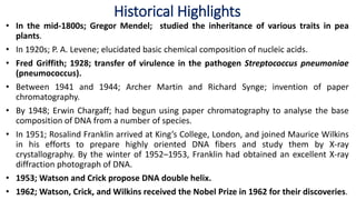
  • 1. • In the mid-1800s; Gregor Mendel; studied the inheritance of various traits in pea plants. • In 1920s; P. A. Levene; elucidated basic chemical composition of nucleic acids. • Fred Griffith; 1928; transfer of virulence in the pathogen Streptococcus pneumoniae (pneumococcus). • Between 1941 and 1944; Archer Martin and Richard Synge; invention of paper chromatography. • By 1948; Erwin Chargaff; had begun using paper chromatography to analyse the base composition of DNA from a number of species. • In 1951; Rosalind Franklin arrived at King’s College, London, and joined Maurice Wilkins in his efforts to prepare highly oriented DNA fibers and study them by X-ray crystallography. By the winter of 1952–1953, Franklin had obtained an excellent X-ray diffraction photograph of DNA. • 1953; Watson and Crick propose DNA double helix. • 1962; Watson, Crick, and Wilkins received the Nobel Prize in 1962 for their discoveries. Historical Highlights
  • 2. The Search for the Genetic Material 1. Some substance must be responsible for passage of traits from parents to offspring. For a substance to do this it must be: a. Stable enough to store information for long periods. b. Able to replicate accurately. c. Capable of change to allow evolution. DNA as Genetic Material
  • 3. In the early 1900s, chromosomes were shown to be the carriers of hereditary information. DNA and Protein Most scientists initially believed that protein must be the genetic material.
  • 4. • Frederick Griffith’s 1928 experiment with Streptococcus pneumoniae bacteria in mice showed that something passed from dead bacteria into nearby living ones, allowing them to change their cell surface. • He called this agent the transforming principle
  • 7. Avery’s Transformation Experiment DNA as Genetic Material: The Avery Experiment 1. In 1944, Avery, MacLeod and McCarty published results of a study that identified the transforming principle from S. pneumoniae. 2. Only the nucleic acid fraction was capable of transforming the bacteria. 3. Critics noted that the nucleic acid fraction was contaminated with proteins.
  • 8. Oswald Avery with his colleagues set out to discover which constituent in the heat-killed virulent pneumococci was responsible for Griffith’s transformation.
  • 9. The Hershey-Chase Bacteriophage Experiment DNA as Genetic Material: The Hershey-Chase Experiment 1. More evidence for DNA as the genetic material came in 1953 with Alfred Hershey and Martha Chase’s work on E. coli infected with bacteriophage T2. 2. In one part of the experiment, T2 proteins were labeled with 35S, and in the other part, T2 DNA was labeled with 32P. Then each group of labeled viruses was mixed separately with the E. coli host. 3. The 35S-labeled protein was found outside the infected cells, while the 32P-labeled DNA was inside the E. coli.
  • 10. The Hershey-Chase Experiment (a) When E. coli was infected with a T2 phage containing 35S protein, most of the radioactivity remained outside the host cell. (b) When a T2 phage containing 32P DNA was mixed with the host bacterium, the radioactive DNA was injected into the cell and phages were produced. Thus DNA was carrying the virus’s genetic information.
  • 11. The Hershey-Chase Waring Blender Martha Chase and Alfred Hershey, 1953
  • 12. The Composition of Nucleic Acids
  • 13. The Composition and Structure of DNA and RNA • DNA and RNA are polymers composed of monomers called nucleotides. • Each nucleotide has three parts: a. A pentose (5-carbon) sugar. b. A nitrogenous base. c. A phosphate group. • The pentose sugar in RNA is ribose, and in DNA it’s deoxyribose. The only difference is at the 2’ position, where RNA has a hydroxyl (OH) group, while DNA has only a hydrogen. Structures of deoxyribose and ribose in DNA and RNA
  • 14. There are two classes of nitrogenous bases: a. Purines (double-ring, nine-membered structures) include adenine (A) and guanine (G). b. Pyrimidines (one-ring, six-membered structures) include cytosine (C), thymine (T) in DNA and uracil (U) in RNA. Structures of the nitrogenous bases in DNA and RNA
  • 15. • The structure of nucleotides has these features: a. The base is always attached by a covalent bond between the 1′ carbon of the pentose sugar and a nitrogen in the base (specifically, the nine nitrogen in purines and the one nitrogen in pyrimidines). b. The sugar-base combination is a nucleoside. When a phosphate is added (always to the 5′ carbon of the pentose sugar), it becomes a nucleoside phosphate, or simply nucleotide. c. Nucleotide examples are shown in Figure 2.11, and naming conventions are given in Table 2.1. • Polynucleotides of both DNA and RNA are formed by stable covalent bonds (phosphodiester linkages) between the phosphate group on the 5′ carbon of one nucleotide, and the 3′ hydroxyl on another nucleotide. This creates the “backbone” of a nucleic acid molecule. • The asymmetry of phosphodiester bonds creates 3′-5′ polarity within the nucleic acid chain.
  • 16. Fig. 2.11 Chemical structures of DNA and RNA
  • 17. The Discovery of the DNA Double Helix • James Watson and Francis Crick published the famous double-helix structure in 1953. • When they began their work, it was known that DNA is composed of nucleotides, but how the nucleotides are assembled into nucleic acid was unknown. • Two additional sources of data assisted Watson and Crick with their model: a. Erwin Chargaff’s ratios obtained for DNA derived from a variety of sources showed that the amount of purine always equals the amount of pyrimidine, and further, that the amount of G equals C, and the amount of A equals T. b. Rosalind Franklin’s X ray diffraction images of DNA showed a helical structure with regularities at 0.34 nm and 3.4 nm along the axis of the molecule (Figure 8.9).
  • 19.
  • 20. Watson and Crick’s three-dimensional model has these main features: a. It is two polynucleotide chains wound around each other in a right-handed helix. b. The two chains are antiparallel. c. The sugar-phosphate backbones are on the outside of the helix, and the bases are on the inside, stacked perpendicularly to the long axis like the steps of a spiral staircase.
  • 21. d. The bases of the two strands are held together by hydrogen bonds between complementary bases (two for A-T pairs and three for G-C pairs). Individual H- bonds are relatively weak and so the strands can be separated (by heating, for example). Complementary base pairing means that the sequence of one strand dictates the sequence of the other strand.
  • 22. e. The base pairs are 0.34 nm apart, and one full turn of the DNA helix takes 3.4 nm, so there are 10 bp in a complete turn. The diameter of a dsDNA helix is 2 nm. f. Because of the way the bases H-bond with each other, the opposite sugar-phosphate backbones are not equally spaced, resulting in a major and minor groove. This feature of DNA structure is important for protein binding. • The 1962 Nobel Prize in Physiology or Medicine was awarded to Francis Crick, James Watson and Maurice Wilkins (the head of the lab in which Franklin worked). Franklin had already died, and so was not eligible.
  • 23. Assignment on – Different DNA Structures
  • 24. The Genetic Code • The final step in the expression of genes that encode proteins is translation. • The mRNA nucleotide sequence is translated into the amino acid sequence of a polypeptide chain. • Protein synthesis is called translation because it is a decoding process. • The information encoded in the language of nucleic acids must be rewritten in the language of proteins. • There is code degeneracy. i.e., there are up to 6 different codons for a given amino acid. • Only 61 codons, the sense codons, direct amino acid incorporation into protein. • The remaining 3 codons (UGA, UAG, and UAA) are involved in the termination of translation and are called stop or nonsense codons.
  • 25. • The 5′ nucleotide in the anticodon can vary, but generally, if the nucleotides in the second and third anticodon positions complement the first two bases of the mRNA codon, an aminoacyl-tRNA with the proper amino acid will bind to the mRNA-ribosome complex. • This pattern is evident on inspection of changes in the amino acid specified with variation in the third position (see table). • This loose base pairing is known as wobble and relieves cells of the need to synthesize so many tRNAs. • Wobble also decreases the effects of DNA mutations.
  • 26. • Genetic codons Discovered by Marshall Nirenberg ,Heinrich Matthaei,philip,Leder and Har Govind Khorana in 1968. • Nirenberg and Khorana the Nobel prize. History and definition of Genetic Code • Genetic code is a triplet of neiutides that coded that coded the specific amino acid. • Ex. AUG-met • UUU-Phe The amino acids encoded by all 64 possible codons were determined.
  • 27. The genetic code required determining how 4 nucleotides (A, T, G, C) could encode more than 20 amino acids. Francis Crick and Sydney Brenner determined that the DNA is read in sets of 3 nucleotides for each amino acid. Each codon consists of three bases (triplet). There are 64 codons. They are all written in the 5' to 3' direction. 61 codons code for amino acids. The other three (UAA, UGA, UAG) are stop codons (or nonsense codons) that terminate translation. There is one start codon (initiation codon), AUG, coding for methionine. Protein synthesis begins with methionine (Met) in eukaryotes, and formylmethionine (fmet) in prokaryotes. The code is unambiguous. The Genetic Code
  • 28. • The code is degenerate. More than one codon can specify a single amino acid. • All amino acids, except Met and tryptophan (Trp), have more than one codon. • For those amino acids having more than one codon, the first two bases in the codon are usually the same. The base in the third position often varies. • The code is almost universal (the same in all organisms). Some minor exceptions to this occur in mitochondria and some organisms. • The code is commaless (contiguous). • There are no spacers or "commas" between codons on an mRNA. • Neighboring codons on a message are non-overlapping.
  • 29. reading frame: the series of nucleotides read in sets of 3 (codon) • only 1 reading frame is correct for encoding the correct sequence UU of amino acids UUC GAG UUU AUG UCG AAU U UCU CGA UGU UUG AGA UU AU CUC GAU GUU UGA GAA U Reading frame 1 Reading frame 2 Reading frame 3
  • 31. stop codons: 3 codons (UUA, UGA, UAG) in the genetic code used to terminate translation. start codon: the codon (AUG) used to signify the start of translation. 31 Types of codons
  • 32. Unambiguous  Each codon specifies a particular amino acid, the codon ACG codes for the amino acid threonine, and only threonine. Non overlapping  This means that successive triplets are read in order. Each nucleotide is part of only one triplet codon. Commanelss which mean there is no punctuation witbeen two codans 32 Characteristics of the Genetic Code
  • 33. DNA Codon RNA Codon
  • 34. • The first two bases of the codon make normal (canonical) H-bond pairs with the 2nd and 3rd bases of the anticodon • At the remaining position, less stringent rules apply and non-canonical pairing may occur • The rules: first base U can recognize A or G, first base G can recognize U or C, and first base I can recognize U, C or A (I comes from deamination of A) • Advantage of wobble: dissociation of tRNA from mRNA is faster and protein synthesis too The Wobble Hypothesis
  • 35. • It is a triplet code. • – Each three-nucleotide codon in the mRNA specifies one amino acid • • It is comma free. • – mRNA is read three bases at a time without skipping any bases. • • It is non-overlapping/non-ambiguous. • • It is almost universal. • – In nearly all organisms, most codons have the same amino acid meaning. • – Of 20 amino acids, 18 are encoded by 2 or more codons. • • The code has start and stop signals. • – AUG is the usual start signal and defines the open reading frame. Points to remember:
  • 36. • • Stop signals are codons with no corresponding tRNA • – the nonsense or chain-terminating codons. • – generally three stop codons: UAG, UAA, and UGA. • More than one codon can code for each amino acid. • Each amino acid can be coded for by more than one codon
  • 37. UNIT 2 Organization and function of genetic material • Bacterial • Viral
  • 38. The Organization of DNA in Chromosomes 1. Cellular DNA is organized into chromosomes. A genome is the chromosome or set of chromosomes that contains all the DNA of an organism. 2. In prokaryotes the genome is usually a single circular chromsome. In eukaryotes, the genome is one complete haploid set of nuclear chromosomes; mitochondrial and chloroplast DNA are not included.
  • 39.
  • 40. Viral Chromosomes 1. A virus is nucleic acid surrounded by a protein coat. The nucleic acid may be dsDNA, ssDNA, dsRNA or ssRNA, and it may be linear or circular, a single molecule or several segments. 2. Bacteriophages are viruses that infect bacteria. Three different types that infect E. coli are good examples of the variety of chromosome structure found in viruses.
  • 41. T-even phage 3. The T-even phages (T2, T4 and T6) have similar structures; all have dsDNA genomes composed of a one linear DNA molecule surrounded by a protein coat. ΦX174 phage 4. ΦX174 is a small, simple virus with one short ssDNA chromosome. In 1959, Robert Sinsheimer found that the DNA of ΦX174 has a base composition that does not fit the complementary base-pair-rules.  single strand DNA rather than dsDNA
  • 42. λ phage 5. Bacteriophage λ is somewhat like the T- even phages in structure. However, its chromosome changes form. A linear molecule of dsDNA is packaged inside the protein head, but after the virus infects its host the chromosome becomes circular due to base-pairing of complementary 12-base single-stranded regions at the ends of the linear molecule (Fig. 2.18)
  • 43. Fig. 2.18  chromosome structure varies at stages of lytic infection of E. coli
  • 44.
  • 45. Prokaryotic Chromosomes 1. The typical prokaryotic genome is one circular dsDNA chromosome, but some prokaryotes are more exotic, with a main chromosome and one or more smaller ones. When a minor chromosome is dispensable to the life of the cell, it is called a plasmid. Some examples: a. Borrelia burgdorferi (Lyme disease in humans) has a 0.91-Mb linear chromosome, plus an additional 0.53-Mb of DNA in 17 different linear and circular molecules. b. Agrobacterium tumefaciens (crown gall disease of plants) has a 3.0-Mb circular chromosome and a 2.1-Mb linear one.
  • 46. 2. Archaebacteria also vary in chromosomal organization, but only circular forms have been found. Examples: a. Methanococcus jannaschii has three chromosomes of 1.66-Mb, 58-kb and 16-kb. b. Archaeoglobus fulgidus has one 2.2-Mb circular chromosome. 3. Both Eubacteria and Archaebacteria lack a membrane-bounded nucleus, hence their classification as prokaryotes. Their DNA is densely arranged in a cytoplasmic region called the nucleoid. 4. In an experiment where E. coli is gently lysed, it releases one 4.6-Mb circular chromosome, highly supercoiled (Figures 2.20). A 4.6-Mb double helix is about 1mm in length, about 103 times longer than an E. coli cell. DNA supercoiling helps it fit into the cell.
  • 47. Fig. 2.20 Illustration of DNA supercoiling
  • 48. 5. A molecule of B-DNA, with 10bp/turn of the helix, is in relaxed conformation. If turns of the helix are removed and the molecule circularized, the DNA will form superhelical turns to compensate for the added tension. 6. Either overwinding or underwinding DNA will create a structure where 10bp/turn of the helix is not the most energetically favored conformation, and supercoils will be induced. Both positive and negative supercoils will condense the DNA. 7. All organisms contain topoisomerase enzymes to supercoil their DNA. 8. Prokaryotes also organize their DNA into looped domains, with the ends of the domains held so that each is supercoiled independently (Figure 2.22). 9. The compaction factor for looped domains is about 10-fold. In E. coli there are about 100 domains of about 40kb each.
  • 49. Fig. 2.22 Model for the structure of a bacterial chromosome
  • 50. UNIT 3 Brief account of plasmids- • Structure • Types
  • 51. • In addition to the genetic material present in the nucleoid, many procaryotes (and some yeasts and other fungi) contain extrachromosomal DNA molecules called plasmids. • In some cases, numerous different plasmids within a single species have been identified. (B. burgdorferi, carries 12 linear and 9 circular plasmids) • Plasmids- small double-stranded DNA molecules can exist independently of the chromosome replicate autonomously mostly circular • Linear plasmids possess special structures or sequences at their ends to prevent their degradation and to permit their replication. • Plasmids have relatively few genes, generally less than 30. Their genetic information is not essential to the host, and cells that lack them usually function normally.
  • 52. • However, many plasmids carry genes that confer a selective advantage to their hosts in certain environments. • Episomes: Plasmids which are able to integrate into the chromosome and are thus replicated with the chromosome. • Plasmids are not always equally apportioned into daughter cells and sometimes are lost. The loss of a plasmid is called curing. • Curing can be induced by acridine mutagens, UV and ionizing radiation, thymine starvation, antibiotics, and growth above optimal temperatures. • Conjugative plasmids have genes for the construction of hair-like structures called pili and can transfer copies of themselves to other bacteria during conjugation. • F plasmid (F factor or fertility factor) of E. coli, was the first conjugative factor to be described; contains genes that direct the formation of sex pili that attach an F+ cell (a cell containing an F plasmid) to an F- cell (a cell lacking an F plasmid). • F factor is also an episome.
  • 53. • R plasmids (Resistance factors or R factors) give antibiotic resistance on the cells that contain them. • R factors typically have genes that code for enzymes capable of destroying or modifying antibiotics. • Bacteriocin-encoding plasmids may give the bacteria that harbor them a competitive advantage in the microbial world. Bacteriocins are bacterial proteins that destroy other bacteria. • Col plasmids contain genes for the synthesis of bacteriocins known as colicins, which are directed against E. coli. Example; cloacins kill Enterobacter species. • Virulence plasmids encode factors that make their hosts more pathogenic. For example, enterotoxigenic strains of E. coli cause traveler’s diarrhea because they contain a plasmid that codes for an enterotoxin. • Metabolic plasmids carry genes for enzymes that degrade substances such as aromatic compounds (toluene), pesticides (2,4-dichlorophenoxyacetic acid), and sugars (lactose). • Metabolic plasmids even carry the genes required for some strains of Rhizobium to induce legume nodulation and carry out nitrogen fixation.
  • 54.
  • 55. UNIT 4 Replication of DNA- • Rolling circle model Replication of RNA- • Reverse transcriptase
  • 56.
  • 57. A process of producing two identical copies from one original DNA molecule Three basic steps: i) Initiation ii) Elongation iii) Termination i) Initiation: During initiation, some enzymes like helicase, cut the hydrogen bonds or produce the nick in the strands of DNA to make it available for the enzymes to carry out further process of replication. Single strand binding proteins restrict the rebinding of hydrogen bonds. Enzymes & proteins reach to the nick; make the complex and replication starts. DNA Replication a general account
  • 58. Real difference is present at the step of elongation. ii) Elongation: Polymerase proceeds on the DNA and formation or elongation starts. In Eukaryotes, the DNA is much larger so the replication forms a linear structure often. But in some Prokaryotes, specially in bacteria who possess the plasmid, a unique type of replication occurs known as “Rolling circle Replication”.
  • 59. • Replication in eukaryotes is bidirectional, this type is unidirectional (i.e. in prokaryotes) • Ideal example of this type is the circular plasmid of bacteria, as it happens only in circular genomes. Initiation • Initiates by the production of nick on one of the two strands producing free 3́-OH and 5́ phosphate ends, by the action of: • Helicase • Topoisomerases • Single stranded binding proteins (SSBPs). Helicase Bacterial Plasmid SSBPs Rolling Circle Model of DNA Replication
  • 60. Elongation • For Elongation, the DNA polymerase III binds to the 3́-OH group of broken strand, using the unbroken strand as a template. • The polymerase will start to move in a circle for elongation, due to which it is named as Rolling circle model. As the elongation proceeds, the 5́end will be displaced and will grow out like a waving thread. DNA Polymerase III Broken Strand Origin Point
  • 61. Termination • At the point of termination, the linear DNA molecule is cleaved from the circle, resulting in a double stranded circular DNA molecule and a single-stranded linear DNA molecule. • The linear single stranded molecule is circularized by the action of ligase and then replication to double stranded circular plasmid molecule. Origin Point
  • 62. Example of Rolling Circle Model: The conjugation between F+ and F- Bacteria
  • 63. Simplified Diagram of Rolling Circle Model of DNA Replication