DNA Replication


         Biochemistry For Medics
         www.namrata.co
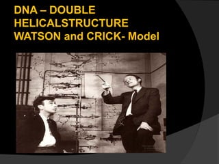
DNA – DOUBLE
HELICALSTRUCTURE
WATSON and CRICK- Model
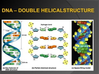
DNA – DOUBLE HELICALSTRUCTURE
Directionality of DNA
                  Nucleotides in DNA backbone
                  are bonded together by
                  phosphodiester linkage
                  between 3′ & 5′ carbons.

                  DNA molecule has “direction.“

                  Complementary strands run in
                  opposite directions.
DNA Replication- Introduction
   Basis for inheritance

   Fundamental process occurring in all
    cells for copying DNA to transfer the
    genetic information to daughter cells

   Each cell must replicate its DNA before
    division.
DNA Replication
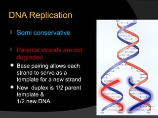
   Semi conservative

   Parental strands are not
    degraded
 Base pairing allows each
  strand to serve as a
  template for a new strand
 New duplex is 1/2 parent
  template &
  1/2 new DNA
Okazaki

DNA Replication
 Semi discontinuous
 Leading & Lagging strands
Leading strand
 continuous synthesis

Lagging strand
 Okazaki fragments

 joined by ligases
DNA Replication
   Energy of Replication
 The nucleotides arrive as
 nucleoside triphosphates
    DNA base, sugar with PPP
      ○ P-P-P = energy for bonding
    DNA bases arrive with their own energy source
     for bonding
    bonded by enzyme: DNA polymerase III
DNA Replication
   Primer is needed
    DNA polymerase can only add nucleotides
     to 3′ end of a growing DNA strand
      ○ need a “starter” nucleotide to make a
        bond
    strand only grows 5′→3′.
    Template is read in the 3′-5′ direction while
     polymerization takes place in the 5′→3′
     direction
Primer
RNA primer
 Synthesized by Primase
 serves as a starter sequence for DNA
  polymerase III
 Only one RNA Primer-required for the
  leading strand
 RNA Primers for the lagging strand depend
  on the number of “OKAZAKI
  FRAGMENTS”
 RNA Primer has a free 3’OH group to
  which the first Nucleotide is bound.
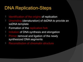
DNA Replication-Steps
   Identification of the origins of replication
   Unwinding (denaturation) of dsDNA to provide an
    ssDNA template
   Formation of the replication fork
   Initiation of DNA synthesis and elongation
   Primer removal and ligation of the newly
    synthesized DNA segments
   Reconstitution of chromatin structure
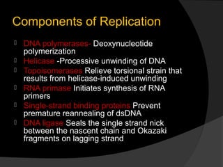
Components of Replication
   DNA polymerases- Deoxynucleotide
    polymerization
   Helicase -Processive unwinding of DNA
   Topoisomerases Relieve torsional strain that
    results from helicase-induced unwinding
   RNA primase Initiates synthesis of RNA
    primers
   Single-strand binding proteins Prevent
    premature reannealing of dsDNA
   DNA ligase Seals the single strand nick
    between the nascent chain and Okazaki
    fragments on lagging strand
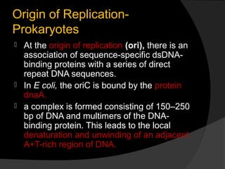
Origin of Replication-
Prokaryotes
   At the origin of replication (ori), there is an
    association of sequence-specific dsDNA-
    binding proteins with a series of direct
    repeat DNA sequences.
   In E coli, the oriC is bound by the protein
    dnaA.
   a complex is formed consisting of 150–250
    bp of DNA and multimers of the DNA-
    binding protein. This leads to the local
    denaturation and unwinding of an adjacent
    A+T-rich region of DNA.
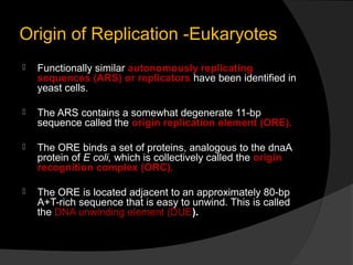
Origin of Replication -Eukaryotes
   Functionally similar autonomously replicating
    sequences (ARS) or replicators have been identified in
    yeast cells.

   The ARS contains a somewhat degenerate 11-bp
    sequence called the origin replication element (ORE).

   The ORE binds a set of proteins, analogous to the dnaA
    protein of E coli, which is collectively called the origin
    recognition complex (ORC).

   The ORE is located adjacent to an approximately 80-bp
    A+T-rich sequence that is easy to unwind. This is called
    the DNA unwinding element (DUE).
Unwinding of DNA
 The interaction of proteins with ori
  defines the start site of replication and
  provides a short region of ssDNA
  essential for initiation of synthesis of the
  nascent DNA strand.
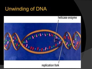
 DNA Helicase allows for processive
  unwinding of DNA.
 Single-stranded DNA-binding proteins
  (SSBs) stabilize this complex.
 In cooperation with SSB, this leads to
  DNA unwinding and active replication.
Unwinding of DNA
Formation of the Replication
Fork
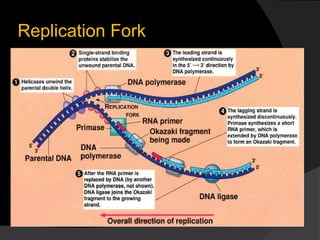
   The polymerase III holoenzyme binds to
    template DNA as part of a multiprotein
    complex
   DNA polymerases only synthesize DNA in the
    5' to 3' direction,
   Because the DNA strands are antiparallel , the
    polymerase functions asymmetrically.
   On the leading (forward) strand, the DNA is
    synthesized continuously.
   On the lagging (retrograde) strand, the DNA is
    synthesized in short (1–5 kb)fragments, the
    so-called Okazaki fragments.
Replication Fork
Formation of Replication
Bubbles
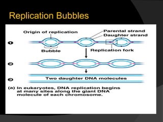
 Replication occurs in both directions
  along the length of DNA and both
  strands are replicated simultaneously.
 This replication process generates
  "replication bubbles"
Replication Bubbles
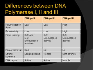
The DNA Polymerase Complex
 A number of different DNA polymerase
  molecules engage in DNA replication. These
  share three important properties: (1) chain
  elongation, (2) Processivity, and (3)
  proofreading.
 Chain elongation accounts for the rate (in
  nucleotides per second) at which
  polymerization occurs.
 Processivity is an expression of the number of
  nucleotides added to the nascent chain before
  the polymerase disengages from the template.
 The proofreading function identifies copying
  errors and corrects them
DNA Polymerase Complex
 In E coli, polymerase III (pol III)
  functions at the replication fork. Of all
  polymerases, it catalyzes the highest
  rate of chain elongation and is the most
  processive.
 Polymerase II (pol II) is mostly involved
  in proofreading and DNA repair.
 Polymerase I (pol I) completes chain
  synthesis between Okazaki fragments
  on the lagging strand.
Differences between DNA
Polymerase I, II and III
Eukaryotic DNA polymerases
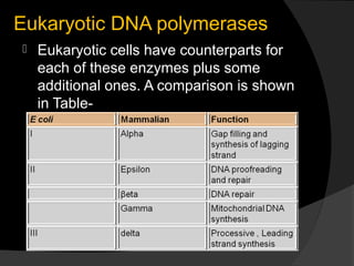
   Eukaryotic cells have counterparts for
    each of these enzymes plus some
    additional ones. A comparison is shown
    in Table-
Initiation & Elongation of DNA
 Synthesis
 Primer-The priming process involves the
  nucleophilic attack by the 3'-hydroxyl
  group of the RNA primer on the
  phosphate of the first entering
  deoxynucleoside triphosphate with the
  splitting off of pyrophosphate.
 Mammalian DNA polymerase Alpha is
  mainly responsible for the synthesis of
  primer.
Initiation & Elongation of DNA
 Synthesis
   Selection of the proper
    deoxyribonucleotide whose terminal 3'-
    hydroxyl group is to be attacked is
    dependent upon proper base pairing
    with the other strand of the DNA
    molecule according to the rules
    proposed originally by Watson and Crick
Initiation & Elongation of DNA
Synthesis
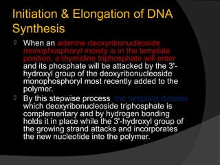
 When an adenine deoxyribonucleoside
  monophosphoryl moiety is in the template
  position, a thymidine triphosphate will enter
  and its phosphate will be attacked by the 3'-
  hydroxyl group of the deoxyribonucleoside
  monophosphoryl most recently added to the
  polymer.
 By this stepwise process, the template dictates
  which deoxyribonucleoside triphosphate is
  complementary and by hydrogen bonding
  holds it in place while the 3'-hydroxyl group of
  the growing strand attacks and incorporates
  the new nucleotide into the polymer.
Base pairing in DNA Replication
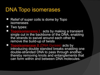
DNA Topo isomerases
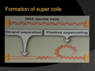
 Relief of super coils is dome by Topo
  isomerases
 Two types:
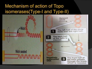
 Topoisomerases I : acts by making a transient
  single cut in the backbone of the DNA, enabling
  the strands to swivel around each other to
  remove the build-up of twists
 Topoisomerase II (DNA Gyrase) acts by
  introducing double standed breaks enabling one
  double-stranded DNA to pass through another,
  thereby removing knots and entanglements that
  can form within and between DNA molecules.
Formation of super coils
Mechanism of action of Topo
isomerases(Type-I and Type-II)
Primer removal and Nick
sealing
 Primers are removed by DNA
  polymerase I by replacing
  ribonucleotides with deoxy
  Ribonucleotides
 Nicks are sealed by DNA ligase
 Multiple primers on the Lagging strand
  while single primer on the leading
  strand.
Proof reading and Editing
   1000 bases/second =
    lots of typos!
   DNA polymerase I
     proofreads & corrects typos
     repairs mismatched bases
     removes abnormal bases
       ○ repairs damage
         throughout life
     reduces error rate from
      1 in 10,000 to
      1 in 100 million bases
Termination of replication
 In prokaryotes:
  DNA replication terminates when
  replication forks reach specific
  “termination sites”.
 the two replication forks meet each
  other on the opposite end of the
  parental circular DNA .
Termination of replication
 This process is completed in about 30
  minutes, a replication rate of 3 x 10⁵
  bp/min in prokaryotes
 The entire mammalian genome
  replicates in approximately 9 hours, the
  average period required for formation of
  a tetraploid genome from a diploid
  genome in a replicating cell.
Reconstitution of Chromatin
Structure
   chromatin structure must be re-formed
    after replication. Newly replicated DNA
    is rapidly assembled into nucleosomes,
    and the preexisting and newly
    assembled histone octamers are
    randomly distributed to each arm of the
    replication fork.
DNA Synthesis and the Cell
Cycle
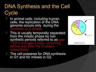
 In animal cells, including human
  cells, the replication of the DNA
  genome occurs only during the
  synthetic or S phase.
 This is usually temporally separated
  from the mitotic phase by non
  synthetic periods referred to as gap
  1 (G1) and gap 2 (G2), occurring
  before and after the S phase,
  respectively
 The cell prepares for DNA synthesis
  in G1 and for mitosis in G2.
Telomeres
 In eukaryotic replication, following
  removal of RNA Primer from the
  5’end of lagging strand; a gap is left.
 This gap exposes DNA strand to
  attack of 5’ exonucleases.
 This problem is overcome by
  Telomerase.
Telomeres


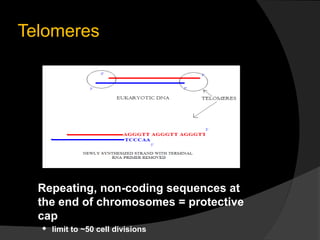
  Repeating, non-coding sequences at
  the end of chromosomes = protective
  cap
     limit to ~50 cell divisions
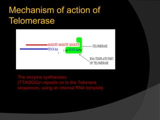
Mechanism of action of
Telomerase


  The enzyme synthesizes
  (TTAGGG)n repeats on to the Telomere
  sequences, using an internal RNA template.
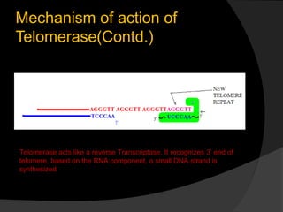
Mechanism of action of
Telomerase(Contd.)


Telomerase acts like a reverse Transcriptase. It recognizes 3’ end of
telomere, based on the RNA component, a small DNA strand is
synthesized
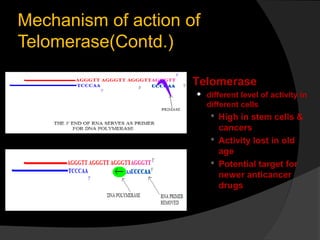
Mechanism of action of
Telomerase(Contd.)

                     Telomerase
                        different level of activity in
                         different cells
                           High in stem cells &
                            cancers
                           Activity lost in old
                            age
                           Potential target for
                            newer anticancer
                            drugs
Inhibitors of DNA replication
  Bacterial DNA Gyrase(Type II
   Topoisomerase)- Inhibited by
   Novobiocin and Nalidixic acid.
  Ciprofloxacin interferes with DNA
   breakage and rejoining process
  Mammalian topoisomerases – inhibited
   by Etoposide and Adriamycin, used as
   anticancer drugs.
  Nucleoside analogues also inhibit
   replication and are used as anticancer
   drugs.
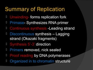
Summary of Replication
   Unwinding- forms replication fork
   Primase- Synthesizes RNA primer
   Continuous synthesis -Leading strand
   Discontinuous synthesis – Lagging
    strand (Okazaki fragments)
   Synthesis 5’-3’ direction
   Primers removed, nick sealed
   Proof reading by DNA polymerases
   Organized in to chromatin structure