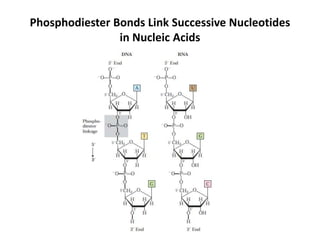
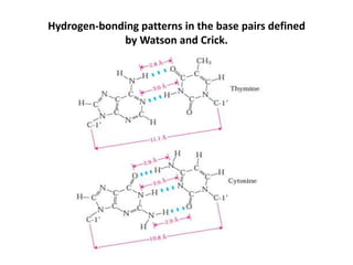
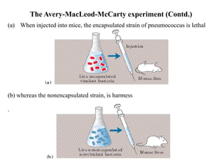
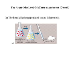
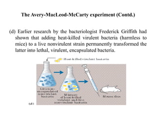
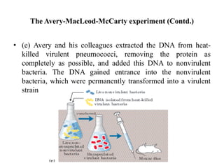
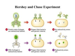
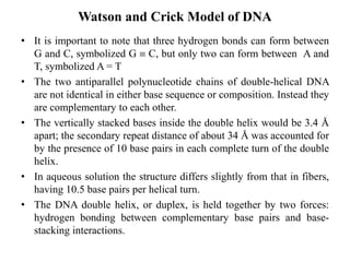
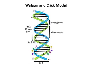
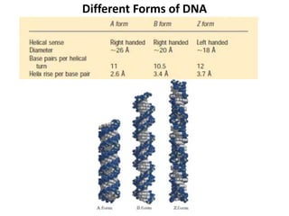
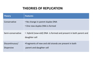
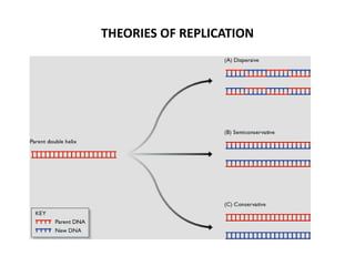
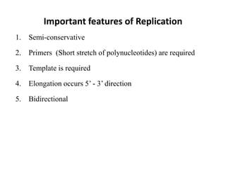
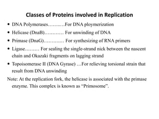
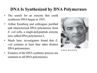
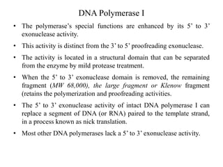
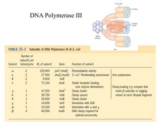
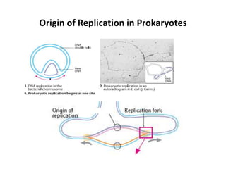
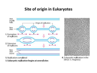
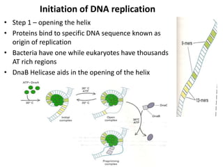
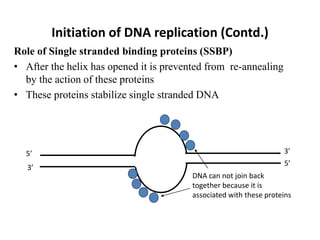
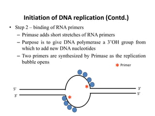
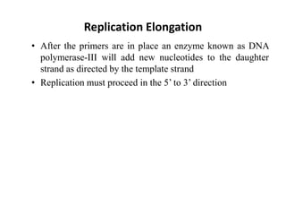
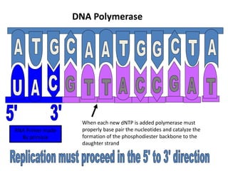
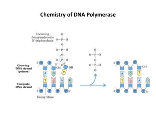
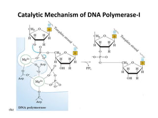
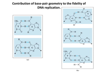
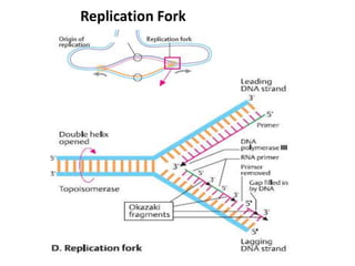
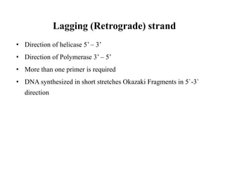
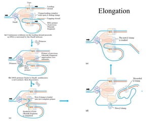
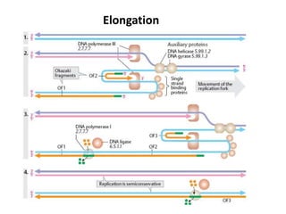
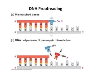
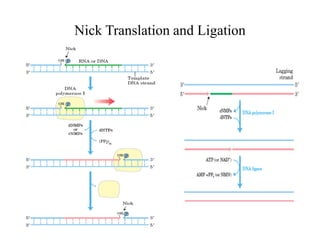
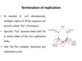
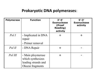
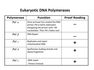
DNA and Replication
Phosphodiester Bonds Link Successive Nucleotides
in Nucleic Acids
Hydrogen-bonding patterns in the base pairs defined
by Watson and Crick.
DNA Stores Genetic Information
• The biochemical investigation of DNA began with Friedrich
Miescher, who carried out the first systematic chemical studies
of cell nuclei.
• In 1868 Miescher isolated a phosphorus-containing substance,
which he called “nuclein,” from the nuclei of pus cells
(leukocytes) obtained from discarded surgical bandages.
• He found nuclein to consist of an acidic portion, which we know
today as DNA, and a basic portion, protein.
• Miescher and many others suspected that nuclein (nucleic acid)
was associated in some way with cell inheritance.
• But the first direct evidence that DNA is the bearer of genetic
information came in 1944 through a discovery made by Oswald
T. Avery, Colin MacLeod, and Maclyn McCarty.
The Avery-MacLeod-McCarty experiment (Contd.)
(a) When injected into mice, the encapsulated strain of pneumococcus is lethal
(b) whereas the nonencapsulated strain, is harmess
.
(c) The heat-killed encapsulated strain, is harmless.
The Avery-MacLeod-McCarty experiment (Contd.)
(d) Earlier research by the bacteriologist Frederick Griffith had
shown that adding heat-killed virulent bacteria (harmless to
mice) to a live nonvirulent strain permanently transformed the
latter into lethal, virulent, encapsulated bacteria.
The Avery-MacLeod-McCarty experiment (Contd.)
• (e) Avery and his colleagues extracted the DNA from heat-
killed virulent pneumococci, removing the protein as
completely as possible, and added this DNA to nonvirulent
bacteria. The DNA gained entrance into the nonvirulent
bacteria, which were permanently transformed into a virulent
strain
The Avery-MacLeod-McCarty experiment (Contd.)
• Avery and his colleagues concluded that the DNA extracted
from the virulent strain carried the inheritable genetic message
for virulence.
• Not everyone accepted these conclusions, because protein
impurities present in the DNA could have been the carrier of
the genetic information.
The Avery-MacLeod-McCarty experiment (Contd.)
Hershey and Chase Experiment
• A second important experiment provided independent evidence that
DNA carries genetic information.
• In 1952 Alfred D. Hershey and Martha Chase used radioactive
phosphorus (32P) and radioactive sulfur (35S) tracers to show that
when the bacteriophage T2 infects its host cell, E. coli, it is the
phosphorus-containing DNA of the viral particle, not the sulfur-
containing protein of the viral coat, that enters the host cell and
furnishes the genetic information for viral replication.
• These important early experiments and many other lines of evidence
have shown that DNA is the exclusive chromosomal component
bearing the genetic information of living cells.
Hershey and Chase Experiment
Chargaff`s Rules
• A most important clue to the structure of DNA came from the work of
Erwin Chargaff and his colleagues in the late 1940s.
• Data collected from DNAs of a great many different species, led
Chargaff to the following conclusions:
1. The base composition of DNA generally varies from one species to another.
2. DNA specimens isolated from different tissues of the same species have the
same base composition.
3. The base composition of DNA in a given species does not change with an
organism’s age, nutritional state, or changing environment.
4. In all cellular DNAs, regardless of the species, the number of adenosine
residues is equal to the number of thymidine residues (that is, A = T), and the
number of guanosine residues is equal to the number of cytidine residues (G
= C).
From these relationships it follows that the sum of the purine residues
equals the sum of the pyrimidine residues; that is,
A + G = T + C
Watson and Crick Model of DNA
• In 1953 Watson and Crick postulated a three dimensional
model of DNA structure that accounted for all the available
data.
• It consists of two helical DNA chains wound around the same
axis to form a right handed double helix.
• The hydrophilic backbones of alternating deoxyribose and
phosphate groups are on the outside of the double helix, facing
the surrounding water.
• The purine and pyrimidine bases of both strands are stacked
inside the double helix, with their hydrophobic and nearly
planar ring structures very close together and perpendicular to
the long axis.
• The counterbalanced pairing of the two strands creates a major
groove and minor groove on the surface of the duplex.
• It is important to note that three hydrogen bonds can form between
G and C, symbolized G  C, but only two can form between A and
T, symbolized A = T
• The two antiparallel polynucleotide chains of double-helical DNA
are not identical in either base sequence or composition. Instead they
are complementary to each other.
• The vertically stacked bases inside the double helix would be 3.4 Å
apart; the secondary repeat distance of about 34 Å was accounted for
by the presence of 10 base pairs in each complete turn of the double
helix.
• In aqueous solution the structure differs slightly from that in fibers,
having 10.5 base pairs per helical turn.
• The DNA double helix, or duplex, is held together by two forces:
hydrogen bonding between complementary base pairs and base-
stacking interactions.
Watson and Crick Model of DNA
Watson and Crick Model
Different Forms of DNA
Replication
Copying of DNA molecule into another DNA molecule
THEORIES OF REPLICATION
Theory Features
Conservative • No change in parent duplex DNA
• One new duplex DNA is formed
Semi-conservative • Hybrid (new+old) DNA is formed and present in both parent and
daughter cell
Discontinuous/
Dispersive
•Fragments of new and old strands are present in both
parent and daughter cell
THEORIES OF REPLICATION
Important features of Replication
1. Semi-conservative
2. Primers (Short stretch of polynucleotides) are required
3. Template is required
4. Elongation occurs 5’ - 3’ direction
5. Bidirectional
Cell Cycle and Time of Replication
Classes of Proteins involved in Replication
 DNA Polymerases……..…For DNA ploymerization
 Helicase (DnaB)………… For unwinding of DNA
 Primase (DnaG)……….… For synthesizing of RNA primers
 Ligase……… For sealing the single-strand nick between the nascent
chain and Okazaki fragments on lagging strand
 Topoisomerase II (DNA Gyrase) …For relieving torsional strain that
result from DNA unwinding
Note: At the replication fork, the helicase is associated with the primase
enzyme. This complex is known as “Primosome”.
DNA Is Synthesized by DNA Polymerases
• The search for an enzyme that could
synthesize DNA began in 1955.
• Arthur Kornberg and colleagues purified
and characterized DNA polymerase from
E. coli cells, a single-polypeptide enzyme
now called DNA polymerase-I.
• Much later, investigators found that E.
coli contains at least four other distinct
DNA polymerases.
• Features of the DNA synthetic process are
common to all DNA polymerases.
DNA Polymerase I
• The polymerase’s special functions are enhanced by its 5’ to 3’
exonuclease activity.
• This activity is distinct from the 3’ to 5’ proofreading exonuclease.
• The activity is located in a structural domain that can be separated
from the enzyme by mild protease treatment.
• When the 5’ to 3’ exonuclease domain is removed, the remaining
fragment (MW 68,000), the large fragment or Klenow fragment
(retains the polymerization and proofreading activities.
• The 5’ to 3’ exonuclease activity of intact DNA polymerase I can
replace a segment of DNA (or RNA) paired to the template strand,
in a process known as nick translation.
• Most other DNA polymerases lack a 5’ to 3’ exonuclease activity.
DNA Polymerase III
Origin of Replication in Prokaryotes
Site of origin in Eukaryotes
Initiation of DNA replication
• Step 1 – opening the helix
• Proteins bind to specific DNA sequence known as
origin of replication
• Bacteria have one while eukaryotes have thousands
AT rich regions
• DnaB Helicase aids in the opening of the helix
Initiation of DNA replication (Contd.)
Role of Single stranded binding proteins (SSBP)
• After the helix has opened it is prevented from re-annealing
by the action of these proteins
• These proteins stabilize single stranded DNA
5’
5’
3’
3’
DNA can not join back
together because it is
associated with these proteins
Initiation of DNA replication (Contd.)
• Step 2 – binding of RNA primers
– Primase adds short stretches of RNA primers
– Purpose is to give DNA polymerase a 3’OH group from
which to add new DNA nucleotides
– Two primers are synthesized by Primase as the replication
bubble opens
5’
5’
3’
3’
*
*
Primer
*
Replication Elongation
• After the primers are in place an enzyme known as DNA
polymerase-III will add new nucleotides to the daughter
strand as directed by the template strand
• Replication must proceed in the 5’ to 3’ direction
RNA Primer made
By primase
DNA Polymerase
When each new dNTP is added polymerase must
properly base pair the nucleotides and catalyze the
formation of the phosphodiester backbone to the
daughter strand
Chemistry of DNA Polymerase
Catalytic Mechanism of DNA Polymerase-I
Contribution of base-pair geometry to the fidelity of
DNA replication.
Replication Fork
Leading (Forward) strand:
• Direction of helicase 3’ – 5’
• Direction of polymerase 3’ – 5’
• One primer at each point of origin
• DNA is synthesized continuously in 5` -3` direction.
Lagging (Retrograde) strand
• Direction of helicase 5’ – 3’
• Direction of Polymerase 3’ – 5’
• More than one primer is required
• DNA synthesized in short stretches Okazaki Fragments in 5`-3`
direction
Elongation
Elongation
Proofreading
“Proofreading” refers to any mechanism for correcting errors in
protein or nucleic acid synthesis that involves scrutiny of
individual units after they have been added to the chain.
DNA Proofreading
Nick Translation and Ligation
Termination of replication
• In circular E. coli chromosome,
multiple copies of 20 bp sequence are
present called “Ter” (Terminus).
• Specific “Tus” proteins bind with Ter
to arrest either of the two replication
forks.
• One Ter-Tus complex functions per
replication cycle.
Polymerase Function 3’-5’
Exonuclease
(Proof
Reading)
activity
5’-3’
Exonuclease
activity
Pol I - Implicated in DNA
repair
- Primer removal
+ +
Pol II - DNA Repair + -
Pol III - Main ploymerase
which synthesizes
leading strands and
Okazai fragments
+ -
Prokaryotic DNA polymerases:
Eukaryotic DNA Polymerases
Polymerase Function Proof Reading
Pol  Once primase has created the RNA
primer, Pol α starts replication
elongating the primer with ~20
nucleotides. Then Pol  takes over

Pol  DNA Repair

Pol  Replicates and repair
mitochondrial DNA +
Pol  Synthesizes leading strands and
okazai fragments +
Pol  -DNA repair
- Primer removal
+
Replication in Eukaryotes is similar but more complex
• Origins of replication, called autonomously replicating
sequences (ARS) or replicators, have been identified and best
studied in yeast.
• Yeast replicators span ~150 bp and contain several essential
conserved sequences. About 400 replicators are distributed
among the 16 chromosomes in a haploid yeast genome.
Replication in Eukaryotes is similar but more complex
• Initiation of replication in all eukaryotes requires a
multisubunit protein, the origin recognition complex (ORC),
which binds to several sequences within the replicator.
• ORC = DnaA in prokaryotes
• CDC6 and CDT1 bind to ORC and mediate the loading of a
heterohexamer of minichromosome maintenance proteins
(MCM2-7)
• MCM2-7 = DnaB helicase
• Hence, the CDC6 and CDT1 proteins have a role comparable
to that of the bacterial DnaC protein, loading the MCM
helicase onto the DNA near the replication origin.
Dna and replication

Dna and replication

  • 1.
  • 2.
    Phosphodiester Bonds LinkSuccessive Nucleotides in Nucleic Acids
  • 3.
    Hydrogen-bonding patterns inthe base pairs defined by Watson and Crick.
  • 4.
    DNA Stores GeneticInformation • The biochemical investigation of DNA began with Friedrich Miescher, who carried out the first systematic chemical studies of cell nuclei. • In 1868 Miescher isolated a phosphorus-containing substance, which he called “nuclein,” from the nuclei of pus cells (leukocytes) obtained from discarded surgical bandages. • He found nuclein to consist of an acidic portion, which we know today as DNA, and a basic portion, protein. • Miescher and many others suspected that nuclein (nucleic acid) was associated in some way with cell inheritance. • But the first direct evidence that DNA is the bearer of genetic information came in 1944 through a discovery made by Oswald T. Avery, Colin MacLeod, and Maclyn McCarty.
  • 5.
    The Avery-MacLeod-McCarty experiment(Contd.) (a) When injected into mice, the encapsulated strain of pneumococcus is lethal (b) whereas the nonencapsulated strain, is harmess .
  • 6.
    (c) The heat-killedencapsulated strain, is harmless. The Avery-MacLeod-McCarty experiment (Contd.)
  • 7.
    (d) Earlier researchby the bacteriologist Frederick Griffith had shown that adding heat-killed virulent bacteria (harmless to mice) to a live nonvirulent strain permanently transformed the latter into lethal, virulent, encapsulated bacteria. The Avery-MacLeod-McCarty experiment (Contd.)
  • 8.
    • (e) Averyand his colleagues extracted the DNA from heat- killed virulent pneumococci, removing the protein as completely as possible, and added this DNA to nonvirulent bacteria. The DNA gained entrance into the nonvirulent bacteria, which were permanently transformed into a virulent strain The Avery-MacLeod-McCarty experiment (Contd.)
  • 9.
    • Avery andhis colleagues concluded that the DNA extracted from the virulent strain carried the inheritable genetic message for virulence. • Not everyone accepted these conclusions, because protein impurities present in the DNA could have been the carrier of the genetic information. The Avery-MacLeod-McCarty experiment (Contd.)
  • 10.
    Hershey and ChaseExperiment • A second important experiment provided independent evidence that DNA carries genetic information. • In 1952 Alfred D. Hershey and Martha Chase used radioactive phosphorus (32P) and radioactive sulfur (35S) tracers to show that when the bacteriophage T2 infects its host cell, E. coli, it is the phosphorus-containing DNA of the viral particle, not the sulfur- containing protein of the viral coat, that enters the host cell and furnishes the genetic information for viral replication. • These important early experiments and many other lines of evidence have shown that DNA is the exclusive chromosomal component bearing the genetic information of living cells.
  • 11.
  • 12.
    Chargaff`s Rules • Amost important clue to the structure of DNA came from the work of Erwin Chargaff and his colleagues in the late 1940s. • Data collected from DNAs of a great many different species, led Chargaff to the following conclusions: 1. The base composition of DNA generally varies from one species to another. 2. DNA specimens isolated from different tissues of the same species have the same base composition. 3. The base composition of DNA in a given species does not change with an organism’s age, nutritional state, or changing environment. 4. In all cellular DNAs, regardless of the species, the number of adenosine residues is equal to the number of thymidine residues (that is, A = T), and the number of guanosine residues is equal to the number of cytidine residues (G = C). From these relationships it follows that the sum of the purine residues equals the sum of the pyrimidine residues; that is, A + G = T + C
  • 13.
    Watson and CrickModel of DNA • In 1953 Watson and Crick postulated a three dimensional model of DNA structure that accounted for all the available data. • It consists of two helical DNA chains wound around the same axis to form a right handed double helix. • The hydrophilic backbones of alternating deoxyribose and phosphate groups are on the outside of the double helix, facing the surrounding water. • The purine and pyrimidine bases of both strands are stacked inside the double helix, with their hydrophobic and nearly planar ring structures very close together and perpendicular to the long axis. • The counterbalanced pairing of the two strands creates a major groove and minor groove on the surface of the duplex.
  • 14.
    • It isimportant to note that three hydrogen bonds can form between G and C, symbolized G  C, but only two can form between A and T, symbolized A = T • The two antiparallel polynucleotide chains of double-helical DNA are not identical in either base sequence or composition. Instead they are complementary to each other. • The vertically stacked bases inside the double helix would be 3.4 Å apart; the secondary repeat distance of about 34 Å was accounted for by the presence of 10 base pairs in each complete turn of the double helix. • In aqueous solution the structure differs slightly from that in fibers, having 10.5 base pairs per helical turn. • The DNA double helix, or duplex, is held together by two forces: hydrogen bonding between complementary base pairs and base- stacking interactions. Watson and Crick Model of DNA
  • 15.
  • 16.
  • 17.
    Replication Copying of DNAmolecule into another DNA molecule
  • 18.
    THEORIES OF REPLICATION TheoryFeatures Conservative • No change in parent duplex DNA • One new duplex DNA is formed Semi-conservative • Hybrid (new+old) DNA is formed and present in both parent and daughter cell Discontinuous/ Dispersive •Fragments of new and old strands are present in both parent and daughter cell
  • 19.
  • 20.
    Important features ofReplication 1. Semi-conservative 2. Primers (Short stretch of polynucleotides) are required 3. Template is required 4. Elongation occurs 5’ - 3’ direction 5. Bidirectional
  • 21.
    Cell Cycle andTime of Replication
  • 22.
    Classes of Proteinsinvolved in Replication  DNA Polymerases……..…For DNA ploymerization  Helicase (DnaB)………… For unwinding of DNA  Primase (DnaG)……….… For synthesizing of RNA primers  Ligase……… For sealing the single-strand nick between the nascent chain and Okazaki fragments on lagging strand  Topoisomerase II (DNA Gyrase) …For relieving torsional strain that result from DNA unwinding Note: At the replication fork, the helicase is associated with the primase enzyme. This complex is known as “Primosome”.
  • 23.
    DNA Is Synthesizedby DNA Polymerases • The search for an enzyme that could synthesize DNA began in 1955. • Arthur Kornberg and colleagues purified and characterized DNA polymerase from E. coli cells, a single-polypeptide enzyme now called DNA polymerase-I. • Much later, investigators found that E. coli contains at least four other distinct DNA polymerases. • Features of the DNA synthetic process are common to all DNA polymerases.
  • 24.
    DNA Polymerase I •The polymerase’s special functions are enhanced by its 5’ to 3’ exonuclease activity. • This activity is distinct from the 3’ to 5’ proofreading exonuclease. • The activity is located in a structural domain that can be separated from the enzyme by mild protease treatment. • When the 5’ to 3’ exonuclease domain is removed, the remaining fragment (MW 68,000), the large fragment or Klenow fragment (retains the polymerization and proofreading activities. • The 5’ to 3’ exonuclease activity of intact DNA polymerase I can replace a segment of DNA (or RNA) paired to the template strand, in a process known as nick translation. • Most other DNA polymerases lack a 5’ to 3’ exonuclease activity.
  • 25.
  • 26.
    Origin of Replicationin Prokaryotes
  • 27.
    Site of originin Eukaryotes
  • 28.
    Initiation of DNAreplication • Step 1 – opening the helix • Proteins bind to specific DNA sequence known as origin of replication • Bacteria have one while eukaryotes have thousands AT rich regions • DnaB Helicase aids in the opening of the helix
  • 29.
    Initiation of DNAreplication (Contd.) Role of Single stranded binding proteins (SSBP) • After the helix has opened it is prevented from re-annealing by the action of these proteins • These proteins stabilize single stranded DNA 5’ 5’ 3’ 3’ DNA can not join back together because it is associated with these proteins
  • 30.
    Initiation of DNAreplication (Contd.) • Step 2 – binding of RNA primers – Primase adds short stretches of RNA primers – Purpose is to give DNA polymerase a 3’OH group from which to add new DNA nucleotides – Two primers are synthesized by Primase as the replication bubble opens 5’ 5’ 3’ 3’ * * Primer *
  • 31.
    Replication Elongation • Afterthe primers are in place an enzyme known as DNA polymerase-III will add new nucleotides to the daughter strand as directed by the template strand • Replication must proceed in the 5’ to 3’ direction
  • 32.
    RNA Primer made Byprimase DNA Polymerase When each new dNTP is added polymerase must properly base pair the nucleotides and catalyze the formation of the phosphodiester backbone to the daughter strand
  • 33.
    Chemistry of DNAPolymerase
  • 34.
    Catalytic Mechanism ofDNA Polymerase-I
  • 35.
    Contribution of base-pairgeometry to the fidelity of DNA replication.
  • 36.
  • 37.
    Leading (Forward) strand: •Direction of helicase 3’ – 5’ • Direction of polymerase 3’ – 5’ • One primer at each point of origin • DNA is synthesized continuously in 5` -3` direction.
  • 38.
    Lagging (Retrograde) strand •Direction of helicase 5’ – 3’ • Direction of Polymerase 3’ – 5’ • More than one primer is required • DNA synthesized in short stretches Okazaki Fragments in 5`-3` direction
  • 39.
  • 40.
  • 41.
    Proofreading “Proofreading” refers toany mechanism for correcting errors in protein or nucleic acid synthesis that involves scrutiny of individual units after they have been added to the chain.
  • 42.
  • 43.
  • 44.
    Termination of replication •In circular E. coli chromosome, multiple copies of 20 bp sequence are present called “Ter” (Terminus). • Specific “Tus” proteins bind with Ter to arrest either of the two replication forks. • One Ter-Tus complex functions per replication cycle.
  • 45.
    Polymerase Function 3’-5’ Exonuclease (Proof Reading) activity 5’-3’ Exonuclease activity PolI - Implicated in DNA repair - Primer removal + + Pol II - DNA Repair + - Pol III - Main ploymerase which synthesizes leading strands and Okazai fragments + - Prokaryotic DNA polymerases:
  • 46.
    Eukaryotic DNA Polymerases PolymeraseFunction Proof Reading Pol  Once primase has created the RNA primer, Pol α starts replication elongating the primer with ~20 nucleotides. Then Pol  takes over  Pol  DNA Repair  Pol  Replicates and repair mitochondrial DNA + Pol  Synthesizes leading strands and okazai fragments + Pol  -DNA repair - Primer removal +
  • 47.
    Replication in Eukaryotesis similar but more complex • Origins of replication, called autonomously replicating sequences (ARS) or replicators, have been identified and best studied in yeast. • Yeast replicators span ~150 bp and contain several essential conserved sequences. About 400 replicators are distributed among the 16 chromosomes in a haploid yeast genome.
  • 48.
    Replication in Eukaryotesis similar but more complex • Initiation of replication in all eukaryotes requires a multisubunit protein, the origin recognition complex (ORC), which binds to several sequences within the replicator. • ORC = DnaA in prokaryotes • CDC6 and CDT1 bind to ORC and mediate the loading of a heterohexamer of minichromosome maintenance proteins (MCM2-7) • MCM2-7 = DnaB helicase • Hence, the CDC6 and CDT1 proteins have a role comparable to that of the bacterial DnaC protein, loading the MCM helicase onto the DNA near the replication origin.