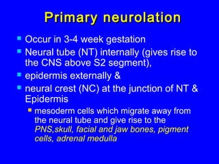
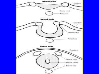
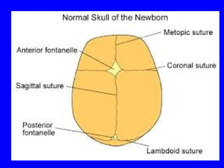
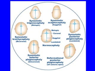
CONGENITALCONGENITAL
ANOMALIES OF CNSANOMALIES OF CNS
ETIOLOGYETIOLOGY
 Hyperthermia
 Drugs
 Malnutretion
 Nutrition: folic acid deficiency
 Disease: diabetes
 Toxins: alcohol, smoking
 Infections: rubella, toxoplasmosis, CMV,
syphilis
 Radiation
 Unknown (most cases)
Primary neurolationPrimary neurolation
 Occur in 3-4 week gestation
 Neural tube (NT) internally (gives rise to
the CNS above S2 segment),
 epidermis externally &
 neural crest (NC) at the junction of NT &
Epidermis
 mesoderm cells which migrate away from
the neural tube and give rise to the
PNS,skull, facial and jaw bones, pigment
cells, adrenal medulla
Neural tube closureNeural tube closure
 Start at the cervical area (cervico-
medularly junction)
 Extend cranially and caudally
 Cephalic neuropore close around day 25
 Caudal neuropore closes around day 29
and correspond to S2 segment
Disorders of primaryDisorders of primary
neurolationneurolation
 Split cord syndrome
 Craniorachischisis
 Neuroenteric cyst
 Anencephaly
 Myeloschisis
 Arnold chiari malformation
 Meningocele, myelomeningocele
 Dermal sinus
 Encephalocele
Dermal sinus tractDermal sinus tract
 result from incomplete disjunction of
neuroectoderm from cutaneous ectoderm
 The sinus tracts extend deep into the
subcutaneous tissues, reaching the spinal
canal in one-half to two-thirds of cases.
 it may be attached to the dura, causing
tenting of the thecal sac.
AnencephalyAnencephaly::
 Failure of anterior neuropore to close 
 Brain and calvarium are absent &
Replaced by a cerebrovasculorum - a
tangle of glial and connective tissue
Spina Bifida OccultaSpina Bifida Occulta
Common anomaly
Midline defect of vertebral bodies without protrusion of
spinal cord or meninges
Asymptomatic
Spine X Ray
MRI
Meningocele andMeningocele and
myelomeningocelemyelomeningocele
 Primary failure of neural tube
closure
 Myelomeningocele
 Meningocele
 Most frequently located in the
thoracolumbar area.
MeningoceleMeningocele
Meninges herniate through a defect in the posterior
vertebral arches
Spinal cord is normal
Fluctuant midline mass
Covered with skin
MeningoceleMeningocele
INVESTIGATIONSINVESTIGATIONS::
Plain X Ray
Ultrasonography
MRI
CT Scan
MyelomeningoceleMyelomeningocele
Severe form of neural tube defectSevere form of neural tube defect
1/40001/4000
--Located anywhere esp. lumbosacralLocated anywhere esp. lumbosacral
--Low sacral region: no motor impairment, anesthesia inLow sacral region: no motor impairment, anesthesia in
perineal areaperineal area
--Mid lumbar region: saclike cystic structure covered withMid lumbar region: saclike cystic structure covered with
thin layer of partially epithelialized tissuethin layer of partially epithelialized tissue..
Remnant of neural tissue are visibleRemnant of neural tissue are visible
Flacid paralysisFlacid paralysis
Clinical MinifestaionClinical Minifestaion
EncephaloceleEncephalocele
 Bony defects in midline
 Brain tissue can protrude through hole
 Most common in occipital region
 Prognosis depends on quantity of
cerebral tissue that herniates into the
defect
 Surgical repair is required
CHIARI MALFORMATIONCHIARI MALFORMATION
Herniation of post. Fossa contents throughHerniation of post. Fossa contents through
foramen magnumforamen magnum..
Normal: upto 5 mm of tonsillar descent throughNormal: upto 5 mm of tonsillar descent through
foramen magnumforamen magnum..
Classified into Chiari I and II based on amountClassified into Chiari I and II based on amount
of descentof descent..
CLINICAL FEATURESCLINICAL FEATURES
Presents in infants or young adultsPresents in infants or young adults..
Headache aggravated by coughing orHeadache aggravated by coughing or
strainingstraining..
Signs of brainstem compressionSigns of brainstem compression
TREATMENTTREATMENT
DECOMPRESSIONDECOMPRESSION
CRANIOSYNOSTOSISCRANIOSYNOSTOSIS
Premature closure of cranial suturesPremature closure of cranial sutures..
May affect single or multiple suturesMay affect single or multiple sutures
Present with abnormal head shapePresent with abnormal head shape..
Associated with rare genetic abnormalitiesAssociated with rare genetic abnormalities..
Surgery is required to correct itSurgery is required to correct it..
THANK YOUTHANK YOU

Cns cong anomalies

  • 1.
  • 2.
    ETIOLOGYETIOLOGY  Hyperthermia  Drugs Malnutretion  Nutrition: folic acid deficiency  Disease: diabetes  Toxins: alcohol, smoking  Infections: rubella, toxoplasmosis, CMV, syphilis  Radiation  Unknown (most cases)
  • 3.
    Primary neurolationPrimary neurolation Occur in 3-4 week gestation  Neural tube (NT) internally (gives rise to the CNS above S2 segment),  epidermis externally &  neural crest (NC) at the junction of NT & Epidermis  mesoderm cells which migrate away from the neural tube and give rise to the PNS,skull, facial and jaw bones, pigment cells, adrenal medulla
  • 5.
    Neural tube closureNeuraltube closure  Start at the cervical area (cervico- medularly junction)  Extend cranially and caudally  Cephalic neuropore close around day 25  Caudal neuropore closes around day 29 and correspond to S2 segment
  • 6.
    Disorders of primaryDisordersof primary neurolationneurolation  Split cord syndrome  Craniorachischisis  Neuroenteric cyst  Anencephaly  Myeloschisis  Arnold chiari malformation  Meningocele, myelomeningocele  Dermal sinus  Encephalocele
  • 7.
    Dermal sinus tractDermalsinus tract  result from incomplete disjunction of neuroectoderm from cutaneous ectoderm  The sinus tracts extend deep into the subcutaneous tissues, reaching the spinal canal in one-half to two-thirds of cases.  it may be attached to the dura, causing tenting of the thecal sac.
  • 8.
    AnencephalyAnencephaly::  Failure ofanterior neuropore to close   Brain and calvarium are absent & Replaced by a cerebrovasculorum - a tangle of glial and connective tissue
  • 9.
    Spina Bifida OccultaSpinaBifida Occulta Common anomaly Midline defect of vertebral bodies without protrusion of spinal cord or meninges Asymptomatic Spine X Ray MRI
  • 10.
    Meningocele andMeningocele and myelomeningocelemyelomeningocele Primary failure of neural tube closure  Myelomeningocele  Meningocele  Most frequently located in the thoracolumbar area.
  • 11.
    MeningoceleMeningocele Meninges herniate througha defect in the posterior vertebral arches Spinal cord is normal Fluctuant midline mass Covered with skin
  • 12.
  • 13.
    MyelomeningoceleMyelomeningocele Severe form ofneural tube defectSevere form of neural tube defect 1/40001/4000
  • 14.
    --Located anywhere esp.lumbosacralLocated anywhere esp. lumbosacral --Low sacral region: no motor impairment, anesthesia inLow sacral region: no motor impairment, anesthesia in perineal areaperineal area --Mid lumbar region: saclike cystic structure covered withMid lumbar region: saclike cystic structure covered with thin layer of partially epithelialized tissuethin layer of partially epithelialized tissue.. Remnant of neural tissue are visibleRemnant of neural tissue are visible Flacid paralysisFlacid paralysis Clinical MinifestaionClinical Minifestaion
  • 15.
    EncephaloceleEncephalocele  Bony defectsin midline  Brain tissue can protrude through hole  Most common in occipital region  Prognosis depends on quantity of cerebral tissue that herniates into the defect  Surgical repair is required
  • 16.
    CHIARI MALFORMATIONCHIARI MALFORMATION Herniationof post. Fossa contents throughHerniation of post. Fossa contents through foramen magnumforamen magnum.. Normal: upto 5 mm of tonsillar descent throughNormal: upto 5 mm of tonsillar descent through foramen magnumforamen magnum.. Classified into Chiari I and II based on amountClassified into Chiari I and II based on amount of descentof descent..
  • 17.
    CLINICAL FEATURESCLINICAL FEATURES Presentsin infants or young adultsPresents in infants or young adults.. Headache aggravated by coughing orHeadache aggravated by coughing or strainingstraining.. Signs of brainstem compressionSigns of brainstem compression
  • 18.
  • 19.
    CRANIOSYNOSTOSISCRANIOSYNOSTOSIS Premature closure ofcranial suturesPremature closure of cranial sutures.. May affect single or multiple suturesMay affect single or multiple sutures Present with abnormal head shapePresent with abnormal head shape.. Associated with rare genetic abnormalitiesAssociated with rare genetic abnormalities.. Surgery is required to correct itSurgery is required to correct it..
  • 24.