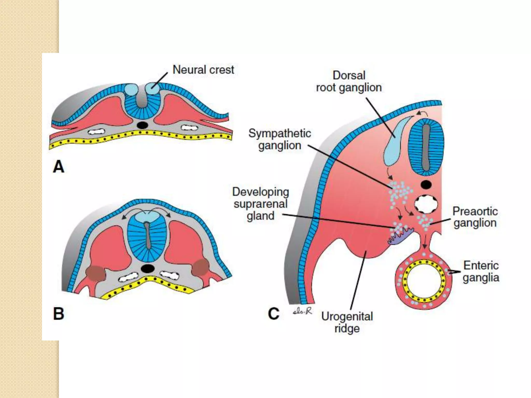
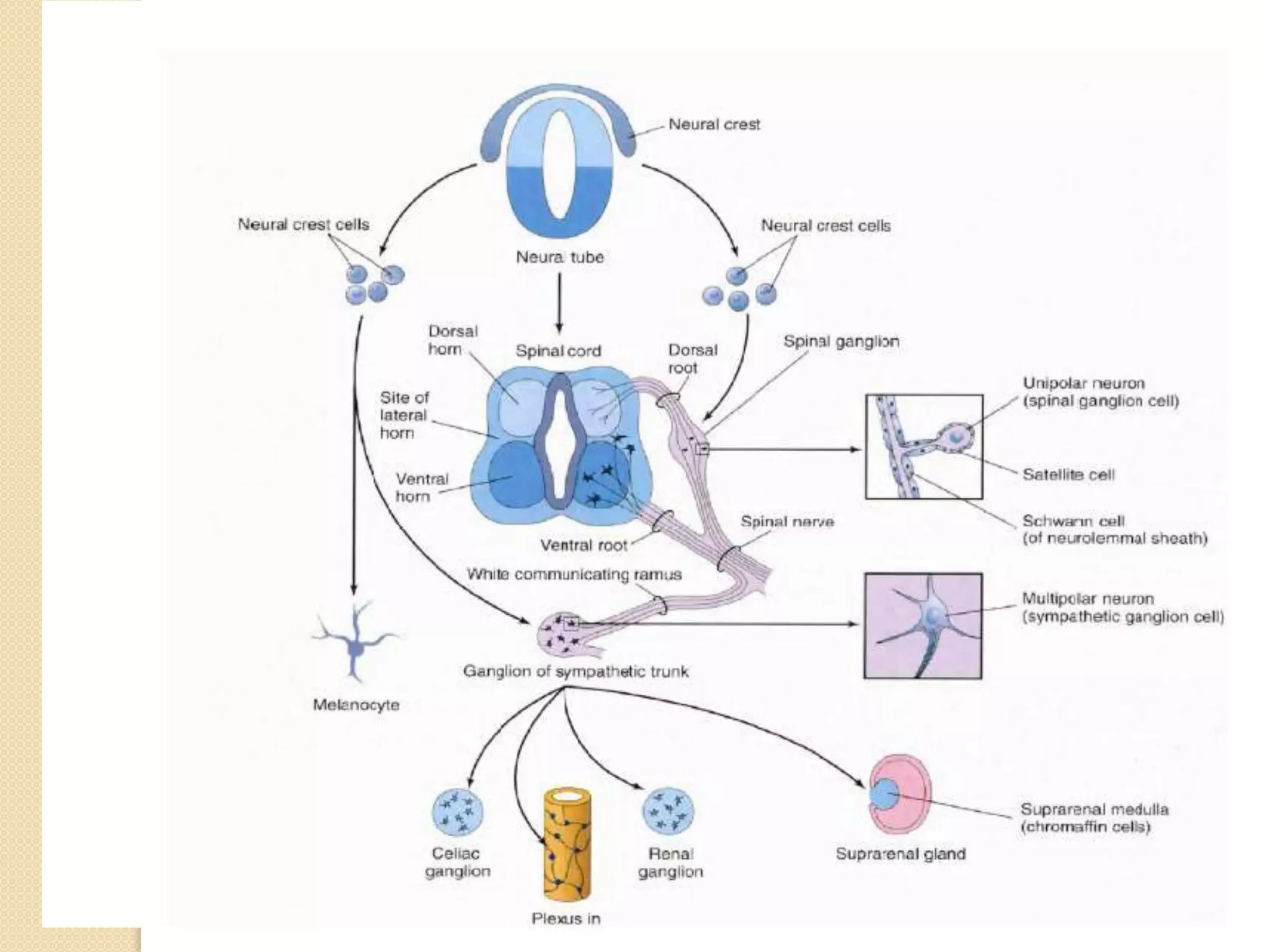
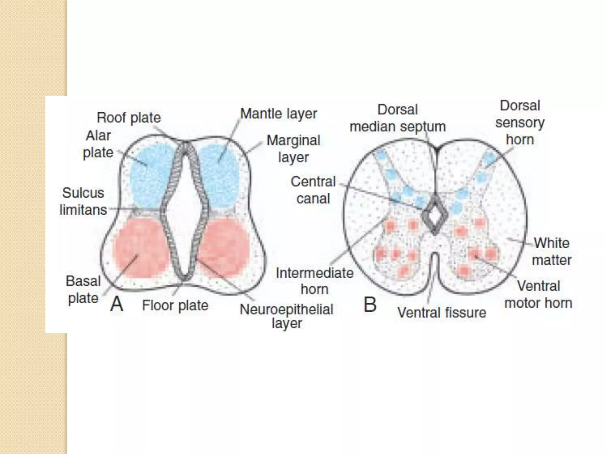
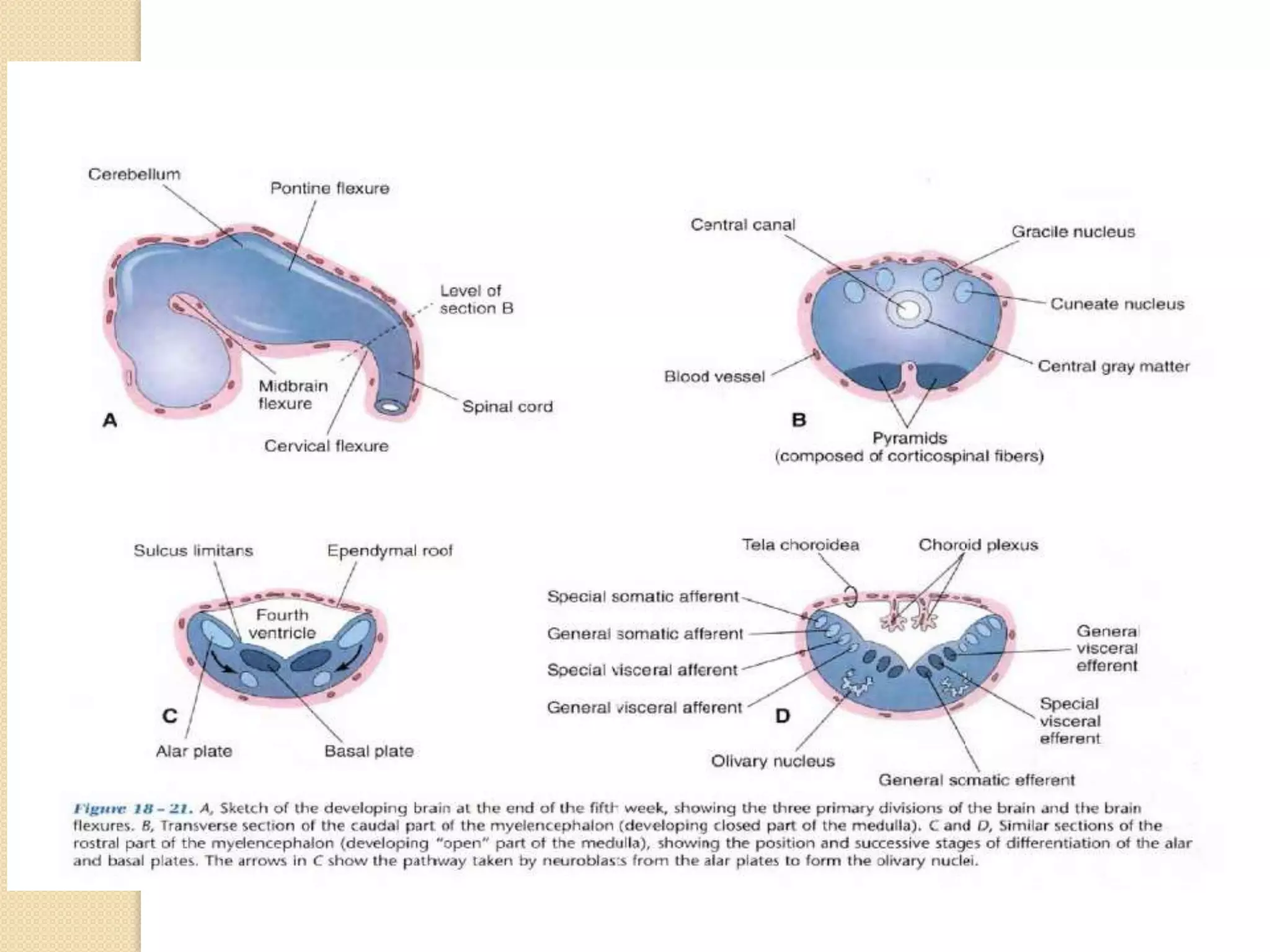
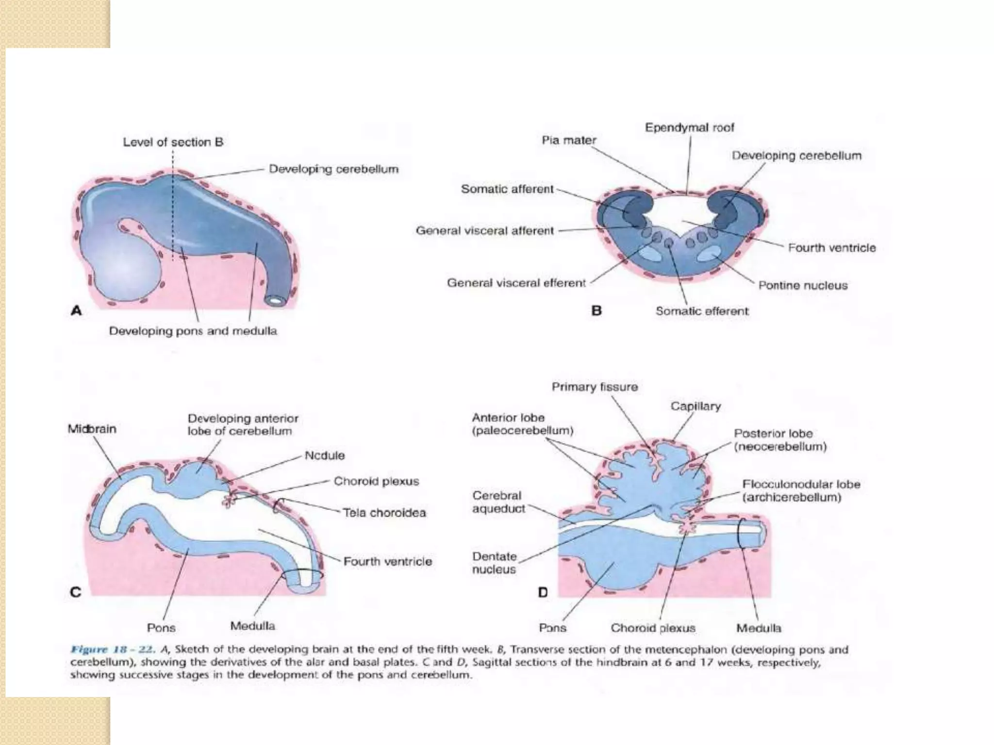
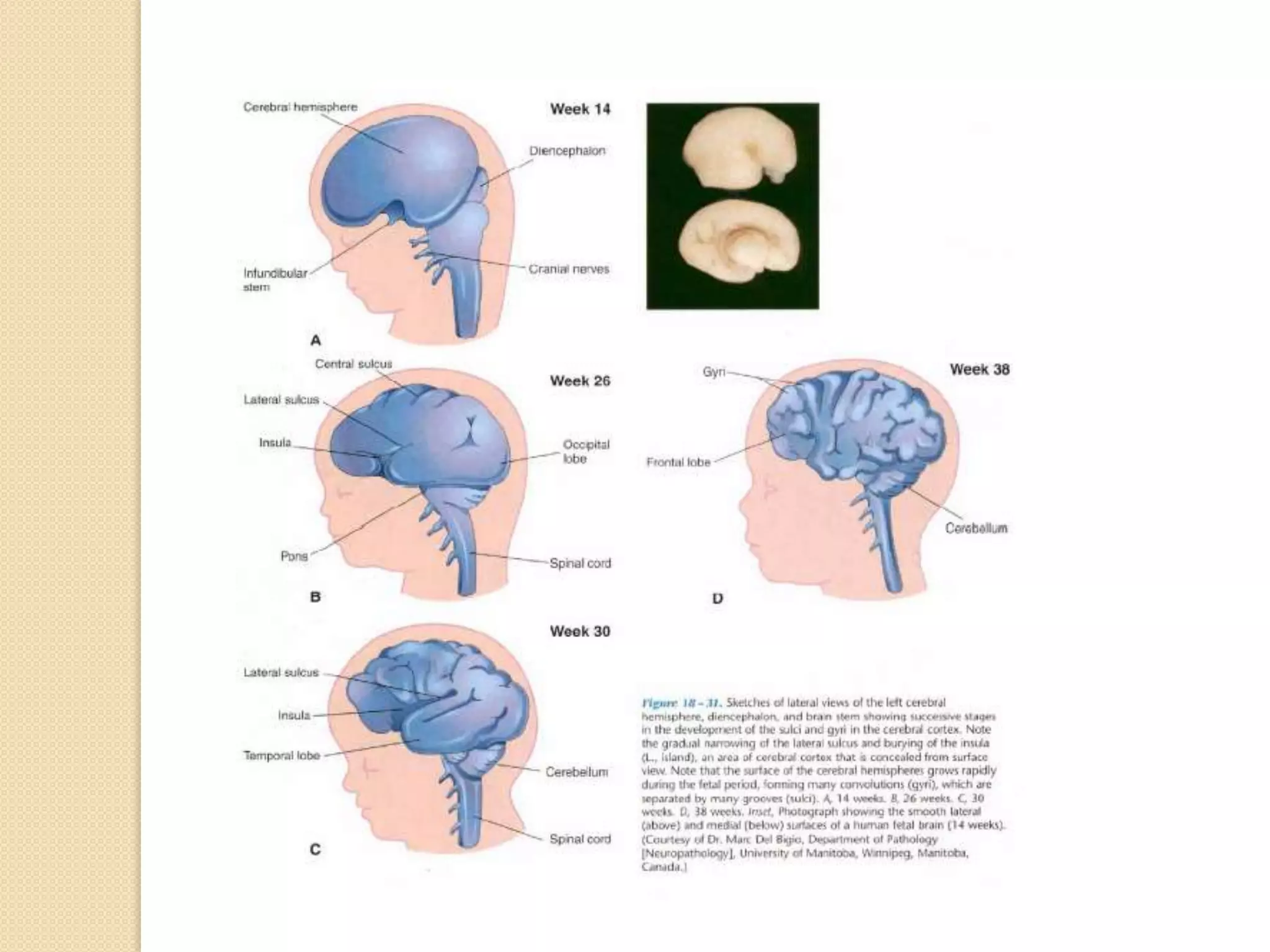
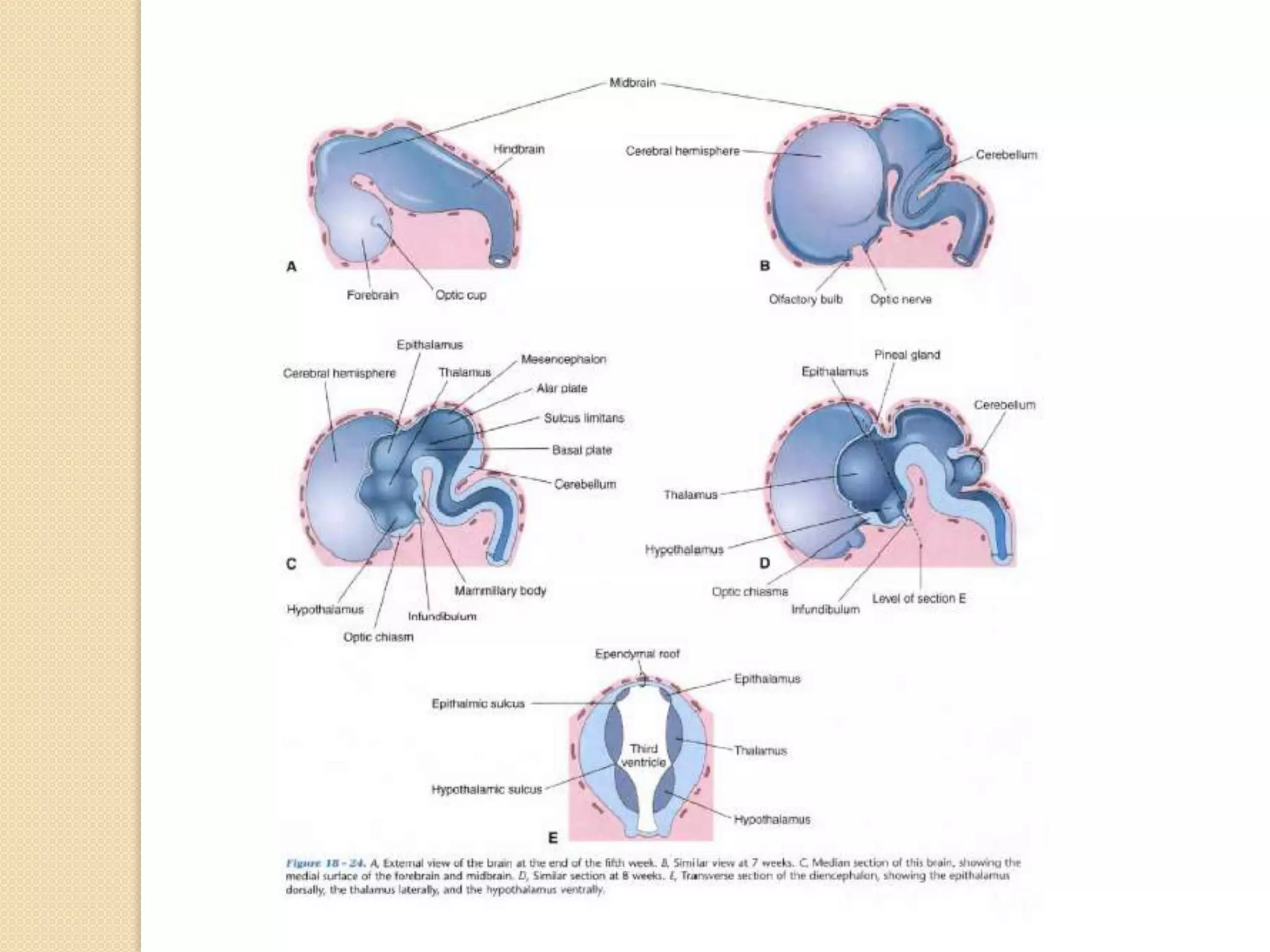
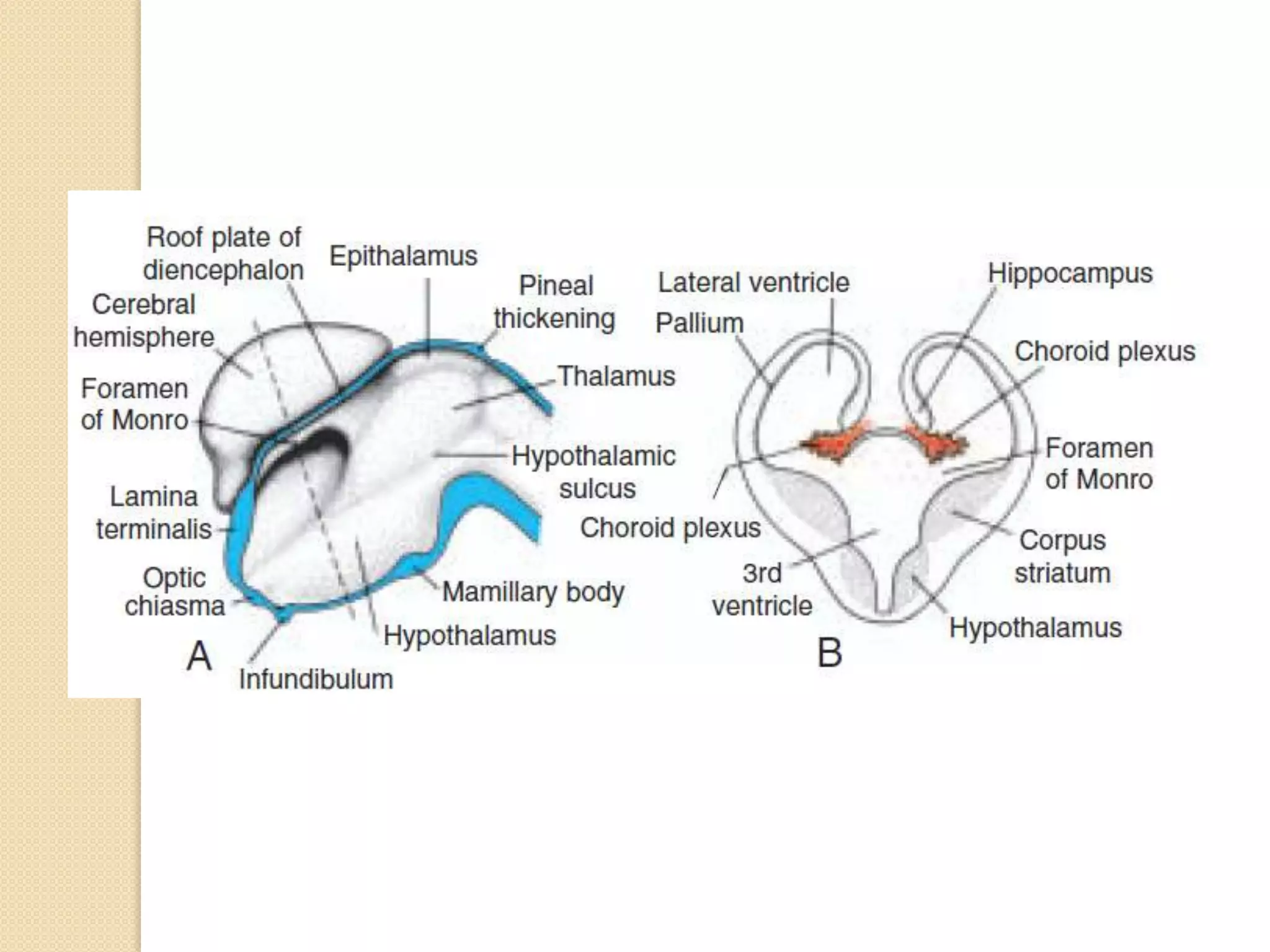
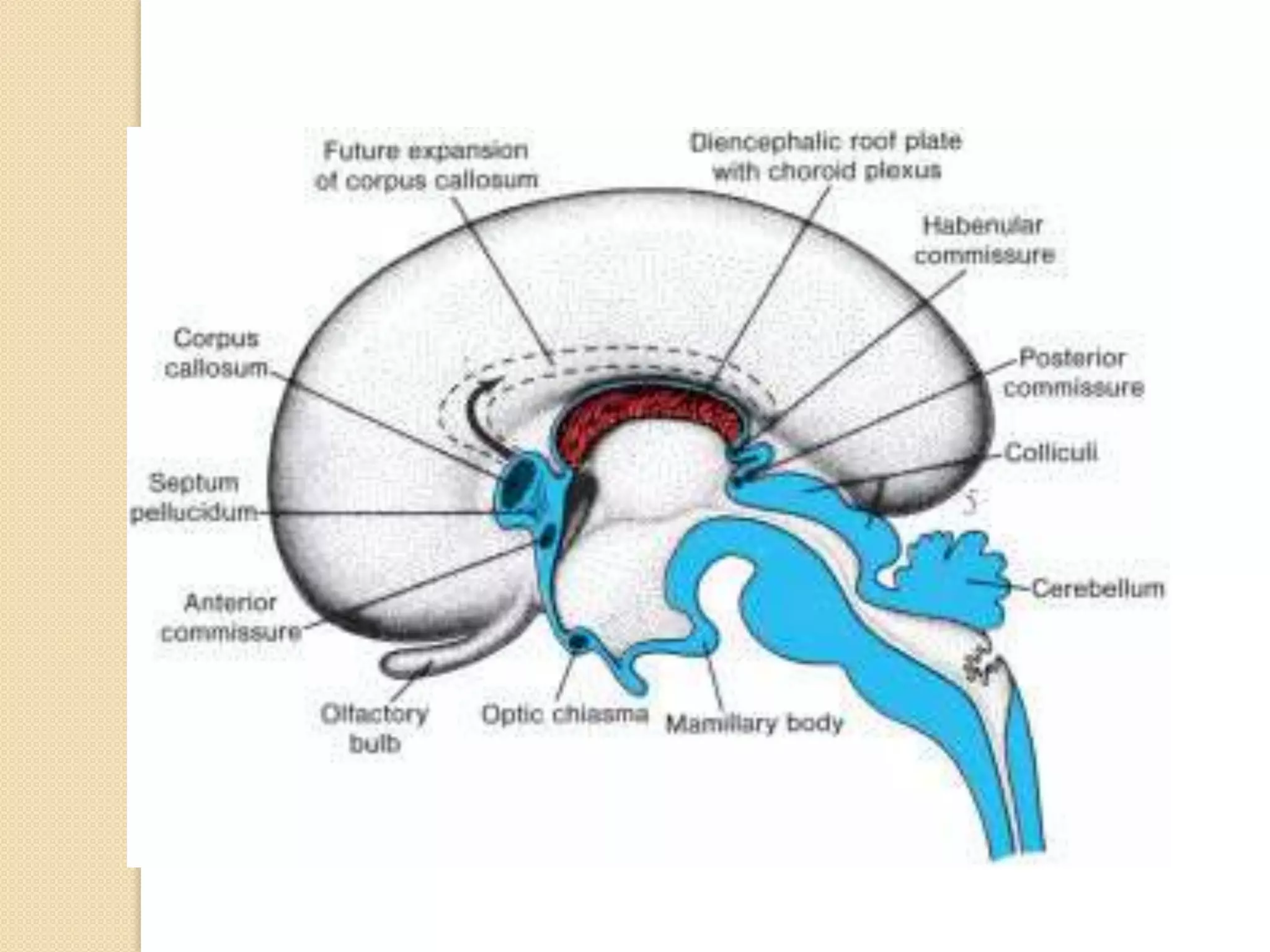
well describes the development of nervous system from basic to advanced concept including neural tube defects. the concepts are presented in graphical form for easy understanding of concepts.
white fibers of the cerebrum, commissural fibers, association fibers and radiation fibers, examples of each types of cerebral fibers, corpus callosum, fornix, habenular commisure, anterior commissure, posterior commissure, superior longitudinal fasciculus, inferior longitudinal fasciculus, occipital fasciculus, uncinate fasciculus, projection fibers, corona radiata, optic radiation
You can watch the video on my you tube channel: https://youtu.be/I0FaX-iQfa0
Medulla oblongata or more simply medulla is part of brain stem which forms base of the brain stem. It contains pyramid, olive and above pyramidal structure, there is decussation of pyramids which explains why each part of brain controls opposite part of body. Adding to that medulla also has several nuclei which controls activity of cardiovascular system and respiratory system. Medulla also has nuclei for controlling reflexes of vomiting, swallowing, hiccuping, coughing and sneezing. It has also nuclei for test, hearing and balance. Medulla also contains nuclei of cranial nerve number VIII, IX, X, XI and XII.
Proof version: Bishop, D., & Rutter, M. (2008). Neurodevelopmental disorders: conceptual approaches. In M. Rutter, D. Bishop, D. Pine, S. Scott, J. Stevenson, E. Taylor & A. Thapar (Eds.), Rutter's Child and Adolescent Psychiatry (pp. 32-41). Oxford: Blackwell.
Neurodevelopmental disorders: are our current diagnostic labels fit for purpose?Dorothy Bishop
Slides from a talk given at University of Western Australia on Tuesday 2nd October 2012, This lecture was co-hosted by the ARC Centre of Excellence in Cognition and
its Disorders and the Institute of Advanced Studies, University of Western Australia
THIS PRESENTATION IS UPLOADED TO HELP THE EDUCATOR OF MEDICAL, NURSING & ALLIE HEALTH SCIENCES TO TEACH THEIR STUDENTS ABOUT THE NERVOUS SYSTEM. IT WILL ALSO CREATE AWARENESS AMONG THE COMMON PEOPLE REGARDING NERVOUS SYSTEM.
this presentation consist of introduction to types of nerves, structure of nerve and cranial nerves. there is a detail description about, origin , course of the trigeminal nerve and its branches and the structures supplying the nerve. it also contains applied anatomy of the nerve and its importance of the nerve in oral and maxillofacial surgeries. a detail description about the examination of the trigeminal nerve is also mentioned in the presentation. hoping that it would be useful to the students and people seeking for knowledge about the trigeminal nerve.
Report Back from SGO 2024: What’s the Latest in Cervical Cancer?bkling
Are you curious about what’s new in cervical cancer research or unsure what the findings mean? Join Dr. Emily Ko, a gynecologic oncologist at Penn Medicine, to learn about the latest updates from the Society of Gynecologic Oncology (SGO) 2024 Annual Meeting on Women’s Cancer. Dr. Ko will discuss what the research presented at the conference means for you and answer your questions about the new developments.
These lecture slides, by Dr Sidra Arshad, offer a quick overview of physiological basis of a normal electrocardiogram.
Learning objectives:
1. Define an electrocardiogram (ECG) and electrocardiography
2. Describe how dipoles generated by the heart produce the waveforms of the ECG
3. Describe the components of a normal electrocardiogram of a typical bipolar leads (limb II)
4. Differentiate between intervals and segments
5. Enlist some common indications for obtaining an ECG
Study Resources:
1. Chapter 11, Guyton and Hall Textbook of Medical Physiology, 14th edition
2. Chapter 9, Human Physiology - From Cells to Systems, Lauralee Sherwood, 9th edition
3. Chapter 29, Ganong’s Review of Medical Physiology, 26th edition
4. Electrocardiogram, StatPearls - https://www.ncbi.nlm.nih.gov/books/NBK549803/
5. ECG in Medical Practice by ABM Abdullah, 4th edition
6. ECG Basics, http://www.nataliescasebook.com/tag/e-c-g-basics
Title: Sense of Smell
Presenter: Dr. Faiza, Assistant Professor of Physiology
Qualifications:
MBBS (Best Graduate, AIMC Lahore)
FCPS Physiology
ICMT, CHPE, DHPE (STMU)
MPH (GC University, Faisalabad)
MBA (Virtual University of Pakistan)
Learning Objectives:
Describe the primary categories of smells and the concept of odor blindness.
Explain the structure and location of the olfactory membrane and mucosa, including the types and roles of cells involved in olfaction.
Describe the pathway and mechanisms of olfactory signal transmission from the olfactory receptors to the brain.
Illustrate the biochemical cascade triggered by odorant binding to olfactory receptors, including the role of G-proteins and second messengers in generating an action potential.
Identify different types of olfactory disorders such as anosmia, hyposmia, hyperosmia, and dysosmia, including their potential causes.
Key Topics:
Olfactory Genes:
3% of the human genome accounts for olfactory genes.
400 genes for odorant receptors.
Olfactory Membrane:
Located in the superior part of the nasal cavity.
Medially: Folds downward along the superior septum.
Laterally: Folds over the superior turbinate and upper surface of the middle turbinate.
Total surface area: 5-10 square centimeters.
Olfactory Mucosa:
Olfactory Cells: Bipolar nerve cells derived from the CNS (100 million), with 4-25 olfactory cilia per cell.
Sustentacular Cells: Produce mucus and maintain ionic and molecular environment.
Basal Cells: Replace worn-out olfactory cells with an average lifespan of 1-2 months.
Bowman’s Gland: Secretes mucus.
Stimulation of Olfactory Cells:
Odorant dissolves in mucus and attaches to receptors on olfactory cilia.
Involves a cascade effect through G-proteins and second messengers, leading to depolarization and action potential generation in the olfactory nerve.
Quality of a Good Odorant:
Small (3-20 Carbon atoms), volatile, water-soluble, and lipid-soluble.
Facilitated by odorant-binding proteins in mucus.
Membrane Potential and Action Potential:
Resting membrane potential: -55mV.
Action potential frequency in the olfactory nerve increases with odorant strength.
Adaptation Towards the Sense of Smell:
Rapid adaptation within the first second, with further slow adaptation.
Psychological adaptation greater than receptor adaptation, involving feedback inhibition from the central nervous system.
Primary Sensations of Smell:
Camphoraceous, Musky, Floral, Pepperminty, Ethereal, Pungent, Putrid.
Odor Detection Threshold:
Examples: Hydrogen sulfide (0.0005 ppm), Methyl-mercaptan (0.002 ppm).
Some toxic substances are odorless at lethal concentrations.
Characteristics of Smell:
Odor blindness for single substances due to lack of appropriate receptor protein.
Behavioral and emotional influences of smell.
Transmission of Olfactory Signals:
From olfactory cells to glomeruli in the olfactory bulb, involving lateral inhibition.
Primitive, less old, and new olfactory systems with different path
- Video recording of this lecture in English language: https://youtu.be/lK81BzxMqdo
- Video recording of this lecture in Arabic language: https://youtu.be/Ve4P0COk9OI
- Link to download the book free: https://nephrotube.blogspot.com/p/nephrotube-nephrology-books.html
- Link to NephroTube website: www.NephroTube.com
- Link to NephroTube social media accounts: https://nephrotube.blogspot.com/p/join-nephrotube-on-social-media.html
TEST BANK for Operations Management, 14th Edition by William J. Stevenson, Ve...kevinkariuki227
TEST BANK for Operations Management, 14th Edition by William J. Stevenson, Verified Chapters 1 - 19, Complete Newest Version.pdf
Couples presenting to the infertility clinic- Do they really have infertility...Sujoy Dasgupta
Dr Sujoy Dasgupta presented the study on "Couples presenting to the infertility clinic- Do they really have infertility? – The unexplored stories of non-consummation" in the 13th Congress of the Asia Pacific Initiative on Reproduction (ASPIRE 2024) at Manila on 24 May, 2024.
Flu Vaccine Alert in Bangalore Karnatakaaddon Scans
As flu season approaches, health officials in Bangalore, Karnataka, are urging residents to get their flu vaccinations. The seasonal flu, while common, can lead to severe health complications, particularly for vulnerable populations such as young children, the elderly, and those with underlying health conditions.
Dr. Vidisha Kumari, a leading epidemiologist in Bangalore, emphasizes the importance of getting vaccinated. "The flu vaccine is our best defense against the influenza virus. It not only protects individuals but also helps prevent the spread of the virus in our communities," he says.
This year, the flu season is expected to coincide with a potential increase in other respiratory illnesses. The Karnataka Health Department has launched an awareness campaign highlighting the significance of flu vaccinations. They have set up multiple vaccination centers across Bangalore, making it convenient for residents to receive their shots.
To encourage widespread vaccination, the government is also collaborating with local schools, workplaces, and community centers to facilitate vaccination drives. Special attention is being given to ensuring that the vaccine is accessible to all, including marginalized communities who may have limited access to healthcare.
Residents are reminded that the flu vaccine is safe and effective. Common side effects are mild and may include soreness at the injection site, mild fever, or muscle aches. These side effects are generally short-lived and far less severe than the flu itself.
Healthcare providers are also stressing the importance of continuing COVID-19 precautions. Wearing masks, practicing good hand hygiene, and maintaining social distancing are still crucial, especially in crowded places.
Protect yourself and your loved ones by getting vaccinated. Together, we can help keep Bangalore healthy and safe this flu season. For more information on vaccination centers and schedules, residents can visit the Karnataka Health Department’s official website or follow their social media pages.
Stay informed, stay safe, and get your flu shot today!
New Directions in Targeted Therapeutic Approaches for Older Adults With Mantl...i3 Health
i3 Health is pleased to make the speaker slides from this activity available for use as a non-accredited self-study or teaching resource.
This slide deck presented by Dr. Kami Maddocks, Professor-Clinical in the Division of Hematology and
Associate Division Director for Ambulatory Operations
The Ohio State University Comprehensive Cancer Center, will provide insight into new directions in targeted therapeutic approaches for older adults with mantle cell lymphoma.
STATEMENT OF NEED
Mantle cell lymphoma (MCL) is a rare, aggressive B-cell non-Hodgkin lymphoma (NHL) accounting for 5% to 7% of all lymphomas. Its prognosis ranges from indolent disease that does not require treatment for years to very aggressive disease, which is associated with poor survival (Silkenstedt et al, 2021). Typically, MCL is diagnosed at advanced stage and in older patients who cannot tolerate intensive therapy (NCCN, 2022). Although recent advances have slightly increased remission rates, recurrence and relapse remain very common, leading to a median overall survival between 3 and 6 years (LLS, 2021). Though there are several effective options, progress is still needed towards establishing an accepted frontline approach for MCL (Castellino et al, 2022). Treatment selection and management of MCL are complicated by the heterogeneity of prognosis, advanced age and comorbidities of patients, and lack of an established standard approach for treatment, making it vital that clinicians be familiar with the latest research and advances in this area. In this activity chaired by Michael Wang, MD, Professor in the Department of Lymphoma & Myeloma at MD Anderson Cancer Center, expert faculty will discuss prognostic factors informing treatment, the promising results of recent trials in new therapeutic approaches, and the implications of treatment resistance in therapeutic selection for MCL.
Target Audience
Hematology/oncology fellows, attending faculty, and other health care professionals involved in the treatment of patients with mantle cell lymphoma (MCL).
Learning Objectives
1.) Identify clinical and biological prognostic factors that can guide treatment decision making for older adults with MCL
2.) Evaluate emerging data on targeted therapeutic approaches for treatment-naive and relapsed/refractory MCL and their applicability to older adults
3.) Assess mechanisms of resistance to targeted therapies for MCL and their implications for treatment selection
HOT NEW PRODUCT! BIG SALES FAST SHIPPING NOW FROM CHINA!! EU KU DB BK substit...GL Anaacs
Contact us if you are interested:
Email / Skype : kefaya1771@gmail.com
Threema: PXHY5PDH
New BATCH Ku !!! MUCH IN DEMAND FAST SALE EVERY BATCH HAPPY GOOD EFFECT BIG BATCH !
Contact me on Threema or skype to start big business!!
Hot-sale products:
NEW HOT EUTYLONE WHITE CRYSTAL!!
5cl-adba precursor (semi finished )
5cl-adba raw materials
ADBB precursor (semi finished )
ADBB raw materials
APVP powder
5fadb/4f-adb
Jwh018 / Jwh210
Eutylone crystal
Protonitazene (hydrochloride) CAS: 119276-01-6
Flubrotizolam CAS: 57801-95-3
Metonitazene CAS: 14680-51-4
Payment terms: Western Union,MoneyGram,Bitcoin or USDT.
Deliver Time: Usually 7-15days
Shipping method: FedEx, TNT, DHL,UPS etc.Our deliveries are 100% safe, fast, reliable and discreet.
Samples will be sent for your evaluation!If you are interested in, please contact me, let's talk details.
We specializes in exporting high quality Research chemical, medical intermediate, Pharmaceutical chemicals and so on. Products are exported to USA, Canada, France, Korea, Japan,Russia, Southeast Asia and other countries.
Pulmonary Thromboembolism - etilogy, types, medical- Surgical and nursing man...VarunMahajani
Disruption of blood supply to lung alveoli due to blockage of one or more pulmonary blood vessels is called as Pulmonary thromboembolism. In this presentation we will discuss its causes, types and its management in depth.
Anti ulcer drugs and their Advance pharmacology ||
Anti-ulcer drugs are medications used to prevent and treat ulcers in the stomach and upper part of the small intestine (duodenal ulcers). These ulcers are often caused by an imbalance between stomach acid and the mucosal lining, which protects the stomach lining.
||Scope: Overview of various classes of anti-ulcer drugs, their mechanisms of action, indications, side effects, and clinical considerations.
2. Introduction
Formation of neurons and neuroglial cells
Neural tube and its sub divisions
Spinal cord
Medulla oblongata
Pons
Midbrain
Cerebral hemisphere
Corpus striatum
Cerebral cortex
Autonomic nervous system
3. Formation of neurons and
neuroglial cells
Neural tube first lined by single layer
of cells
Proliferate to form several layers
◦ Matrix layer
◦ Mantle layer
◦ Marginal layer
4. Contd..
Stages in the formation of a nerve cell
are:
◦ Apolar neuroblast
◦ Bipolar neuroblast
◦ Unipolar neuroblast
◦ Multipolar neuroblast
◦ Axon and dendrites
5. Neuroglial cells are also formed from
germinal cells of the ependymal layer
Glioblasts migrate in to mantle and
marginal layer as medulloblasts
They differentiate either into
astroblasts or oligodendroblasts
Microglial cells are mesodermal in
origin
6.
7. Myelination of fibers
Nerve fibers which remain with in the
brain and spinal cord receives support
from and are ensheathed by neuroglial
cells
Peripheral nerves special sheath
called the neurolemma derived from
schwann cells
Myelin of the CNS derived from
oligodendrocytes
8.
9. Neural tube and its
subdivisions
Whole of the nervous system is
derived from ectoderm except blood
vessels and neuroglial elements
Ectoderm situated on the dorsal
aspect of embryonic disc forms the
neural plate
Neural groove
Neural tube
Enlarges cranial part and caudal
tubular part
10.
11.
12. Contd..
The cavity of the brain shows three
dilatations
Prosencephalon, mesencephalon,
rhombencephalon
Prosencephalon
◦ Diencephalon
◦ Telencephalon
Rhombencephalon
◦ Metencephalon
◦ Myelencephalon
13.
14. Contd..
The relative position is altered by
number of flexures
◦ Cervical flexure
◦ Mesencephalic flexure
◦ Pontine flexure
◦ Telencephalic flexure
15.
16. Neural crest
Cells between the neural plate and the
rest of the ectoderm form primordia of
the neural crest
◦ DRG
◦ Sensory ganglia of 5, 7, 9, 10
◦ Neurons and satellite cells of sympathetic
ganglia
◦ Parasymathetic ganglia
◦ Schwann cells
20. Spinal cord
Devolops from caudal cylindrical part of
neural tube
Cavity of the tube bounded by thick
lateral wall, thin roof and floor
Tube subdivides in to three layers matrix
layer, mantle layer and marginal layer
Ventral layer of the mantle layer grows
faster than dorsal layer
Line seperating the compressed ventral
part from the dorsal part is called sulcus
limtans
21.
22. Contd..
Dorsal or alar lamina
Ventral or basal lamina
Posteriomedeian fissure
Anteriomedian fissure
Nerve cells that devolop in mantle
zone of the basal lamina becomes the
neuron of anterior grey column and
from the alar lamina the neuron of the
posterior grey column
23.
24. Contd..
Dorsal nerve root ganglia are formed
by axons of the cells that develop from
neural crest
Axons from the post grey column
enter the marginal layer to form the
ascending tract
Descending tracts are formed by the
axons projecting from the brain
Grey columns divide the white matter
in to anterior, posterior and lateral
columns
25.
26. POSITIONAL CHANGES OF
THE CORD
In the third month of development the spinal cord extends
the entire length of the embryo, and spinal nerves pass
through the intervertebral foramina at their level of origin.
With increasing age, the vertebral column and dura
lengthen more rapidly than the neural tube, and the terminal
end of the spinal cord gradually shifts to a higher level.
At birth, this end is at the level of the third lumbar
vertebra.
As a result of this disproportionate growth, spinal nerves run
obliquely from their segment of origin in the spinal cord to
the corresponding level of the vertebral column.
The dura remains attached to the vertebral column at the
coccygeal level.
In the adult, the spinal cord terminates at the level of L2 to
L3,
The dural sac and subarachnoid space extend to S2.
Below L2 to L3, a threadlike extension of the pia mater
forms the filum terminale, which is attached to the
periosteum of the first coccygeal vertebra and which marks
the tract of regression of the spinal cord.
Nerve fibers below the terminal end of the cord collectively
constitute the cauda equina.
27. Medulla oblongata
Myelencephalon
Early devolopment simillar to spinal
cord sulcus limitans divides in to alar
and basal lamina
Roof plate becomes greatly widened
resuting which alar plate comes
dorsolateral to basal plate
Alar plate forms olivary nuclei and
cranial nerve nuclei
29. Pons
Ventral part of metencephalon
Contribution alar lamina of the
myelencephalon and gives rise to
pontine nuclei axons arising from them
forms MCP
Lateral part of alar lamina becomes
rhombic lips to form the cerebellum
Nuclei arising from basal and alar
plate lie in dorsal or tegmental part of
pons
30. Contd..
Ventral part of pons constitutes:
◦ Middle cerebellar peduncle
◦ Corticospinal, corticobulbar and
corticopontine fibers
31.
32. Mid brain
Nuclei of the basal lamina
◦ Occulomotor nuclei
◦ Trochlear nuclei
◦ Edinger Westphal nuclei
Alar lamina gives rise to
◦ Colliculi
◦ Red nucleus
◦ Substantia nigra
33. Contd..
Marginal layer ventral part of
mesencephalon invaded by down
growing fibers of CST, corticobulbar
and cortico pontine pathway and
forms crus cerebri
34.
35. Cerebellum
Devolops from dorsolateral part of alar
lamina of the metencephalon
Rhombic lips on either side grows and
fuses medially to form cerebellum
Consists of usual matrix, mantle and
marginal layer
Cells of the mantle layer migrating in to
marginal layer forms cortex and those
don’t forms dentate, emboliform, fastigial
and globose nuclei
38. Cerebral hemispheres
Median diencephalon and lateral
telencephalon
Telancephalon
◦ Cerebral cortex and corpus striatum
Diencephalon
◦ Epithalamus, thalamus and hypothalamus
Telencephalic vesicles are small
initially rapidly grows upwards,
forwards and backwards completely
covers the diencephalon
39. Contd..
Cavity of diencephalon form third
ventricle and of telencephalic vesicles
form lateral ventricles
Each lateral is at first a small spherical
space
◦ Forward and backward growth elongates
anterioposteriorly
◦ Grows downwards and forwards to form the
temporal lobe and horn
◦ Backward growth to form occipital pole and
posterior horn
40.
41.
42.
43. Thalamus and hypothalamus
Devolops from diencephalon
Lateral wall of diencephalon becomes
thickened
Divided by epithalamic and
hypothalamic sulci
Epithalamus represented by
habenular and pineal body
44.
45. Corpus striatum
Derivative of telencephalon
Telencephalic vesicle can be
subdivided in to basal part which is
thick and superior part is thin
Some cells migrate in to marginal
layer and forms cortex and remaining
cells form copus striatum
Corpus striatum subdivided into
medial and lateral divisions
46. Contd..
Fibers descending from cerebral
cortex passes through medial and
lateral parts constitutes internal
capsule
Medial or deeper part form caudate
nucleus and superficial or lateral part
forms lentiform nucleus
47.
48.
49.
50. Cerebral cortex
Formed by migration of cells from the
mantle layer in to overlying marginal
layer
Region of insula relatively slow in
growth and gradually overgrown by
adjacent area forms the opercula
Cortex divided in to
◦ Hippocampal
◦ Pyriform
◦ neocortex
51. Contd..
Pyriform cortex that recives olfactory
sensation constitutes uncus,
parahippocampal gyrus and anterior
perforated substance
Telencephalon has a medial wall
opposing each other gives rise to
hippocampal cortex
Pyriform cortex arises from marginal
layer superficial to corpus striatum
With formation of inferior horn
hippocampus follows the curve and
assumes aring shaped configuration
52. Contd..
Superior part of the hippocampus
becomes ridimentary and forms
indesium gresium
White matter of the cerebral cortex is
formed by projection, association,
commisural and ascending fibers
53.
54.
55.
56. Cerebral commisures
Part of the neural tube that closes the
cranial end of prosencephalon is
lamina terminalis
Anterior commisure
Hippocampal commisure
Corpus collosum
Optic chiasma, habenular commisure,
posterior commisure.
59. Sympathetic nervous system
Preganglionic neurons devolop from
mantle layer of thoracolumbar region
of the spinal cord
Located in lateral horn of the cord
After short course in the spinal
nervethey leave them and grows
towards the postganglionic neurons
60. Contd..
Postganglionic neurons are of two
types
◦ Sympathetic trunk
◦ Visceral ganglia
61.
62. Parasympathetic nervous
system
Cranial parasympathetic out flow
◦ Derived GVE nuclear column of the brain
stem
Edinger westphal nucleus
Salivatory nucleus
Lacrimatory nucleus
Dorsal nucleus of vagus
Sacral parasympathetic out flow
◦ Preganglionic cells are formed in the
mantle layer of the spinal cord near the
sulcus limitans S2-S4
63. Contd..
Postganglionic neurons lies near the
viscera