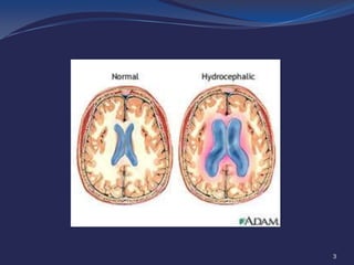
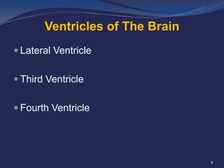
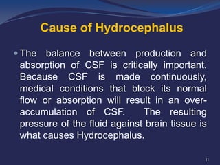
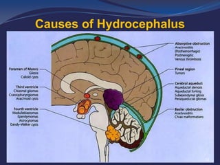
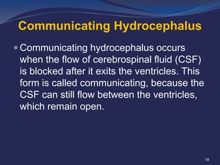
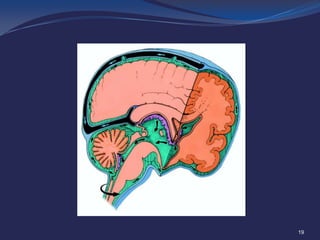
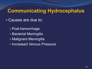
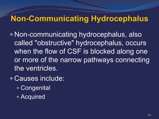
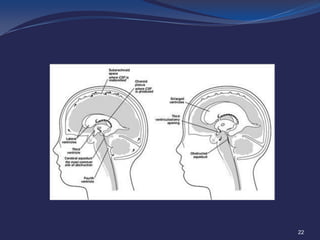
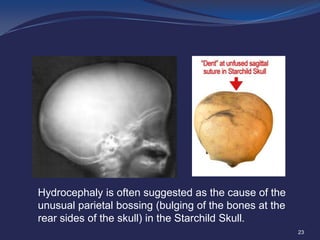
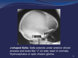
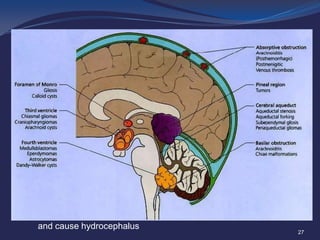
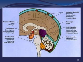
HYDROCEPHALUS
By

Dr Khufsa

1
Introduction
 Derived from Greek word “Hydro” meaning

“Water”, and “Cephalus”, meaning “Head”.
 Excessive accumulation of cerebrospinal

fluid (CSF) resulting in abnormal widening
of the spaces in the brain.

2
3
Ventricles of The Brain
 Lateral Ventricle
 Third Ventricle
 Fourth Ventricle

4
Anatomy of Ventricles

5
Anatomy of Ventricles

6
Circulation of CSF
 Normally, CSF flows continually from the

interior cavities in the brain (ventricles) to
the thin subarachnoid space that
surrounds the brain and spinal cord.

7
Circulation of CSF

8
Circulation of CSF
 Normally, CSF flows through the ventricles,

exits into cisterns (closed spaces that
serve as reservoirs) at the base of the
brain, bathes the surfaces of the brain and
spinal cord, and then reabsorbs into the
bloodstream.

9
Circulation of CSF

10
Cause of Hydrocephalus
 The

balance between production and
absorption of CSF is critically important.
Because CSF is made continuously,
medical conditions that block its normal
flow or absorption will result in an overaccumulation of CSF.
The resulting
pressure of the fluid against brain tissue is
what causes Hydrocephalus.
11
Causes of Hydrocephalus

12
Types of Hydrocephalus
 There are several different types of

Hydrocephalus:
 Congenital hydrocephalus

 Acquired hydrocephalus
 Communicating hydrocephalus
 Non-communicating hydrocephalus

13
Congenital Hydrocephalus
 Congenital hydrocephalus is present at

birth and may be caused by environmental
influences during fetal development or by
genetic factors.
 Causes are:
 Aqueduct Stenosis
 Colloid Cyst

14
Thinning of the choroid plexus.
Displacement of the medial and lateral walls of the lateral ventricle.
15
Acquired Hydrocephalus
 Develops at the time of birth or at some

point afterward. This type of hydrocephalus
can affect individuals of all ages and may
be caused by injury or disease.

16
Acquired Hydrocephalus
 Acquired Hydrocephalus is due to:
 Tumors
 Hemorrhage
 Ventriculitis

17
Communicating Hydrocephalus
 Communicating hydrocephalus occurs

when the flow of cerebrospinal fluid (CSF)
is blocked after it exits the ventricles. This
form is called communicating, because the
CSF can still flow between the ventricles,
which remain open.

18
19
Communicating Hydrocephalus
 Causes are due to:
 Post-hemorrhage
 Bacterial Meningitis

 Malignant Meningitis
 Increased Venous Pressure

20
Non-Communicating Hydrocephalus
 Non-communicating hydrocephalus, also

called "obstructive" hydrocephalus, occurs
when the flow of CSF is blocked along one
or more of the narrow pathways connecting
the ventricles.
 Causes include:
 Congenital

 Acquired

21
22
Hydrocephaly is often suggested as the cause of the
unusual parietal bossing (bulging of the bones at the
rear sides of the skull) in the Starchild Skull.
23
J-shaped Sella: Sella extends under anterior clinoid
process and looks like “J” on side; seen in normals,
Hydrocephalus or optic chiasm glioma.
24
Spot Diagnosis ?

VP Shunt
25
?

26
?

Colloid Cysts are usually located in the anterior (front) portion
of the third ventricle, resting against the hypothalamus, where
they may block the outflow pathways for the cerebrospinal fluid
and cause hydrocephalus

27
?

Colloid cyst with obstructive hydrocephalus. Sagittal T1-weighted
MR image shows a hyperintense nodular mass at the foramen of
Monro. Dilatation of both lateral ventricles is due to obstruction of
CSF flow at the foramen of Monro.
28
?
Axial T2 weighted image demonstrates a
well-marginated intraventricular mass near
the foramen of Monro. The lesion is
lobulated in appearance and is causing
obstructive hydrocephalus. Associated
vasogenic edema is noted extending into
the left frontal lobe. Coronal T1 contrastenhanced MR sequences demonstrate
intense, heterogeneous enhancement of
the mass.
Diagnosis: Subependymal
astrocytoma

giant

cell

29
?

A sagittal T1-weighted MRI shows a large
posterior fossa cyst. The hypoplastic
vermis is everted over the posterior fossa
cyst (long arrow). The cerebellar
hemispheres and brainstem (b) are
hypoplastic. Thinned occipital squama is
seen (arrowheads).

An axial T1-weighted MRI showing
ventriculomegaly and a superiorly
displaced posterior fossa cyst
30
?

MRI sagittal view showing Aqueduct Stenosis causing Hydrocephalus
31
?

Non contrast CT of the brain demonstrates marked hydrocephalus involving
the lateral and third ventricles but sparing the fourth ventricle. .

32
?

Shunt Catheter in Situ
33
?

(C) CT postcontrast imaging showed a tumor in midline causing a
bilateral obstructive hydrocephalus.
(D) Tumor was removed in this postoperative image.

34
?

Medulloblastoma: sagittal view MRI showing an avidly enhancing solid and
cystic lesion filling the fourth ventricle; obstructive hydrocephalus present
35
?

Brain CT at level of Vein of Galen demonstrating thrombus.
36
?

Sagittal view MR T1 post contrast showing Pineocytoma
causing hydrocephalus

37
THANK YOU

Hydrocephalus

  • 1.
  • 2.
    Introduction  Derived fromGreek word “Hydro” meaning “Water”, and “Cephalus”, meaning “Head”.  Excessive accumulation of cerebrospinal fluid (CSF) resulting in abnormal widening of the spaces in the brain. 2
  • 3.
  • 4.
    Ventricles of TheBrain  Lateral Ventricle  Third Ventricle  Fourth Ventricle 4
  • 5.
  • 6.
  • 7.
    Circulation of CSF Normally, CSF flows continually from the interior cavities in the brain (ventricles) to the thin subarachnoid space that surrounds the brain and spinal cord. 7
  • 8.
  • 9.
    Circulation of CSF Normally, CSF flows through the ventricles, exits into cisterns (closed spaces that serve as reservoirs) at the base of the brain, bathes the surfaces of the brain and spinal cord, and then reabsorbs into the bloodstream. 9
  • 10.
  • 11.
    Cause of Hydrocephalus The balance between production and absorption of CSF is critically important. Because CSF is made continuously, medical conditions that block its normal flow or absorption will result in an overaccumulation of CSF. The resulting pressure of the fluid against brain tissue is what causes Hydrocephalus. 11
  • 12.
  • 13.
    Types of Hydrocephalus There are several different types of Hydrocephalus:  Congenital hydrocephalus  Acquired hydrocephalus  Communicating hydrocephalus  Non-communicating hydrocephalus 13
  • 14.
    Congenital Hydrocephalus  Congenitalhydrocephalus is present at birth and may be caused by environmental influences during fetal development or by genetic factors.  Causes are:  Aqueduct Stenosis  Colloid Cyst 14
  • 15.
    Thinning of thechoroid plexus. Displacement of the medial and lateral walls of the lateral ventricle. 15
  • 16.
    Acquired Hydrocephalus  Developsat the time of birth or at some point afterward. This type of hydrocephalus can affect individuals of all ages and may be caused by injury or disease. 16
  • 17.
    Acquired Hydrocephalus  AcquiredHydrocephalus is due to:  Tumors  Hemorrhage  Ventriculitis 17
  • 18.
    Communicating Hydrocephalus  Communicatinghydrocephalus occurs when the flow of cerebrospinal fluid (CSF) is blocked after it exits the ventricles. This form is called communicating, because the CSF can still flow between the ventricles, which remain open. 18
  • 19.
  • 20.
    Communicating Hydrocephalus  Causesare due to:  Post-hemorrhage  Bacterial Meningitis  Malignant Meningitis  Increased Venous Pressure 20
  • 21.
    Non-Communicating Hydrocephalus  Non-communicatinghydrocephalus, also called "obstructive" hydrocephalus, occurs when the flow of CSF is blocked along one or more of the narrow pathways connecting the ventricles.  Causes include:  Congenital  Acquired 21
  • 22.
  • 23.
    Hydrocephaly is oftensuggested as the cause of the unusual parietal bossing (bulging of the bones at the rear sides of the skull) in the Starchild Skull. 23
  • 24.
    J-shaped Sella: Sellaextends under anterior clinoid process and looks like “J” on side; seen in normals, Hydrocephalus or optic chiasm glioma. 24
  • 25.
  • 26.
  • 27.
    ? Colloid Cysts areusually located in the anterior (front) portion of the third ventricle, resting against the hypothalamus, where they may block the outflow pathways for the cerebrospinal fluid and cause hydrocephalus 27
  • 28.
    ? Colloid cyst withobstructive hydrocephalus. Sagittal T1-weighted MR image shows a hyperintense nodular mass at the foramen of Monro. Dilatation of both lateral ventricles is due to obstruction of CSF flow at the foramen of Monro. 28
  • 29.
    ? Axial T2 weightedimage demonstrates a well-marginated intraventricular mass near the foramen of Monro. The lesion is lobulated in appearance and is causing obstructive hydrocephalus. Associated vasogenic edema is noted extending into the left frontal lobe. Coronal T1 contrastenhanced MR sequences demonstrate intense, heterogeneous enhancement of the mass. Diagnosis: Subependymal astrocytoma giant cell 29
  • 30.
    ? A sagittal T1-weightedMRI shows a large posterior fossa cyst. The hypoplastic vermis is everted over the posterior fossa cyst (long arrow). The cerebellar hemispheres and brainstem (b) are hypoplastic. Thinned occipital squama is seen (arrowheads). An axial T1-weighted MRI showing ventriculomegaly and a superiorly displaced posterior fossa cyst 30
  • 31.
    ? MRI sagittal viewshowing Aqueduct Stenosis causing Hydrocephalus 31
  • 32.
    ? Non contrast CTof the brain demonstrates marked hydrocephalus involving the lateral and third ventricles but sparing the fourth ventricle. . 32
  • 33.
  • 34.
    ? (C) CT postcontrastimaging showed a tumor in midline causing a bilateral obstructive hydrocephalus. (D) Tumor was removed in this postoperative image. 34
  • 35.
    ? Medulloblastoma: sagittal viewMRI showing an avidly enhancing solid and cystic lesion filling the fourth ventricle; obstructive hydrocephalus present 35
  • 36.
    ? Brain CT atlevel of Vein of Galen demonstrating thrombus. 36
  • 37.
    ? Sagittal view MRT1 post contrast showing Pineocytoma causing hydrocephalus 37
  • 38.