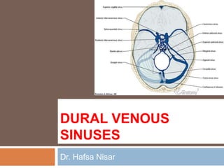
1) The document describes the dural venous sinuses, which drain blood from the brain into the internal jugular veins. They include the superior and inferior sagittal sinuses, straight sinus, transverse sinus, sigmoid sinus, occipital sinus, cavernous sinus, and superior and inferior petrosal sinuses.
2) The dural sinuses have no valves or muscle in their walls and are located between the layers of the dura mater. They drain blood from the brain and cerebrospinal fluid from the subarachnoid space.
3) The blood in the dural sinuses ultimately drains into the internal jugular veins in the neck through a series of sinuses and
Lateral ventricle of Brain. By Dr.N.Mugunthan.M.Smgmcri1234
Lateral ventricle of brain. Lecture by Dr.N.Mugunthan.
Associate Professor,
Mahatma Gandhi Medical College & Research Institute,
Sri Balaji Vidyapeeth, Pondicherry.
Lateral ventricle of Brain. By Dr.N.Mugunthan.M.Smgmcri1234
Lateral ventricle of brain. Lecture by Dr.N.Mugunthan.
Associate Professor,
Mahatma Gandhi Medical College & Research Institute,
Sri Balaji Vidyapeeth, Pondicherry.
introduction to skull, parts of skull, bones involved forming skull, different views of skull, norma basalis, anterio cranial middle cranial and posterior cranial fossa, clinical aspects of cranial fossa, foramens present in the cranial fossa
hypoglossal nerve, origin course an termination of hypoglossal nerve, function of hypoglossal nerve, clinical examination of hypoglossal nerve, hypoglossal nerve palsy
4 th ventricle- Anatomical and surgical perspectivesuresh Bishokarma
4th ventricle connects the entire ventricular system of brain. Its connection with cisterns magna and cerebella pontine cistern via foramen of magenta and Luschka. CSF absorbs into the arachnoid granulation.
Referred from different sources , here i present a very concise presentation on CRANIAL CAVITY . This presentation will give you complete knowledge of the topic cranial cavity with well elaborated and intellectual diagrams hand picked from F. Netter. ......... Do like and share , Leave your comments so as to get more stuff like this in future.
Anatomy and function of the dural venous sinusesSaad Salih
Dural Venous Sinuses
The dural venous sinuses lie between the periosteal and meningeal layers of the dura mater. They are best thought of as collecting pools of blood, which drain the central nervous system, the face, and the scalp. All the dural venous sinuses ultimately drain into the internal jugular vein. Unlike most veins of the body, the dural venous sinuses do not have valves.
There are eleven venous sinuses in total. The straight, superior, and inferior sagittal sinuses are found in the falx cerebri of the dura mater. They converge at the confluence of sinuses (overlying the internal occipital protuberance). The straight sinus is a continuation of the great cerebral vein and the inferior sagittal sinus.
From the confluence, the transverse sinus continues bi-laterally and curves into the sigmoid sinus to meet the opening of the internal jugular vein.
The cavernous sinus drains the ophthalmic veins and can be found on either side of the sella turcica. From here, the blood returns to the internal jugular vein via the superior or inferior petrosal sinuses.
venous drainage of head and neck and its branches are described in detail along with applied anatomy for better understanding of the anatomy and its application in oral and maxillary surgeries. knowing the anatomy and the course of the veins is crucial and helps in better locating the vein and ligating it to avoid further complications while performing a oral and maxillofacial surgeries such as in trauma fixation, tumor resection and as well as reconstruction of the defect pertaining to the maxillofacial region.
VENOUS DRAINAGE OF HEAD, FACE, NECK AND BRAINDrVishal2
THIS SEMINAR ON VENOUS DRAINAGE OF HEAD, FACE, NECK AND BRAIN ENCOMPASSES ALL THE POSSIBLE DETAILED EXPLANATION ALONG WITH DIAGRAMMATIC ILLUSTRATIONS OF THE SAME. APPLIED AND SURGICAL ANATOMY ALONG WITH RECENT MODALITIES HAS BEEN ADDED HEREIN..
Instructions for Submissions thorugh G- Classroom.pptxJheel Barad
This presentation provides a briefing on how to upload submissions and documents in Google Classroom. It was prepared as part of an orientation for new Sainik School in-service teacher trainees. As a training officer, my goal is to ensure that you are comfortable and proficient with this essential tool for managing assignments and fostering student engagement.
Model Attribute Check Company Auto PropertyCeline George
In Odoo, the multi-company feature allows you to manage multiple companies within a single Odoo database instance. Each company can have its own configurations while still sharing common resources such as products, customers, and suppliers.
The Roman Empire A Historical Colossus.pdfkaushalkr1407
The Roman Empire, a vast and enduring power, stands as one of history's most remarkable civilizations, leaving an indelible imprint on the world. It emerged from the Roman Republic, transitioning into an imperial powerhouse under the leadership of Augustus Caesar in 27 BCE. This transformation marked the beginning of an era defined by unprecedented territorial expansion, architectural marvels, and profound cultural influence.
The empire's roots lie in the city of Rome, founded, according to legend, by Romulus in 753 BCE. Over centuries, Rome evolved from a small settlement to a formidable republic, characterized by a complex political system with elected officials and checks on power. However, internal strife, class conflicts, and military ambitions paved the way for the end of the Republic. Julius Caesar’s dictatorship and subsequent assassination in 44 BCE created a power vacuum, leading to a civil war. Octavian, later Augustus, emerged victorious, heralding the Roman Empire’s birth.
Under Augustus, the empire experienced the Pax Romana, a 200-year period of relative peace and stability. Augustus reformed the military, established efficient administrative systems, and initiated grand construction projects. The empire's borders expanded, encompassing territories from Britain to Egypt and from Spain to the Euphrates. Roman legions, renowned for their discipline and engineering prowess, secured and maintained these vast territories, building roads, fortifications, and cities that facilitated control and integration.
The Roman Empire’s society was hierarchical, with a rigid class system. At the top were the patricians, wealthy elites who held significant political power. Below them were the plebeians, free citizens with limited political influence, and the vast numbers of slaves who formed the backbone of the economy. The family unit was central, governed by the paterfamilias, the male head who held absolute authority.
Culturally, the Romans were eclectic, absorbing and adapting elements from the civilizations they encountered, particularly the Greeks. Roman art, literature, and philosophy reflected this synthesis, creating a rich cultural tapestry. Latin, the Roman language, became the lingua franca of the Western world, influencing numerous modern languages.
Roman architecture and engineering achievements were monumental. They perfected the arch, vault, and dome, constructing enduring structures like the Colosseum, Pantheon, and aqueducts. These engineering marvels not only showcased Roman ingenuity but also served practical purposes, from public entertainment to water supply.
Synthetic Fiber Construction in lab .pptxPavel ( NSTU)
Synthetic fiber production is a fascinating and complex field that blends chemistry, engineering, and environmental science. By understanding these aspects, students can gain a comprehensive view of synthetic fiber production, its impact on society and the environment, and the potential for future innovations. Synthetic fibers play a crucial role in modern society, impacting various aspects of daily life, industry, and the environment. ynthetic fibers are integral to modern life, offering a range of benefits from cost-effectiveness and versatility to innovative applications and performance characteristics. While they pose environmental challenges, ongoing research and development aim to create more sustainable and eco-friendly alternatives. Understanding the importance of synthetic fibers helps in appreciating their role in the economy, industry, and daily life, while also emphasizing the need for sustainable practices and innovation.
Biological screening of herbal drugs: Introduction and Need for
Phyto-Pharmacological Screening, New Strategies for evaluating
Natural Products, In vitro evaluation techniques for Antioxidants, Antimicrobial and Anticancer drugs. In vivo evaluation techniques
for Anti-inflammatory, Antiulcer, Anticancer, Wound healing, Antidiabetic, Hepatoprotective, Cardio protective, Diuretics and
Antifertility, Toxicity studies as per OECD guidelines
2024.06.01 Introducing a competency framework for languag learning materials ...Sandy Millin
http://sandymillin.wordpress.com/iateflwebinar2024
Published classroom materials form the basis of syllabuses, drive teacher professional development, and have a potentially huge influence on learners, teachers and education systems. All teachers also create their own materials, whether a few sentences on a blackboard, a highly-structured fully-realised online course, or anything in between. Despite this, the knowledge and skills needed to create effective language learning materials are rarely part of teacher training, and are mostly learnt by trial and error.
Knowledge and skills frameworks, generally called competency frameworks, for ELT teachers, trainers and managers have existed for a few years now. However, until I created one for my MA dissertation, there wasn’t one drawing together what we need to know and do to be able to effectively produce language learning materials.
This webinar will introduce you to my framework, highlighting the key competencies I identified from my research. It will also show how anybody involved in language teaching (any language, not just English!), teacher training, managing schools or developing language learning materials can benefit from using the framework.
Read| The latest issue of The Challenger is here! We are thrilled to announce that our school paper has qualified for the NATIONAL SCHOOLS PRESS CONFERENCE (NSPC) 2024. Thank you for your unwavering support and trust. Dive into the stories that made us stand out!
Introduction to AI for Nonprofits with Tapp NetworkTechSoup
Dive into the world of AI! Experts Jon Hill and Tareq Monaur will guide you through AI's role in enhancing nonprofit websites and basic marketing strategies, making it easy to understand and apply.
Embracing GenAI - A Strategic ImperativePeter Windle
Artificial Intelligence (AI) technologies such as Generative AI, Image Generators and Large Language Models have had a dramatic impact on teaching, learning and assessment over the past 18 months. The most immediate threat AI posed was to Academic Integrity with Higher Education Institutes (HEIs) focusing their efforts on combating the use of GenAI in assessment. Guidelines were developed for staff and students, policies put in place too. Innovative educators have forged paths in the use of Generative AI for teaching, learning and assessments leading to pockets of transformation springing up across HEIs, often with little or no top-down guidance, support or direction.
This Gasta posits a strategic approach to integrating AI into HEIs to prepare staff, students and the curriculum for an evolving world and workplace. We will highlight the advantages of working with these technologies beyond the realm of teaching, learning and assessment by considering prompt engineering skills, industry impact, curriculum changes, and the need for staff upskilling. In contrast, not engaging strategically with Generative AI poses risks, including falling behind peers, missed opportunities and failing to ensure our graduates remain employable. The rapid evolution of AI technologies necessitates a proactive and strategic approach if we are to remain relevant.
7. Dural Venous Sinuses
Situated between the layers of
the dura mater .
Dural sinuses are lined by
endothelium, and their walls
are thick but devoid of muscular
tissue. They have no valves.
Function. to receive blood from
the brain through the cerebral
veins and the cerebrospinal fluid
from the subarachnoid space
through the arachnoid villi
.
8.
9. The blood in the dural
sinuses ultimately drains
into the internal jugular
veins in the neck.
Emissary veins, which
are also valveless,
connect the dural venous
sinuses with the diploic
veins of the skull and with
the veins of the scalp
14. It begins anteriorly at the foramen
cecum, where it occasionally
receives a vein from the nasal
cavity.
It runs posteriorly, grooving the
vault of the skull.
it deviates to one or the other
side (usually the right) at the
internal occipital protuberance
and becomes continuous with the
corresponding transverse sinus.
15. The superior sagittal sinus
The confluence of
the sinuses
is formed at the
internal occipital
protuberance.
sinus usually dilates
and becomes
continuous with the right
transverse sinus; it is
connected to the
opposite transverse
16. Numerous arachnoid
villi and granulations
project into the
lacunae, which also
receive the diploic
and meningeal veins .
The superior sagittal
sinus in its course
receives the superior
cerebral veins
17.
18. 2. The inferior sagittal sinus
occupies the free lower margin
of the falx cerebri.
It runs backward and joins the
great cerebral vein at the free
margin of the tentorium
cerebelli to form the straight
sinus.
19.
20. The inferior sagittal sinus receives a few cerebral veins
from the medial surface of the cerebral hemispheres.
21. The straight sinus
It is formed by the union
of the inferior sagittal
sinus with the great
cerebral vein.
occupies the line of
junction of the falx cerebri
with the tentorium
cerebelli.
It ends by turning to the
left (sometimes to the
right) to form the
22. 1. The transverse sinuses
are paired structures that begin at the internal
occipital protuberance
The right sinus is usually continuous with the
superior sagittal sinus, and the left is
continuous with the straight sinus.
The transverse sinuses receive the superior
petrosal sinuses, the inferior cerebral and
cerebellar veins, and the diploic veins.
23. Each sinus occupies the
attached margin of the
tentorium cerebelli, grooving
the occipital bone and the
posteroinferior angle of the
parietal bone.
They end by turning
downward as the sigmoid
sinuses.
24. The sigmoid sinuses
are a direct continuation of the transverse
sinuses.
Each sinus turns downward and medially and
grooves the mastoid part of the temporal bone.
pass inferiorly through the posterior part of the
jugular foramen to become continuous with the
superior bulb of the internal jugular vein.
25.
26.
27. The sigmoid sinuses become continuous with the
superior bulb of the internal jugular vein.
28. The occipital sinus
is a small sinus occupying
the attached margin of the
falx cerebelli.
It commences near the
foramen magnum, where
it communicates with the
vertebral veins and drains
into the confluence of
sinuses.
29. The cavernous sinuses
Situated in the middle
cranial fossa on each side
of the body of the sphenoid
bone.
Each sinus extends from
the superior orbital fissure
to the apex of the petrous
part of the temporal bone.
30. Numerous trabeculae cross their
interior, giving them a spongy
appearance.
The two sinuses communicate with
each other by means of the anterior
and posterior intercavernous
sinuses,
31. Tributaries : the superior and
inferior ophthalmic veins, the
inferior cerebral veins, the
sphenoparietal sinus, and the
central vein of the retina.
The sinus drains posteriorly
into the superior and inferior
petrosal sinuses and inferiorly
into the pterygoid venous plexus.
32.
33.
34. Each sinus has an
important
communication with the
facial vein through the
superior ophthalmic
vein.
This is a route by which
infection can travel from
the facial skin to the
cavernous sinus.
35.
36.
37.
38.
39. Cavernous sinus Thrombosis
Causes:
Bacterial infection that has spread
from the sinuses, teeth, ears,
eyes, nose, or skin of the face
Symptoms:
Bulging eyeball, usually on one
side of face
Cannot move the eye in a
particular direction
Drooping eyelids
Headaches
Vision loss
40. Tests include:
CT scan of the head
Magnetic resonance imaging (MRI) of the
brain
Magnetic resonance venogram
Sinus x-ray
Treatment:
Cavernous sinus thrombosis is treated
with high-dose antibiotics given through a
vein (IV).
Sometimes surgery is needed to drain
41. Petrosal sinuses
The superior and inferior petrosal sinuses are
small sinuses situated on the superior and
inferior borders of the petrous part of the
temporal bone on each side of the skull.
42. Each superior
sinus drains the
cavernous sinus into
the transverse
sinus
Each inferior sinus
drains the
cavernous sinus into
the internal jugular
vein.
43. The flowing of the blood in dural
sinus
Sup. sagittal sinus
Inf. sagittal sinus Straight sinus Confluence of sinus Transverse sinus
Cavernous sinus
Sup. petrosal sinus
Inf. petrosal sinus Internal jugular vein
Sigmoid sinus