SlideShare a Scribd company logo
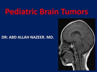
Pediatric Brain Tumors
DR: ABD ALLAH NAZEER. MD.
Juvenile pilocytic astrocytoma (JPA).
Intra-ventricular ependymoma
Bilateral acoustic neuroma of NF type 11
Epidermoid cysts, also known as "pearly tumors”
because of their bright white appearance at intraoperative resection, developed from epithelial
inclusions in basal cisterns, diploe of the skull and,
very rarely in the intrinsic brainstem or pineal region,
during neural tube closure or formation of secondary
cerebral vesicles. They are not really tumors, their
growth is due to the continuous formation of the
keratohyalin from continued desquamation by the
epithelium tissue. Their growth is very slow, so the
onset of their clinical manifestations is variable
between 20 and
40 years in most of the cases.
CP angle epidermoid cysts
Cerebellopontine angle epidermoid cyst.
Cerebral Pilocytic Astrocytoma with
Spontaneous intracranial Hemorrhage
Supratentorial ependymoma in a 10-year-old girl on a noncontrast CT, b T2-weighted, c
FLAIR, and d–f postgadolinium T1-weighted MRI. Unlike posterior fossa ependymomas,
most supratentorial ependymomas (70%) are extraventricular in origin.
Supra-tentorial grade 2 ependymoma in a 12-year-old girl. a Axial
FLAIR, b axial perfusion MRI, c post-contrast axial T1, d axial cerebral
blood volume map derived from perfusion MRI, e axial ADC coefficient,
and f perfusion MRI T2*-weighted dynamic susceptibility .
Presentation2.pptx pediatric brain tumour

More Related Content

What's hot

Pediatric brain tumors Dr. Muhammad Bin Zulfiqar
Pediatric brain tumors Dr. Muhammad Bin Zulfiqar Pediatric brain tumors Dr. Muhammad Bin Zulfiqar
Pediatric brain tumors Dr. Muhammad Bin Zulfiqar
Dr. Muhammad Bin Zulfiqar
 
Neuroblastoma and Nephroblastoma
Neuroblastoma and NephroblastomaNeuroblastoma and Nephroblastoma
Neuroblastoma and NephroblastomaThe Medical Post
 
Rhabdomyosarcoma
RhabdomyosarcomaRhabdomyosarcoma
Rhabdomyosarcoma
damuluri ramu
 
Pediatric cental nervous system tumors
Pediatric cental nervous system tumorsPediatric cental nervous system tumors
Pediatric cental nervous system tumors
Myatsu Aung
 
Imaging in pediatric brain tumors
Imaging in pediatric brain tumorsImaging in pediatric brain tumors
Imaging in pediatric brain tumors
Dr.Suhas Basavaiah
 
Pediatric hypertension
Pediatric hypertensionPediatric hypertension
Pediatric hypertension
Tauhid Iqbali
 
Gliomas - Brain Tumor
Gliomas - Brain TumorGliomas - Brain Tumor
Gliomas - Brain Tumor
suresh Bishokarma
 
Common Pediatric Solid Tumors
Common Pediatric Solid TumorsCommon Pediatric Solid Tumors
Common Pediatric Solid Tumors
Abdullatif Al-Rashed
 
Wilms tumor
Wilms tumorWilms tumor
Wilms tumor
sheena bhatia
 
Central nervous system tumors in children
Central nervous system tumors in childrenCentral nervous system tumors in children
Central nervous system tumors in children
Sasikumar Sambasivam
 
2021 WHO Classification of brain tumours.pptx
2021 WHO Classification of brain tumours.pptx2021 WHO Classification of brain tumours.pptx
2021 WHO Classification of brain tumours.pptx
RejoyceAnto
 
Pineal gland tumors
Pineal gland tumorsPineal gland tumors
Pineal gland tumors
airwave12
 
Childhood cancer / Oncological diseases in Pediatric groups
Childhood cancer / Oncological diseases in Pediatric groupsChildhood cancer / Oncological diseases in Pediatric groups
Childhood cancer / Oncological diseases in Pediatric groups
Monisha Sekar
 
Imaging in head trauma
Imaging in head traumaImaging in head trauma
Imaging in head trauma
SCGH ED CME
 
Overview of brain tumors
Overview of brain tumorsOverview of brain tumors
Overview of brain tumors
DrAyush Garg
 
Imaging of Traumatic Brain Injury
Imaging of Traumatic Brain InjuryImaging of Traumatic Brain Injury
Imaging of Traumatic Brain Injury
Rathachai Kaewlai
 
Lymphoma in children Dr G.Rajkumar
Lymphoma in children Dr G.RajkumarLymphoma in children Dr G.Rajkumar
Lymphoma in children Dr G.Rajkumar
grkmedico
 
Wilms tumor
Wilms tumorWilms tumor
Wilms tumor
GAURAV NAHAR
 
Solid tumors in children 2021
Solid tumors in children 2021Solid tumors in children 2021
Solid tumors in children 2021
Imran Iqbal
 

What's hot (20)

Pediatric brain tumors Dr. Muhammad Bin Zulfiqar
Pediatric brain tumors Dr. Muhammad Bin Zulfiqar Pediatric brain tumors Dr. Muhammad Bin Zulfiqar
Pediatric brain tumors Dr. Muhammad Bin Zulfiqar
 
Neuroblastoma and Nephroblastoma
Neuroblastoma and NephroblastomaNeuroblastoma and Nephroblastoma
Neuroblastoma and Nephroblastoma
 
Rhabdomyosarcoma
RhabdomyosarcomaRhabdomyosarcoma
Rhabdomyosarcoma
 
Pediatric cental nervous system tumors
Pediatric cental nervous system tumorsPediatric cental nervous system tumors
Pediatric cental nervous system tumors
 
Imaging in pediatric brain tumors
Imaging in pediatric brain tumorsImaging in pediatric brain tumors
Imaging in pediatric brain tumors
 
Paediatric oncology
Paediatric oncologyPaediatric oncology
Paediatric oncology
 
Pediatric hypertension
Pediatric hypertensionPediatric hypertension
Pediatric hypertension
 
Gliomas - Brain Tumor
Gliomas - Brain TumorGliomas - Brain Tumor
Gliomas - Brain Tumor
 
Common Pediatric Solid Tumors
Common Pediatric Solid TumorsCommon Pediatric Solid Tumors
Common Pediatric Solid Tumors
 
Wilms tumor
Wilms tumorWilms tumor
Wilms tumor
 
Central nervous system tumors in children
Central nervous system tumors in childrenCentral nervous system tumors in children
Central nervous system tumors in children
 
2021 WHO Classification of brain tumours.pptx
2021 WHO Classification of brain tumours.pptx2021 WHO Classification of brain tumours.pptx
2021 WHO Classification of brain tumours.pptx
 
Pineal gland tumors
Pineal gland tumorsPineal gland tumors
Pineal gland tumors
 
Childhood cancer / Oncological diseases in Pediatric groups
Childhood cancer / Oncological diseases in Pediatric groupsChildhood cancer / Oncological diseases in Pediatric groups
Childhood cancer / Oncological diseases in Pediatric groups
 
Imaging in head trauma
Imaging in head traumaImaging in head trauma
Imaging in head trauma
 
Overview of brain tumors
Overview of brain tumorsOverview of brain tumors
Overview of brain tumors
 
Imaging of Traumatic Brain Injury
Imaging of Traumatic Brain InjuryImaging of Traumatic Brain Injury
Imaging of Traumatic Brain Injury
 
Lymphoma in children Dr G.Rajkumar
Lymphoma in children Dr G.RajkumarLymphoma in children Dr G.Rajkumar
Lymphoma in children Dr G.Rajkumar
 
Wilms tumor
Wilms tumorWilms tumor
Wilms tumor
 
Solid tumors in children 2021
Solid tumors in children 2021Solid tumors in children 2021
Solid tumors in children 2021
 

Viewers also liked

Ocular anaesthesia by dr.roopashree.c .r
Ocular anaesthesia by dr.roopashree.c .rOcular anaesthesia by dr.roopashree.c .r
Ocular anaesthesia by dr.roopashree.c .r
Dr ROOPASHREE C R
 
Cerebro vascular lesions
Cerebro vascular lesionsCerebro vascular lesions
Cerebro vascular lesionsAbdellah Nazeer
 
Presentation1.pptx, radiological anatomy of the chest.
Presentation1.pptx, radiological anatomy of the chest.Presentation1.pptx, radiological anatomy of the chest.
Presentation1.pptx, radiological anatomy of the chest.Abdellah Nazeer
 
Anesthesia in ophthalmic surgery
Anesthesia in ophthalmic surgeryAnesthesia in ophthalmic surgery
Anesthesia in ophthalmic surgery
Panit Cherdchu
 
OCULAR Anesthesia
OCULAR AnesthesiaOCULAR Anesthesia
OCULAR Anesthesia
DARSHAN S M
 
Complications of anaesthesia in opthalmic surgery
Complications of anaesthesia in opthalmic surgeryComplications of anaesthesia in opthalmic surgery
Complications of anaesthesia in opthalmic surgeryDevdutta Nayak
 

Viewers also liked (7)

Ocular anaesthesia by dr.roopashree.c .r
Ocular anaesthesia by dr.roopashree.c .rOcular anaesthesia by dr.roopashree.c .r
Ocular anaesthesia by dr.roopashree.c .r
 
Cerebro vascular lesions
Cerebro vascular lesionsCerebro vascular lesions
Cerebro vascular lesions
 
Presentation1.pptx, radiological anatomy of the chest.
Presentation1.pptx, radiological anatomy of the chest.Presentation1.pptx, radiological anatomy of the chest.
Presentation1.pptx, radiological anatomy of the chest.
 
Anesthesia in ophthalmic surgery
Anesthesia in ophthalmic surgeryAnesthesia in ophthalmic surgery
Anesthesia in ophthalmic surgery
 
OCULAR Anesthesia
OCULAR AnesthesiaOCULAR Anesthesia
OCULAR Anesthesia
 
Complications of anaesthesia in opthalmic surgery
Complications of anaesthesia in opthalmic surgeryComplications of anaesthesia in opthalmic surgery
Complications of anaesthesia in opthalmic surgery
 
Cataract surgery complications
Cataract surgery complicationsCataract surgery complications
Cataract surgery complications
 

Similar to Presentation2.pptx pediatric brain tumour

Neural tube Defect & Hydrocephalus
Neural tube Defect & HydrocephalusNeural tube Defect & Hydrocephalus
Neural tube Defect & Hydrocephalus
Jannatul Nayeema Tonny
 
intracranial space occupying lession (pediatrics)
intracranial space occupying lession (pediatrics)intracranial space occupying lession (pediatrics)
intracranial space occupying lession (pediatrics)
Jimmy Sunda
 
NON GLIAL TUMORS
NON GLIAL TUMORSNON GLIAL TUMORS
NON GLIAL TUMORS
AvinashDahatre
 
Neural tube defects
Neural tube defectsNeural tube defects
Neural tube defects
ALIYU USMAN MUHAMMAD
 
Diagnostic Neuroradiology
Diagnostic NeuroradiologyDiagnostic Neuroradiology
Diagnostic NeuroradiologySumit Prajapati
 
craniopharyngioma
 craniopharyngioma craniopharyngioma
craniopharyngioma
Md Roohia
 
Radiological findings of congenital anomalies of the spine and spinal cord
Radiological findings of congenital anomalies of the spine and spinal cordRadiological findings of congenital anomalies of the spine and spinal cord
Radiological findings of congenital anomalies of the spine and spinal cord
Dr. Armaan Singh
 
Imaging of congenital CNS lesions
Imaging of congenital CNS lesionsImaging of congenital CNS lesions
Imaging of congenital CNS lesions
Milan Silwal
 
Ophthalmology 5th year, 7th lecture (Dr. Bakhtyar)
Ophthalmology 5th year, 7th lecture (Dr. Bakhtyar)Ophthalmology 5th year, 7th lecture (Dr. Bakhtyar)
Ophthalmology 5th year, 7th lecture (Dr. Bakhtyar)
College of Medicine, Sulaymaniyah
 
Imaging of congenital cns lesions
Imaging of congenital cns lesionsImaging of congenital cns lesions
Imaging of congenital cns lesions
Gobardhan Thapa
 
Birth Injuries of Newborn
Birth Injuries of NewbornBirth Injuries of Newborn
Birth Injuries of Newborn
Lipi Mondal
 
Spotters ppt 1
Spotters ppt 1Spotters ppt 1
Spotters ppt 1
Chandni Wadhwani
 
Brain abscess
Brain abscessBrain abscess
Brain abscess
joemdas
 
Brain abscess and it course of treatment
Brain abscess and it course of treatmentBrain abscess and it course of treatment
Brain abscess and it course of treatment
AnnaSingh32
 
Pediatric stroke modified
Pediatric stroke modifiedPediatric stroke modified
Pediatric stroke modified
Anish Choudhary
 
Intracranial Calcification in Cone Beam CT & Medical CT
Intracranial Calcification in Cone Beam CT & Medical CTIntracranial Calcification in Cone Beam CT & Medical CT
Intracranial Calcification in Cone Beam CT & Medical CT
Judy Oh, D.D.S.
 
Cns tumors bikash
Cns tumors  bikashCns tumors  bikash
Cns tumors bikash
Bikash Praharaj
 
Oral surgery i report
Oral surgery i reportOral surgery i report
Oral surgery i reportHgedas Imezak
 
Birth injuries and icterus neonatarum
Birth injuries and icterus neonatarumBirth injuries and icterus neonatarum
Birth injuries and icterus neonatarum
Deepthy Philip Thomas
 

Similar to Presentation2.pptx pediatric brain tumour (20)

Neural tube Defect & Hydrocephalus
Neural tube Defect & HydrocephalusNeural tube Defect & Hydrocephalus
Neural tube Defect & Hydrocephalus
 
intracranial space occupying lession (pediatrics)
intracranial space occupying lession (pediatrics)intracranial space occupying lession (pediatrics)
intracranial space occupying lession (pediatrics)
 
NON GLIAL TUMORS
NON GLIAL TUMORSNON GLIAL TUMORS
NON GLIAL TUMORS
 
Neural tube defects
Neural tube defectsNeural tube defects
Neural tube defects
 
Diagnostic Neuroradiology
Diagnostic NeuroradiologyDiagnostic Neuroradiology
Diagnostic Neuroradiology
 
craniopharyngioma
 craniopharyngioma craniopharyngioma
craniopharyngioma
 
Radiological findings of congenital anomalies of the spine and spinal cord
Radiological findings of congenital anomalies of the spine and spinal cordRadiological findings of congenital anomalies of the spine and spinal cord
Radiological findings of congenital anomalies of the spine and spinal cord
 
Imaging of congenital CNS lesions
Imaging of congenital CNS lesionsImaging of congenital CNS lesions
Imaging of congenital CNS lesions
 
Ophthalmology 5th year, 7th lecture (Dr. Bakhtyar)
Ophthalmology 5th year, 7th lecture (Dr. Bakhtyar)Ophthalmology 5th year, 7th lecture (Dr. Bakhtyar)
Ophthalmology 5th year, 7th lecture (Dr. Bakhtyar)
 
Brain tumors 2
Brain tumors 2Brain tumors 2
Brain tumors 2
 
Imaging of congenital cns lesions
Imaging of congenital cns lesionsImaging of congenital cns lesions
Imaging of congenital cns lesions
 
Birth Injuries of Newborn
Birth Injuries of NewbornBirth Injuries of Newborn
Birth Injuries of Newborn
 
Spotters ppt 1
Spotters ppt 1Spotters ppt 1
Spotters ppt 1
 
Brain abscess
Brain abscessBrain abscess
Brain abscess
 
Brain abscess and it course of treatment
Brain abscess and it course of treatmentBrain abscess and it course of treatment
Brain abscess and it course of treatment
 
Pediatric stroke modified
Pediatric stroke modifiedPediatric stroke modified
Pediatric stroke modified
 
Intracranial Calcification in Cone Beam CT & Medical CT
Intracranial Calcification in Cone Beam CT & Medical CTIntracranial Calcification in Cone Beam CT & Medical CT
Intracranial Calcification in Cone Beam CT & Medical CT
 
Cns tumors bikash
Cns tumors  bikashCns tumors  bikash
Cns tumors bikash
 
Oral surgery i report
Oral surgery i reportOral surgery i report
Oral surgery i report
 
Birth injuries and icterus neonatarum
Birth injuries and icterus neonatarumBirth injuries and icterus neonatarum
Birth injuries and icterus neonatarum
 

More from Abdellah Nazeer

Muculoskeletal Pediatic Imaging..pptx
Muculoskeletal Pediatic Imaging..pptxMuculoskeletal Pediatic Imaging..pptx
Muculoskeletal Pediatic Imaging..pptx
Abdellah Nazeer
 
Presentation1, Ultrasound of the bowel loops and the lymph nodes..pptx
Presentation1, Ultrasound of the bowel loops and the lymph nodes..pptxPresentation1, Ultrasound of the bowel loops and the lymph nodes..pptx
Presentation1, Ultrasound of the bowel loops and the lymph nodes..pptx
Abdellah Nazeer
 
Presentation1 Short cases MD..pptx
Presentation1 Short cases MD..pptxPresentation1 Short cases MD..pptx
Presentation1 Short cases MD..pptx
Abdellah Nazeer
 
Presentation1, MD MCQ Cases..pptx
Presentation1, MD MCQ Cases..pptxPresentation1, MD MCQ Cases..pptx
Presentation1, MD MCQ Cases..pptx
Abdellah Nazeer
 
Presentation1, Short Cases Quiz..pptx
Presentation1, Short Cases Quiz..pptxPresentation1, Short Cases Quiz..pptx
Presentation1, Short Cases Quiz..pptx
Abdellah Nazeer
 
Presentation1, radiological imaging of lateral hindfoot impingement.
Presentation1, radiological imaging of lateral hindfoot impingement.Presentation1, radiological imaging of lateral hindfoot impingement.
Presentation1, radiological imaging of lateral hindfoot impingement.
Abdellah Nazeer
 
Presentation2, radiological anatomy of the liver and spleen.
Presentation2, radiological anatomy of the liver and spleen.Presentation2, radiological anatomy of the liver and spleen.
Presentation2, radiological anatomy of the liver and spleen.
Abdellah Nazeer
 
Presentation1, artifacts and pitfalls of the wrist and elbow joints.
Presentation1, artifacts and pitfalls of the wrist and elbow joints.Presentation1, artifacts and pitfalls of the wrist and elbow joints.
Presentation1, artifacts and pitfalls of the wrist and elbow joints.
Abdellah Nazeer
 
Presentation1, artifact and pitfalls of the knee, hip and ankle joints.
Presentation1, artifact and pitfalls of the knee, hip and ankle joints.Presentation1, artifact and pitfalls of the knee, hip and ankle joints.
Presentation1, artifact and pitfalls of the knee, hip and ankle joints.
Abdellah Nazeer
 
Presentation1, radiological imaging of artifact and pitfalls in shoulder join...
Presentation1, radiological imaging of artifact and pitfalls in shoulder join...Presentation1, radiological imaging of artifact and pitfalls in shoulder join...
Presentation1, radiological imaging of artifact and pitfalls in shoulder join...
Abdellah Nazeer
 
Presentation1, radiological imaging of internal abdominal hernia.
Presentation1, radiological imaging of internal abdominal hernia.Presentation1, radiological imaging of internal abdominal hernia.
Presentation1, radiological imaging of internal abdominal hernia.
Abdellah Nazeer
 
Presentation11, radiological imaging of ovarian torsion.
Presentation11, radiological imaging of ovarian torsion.Presentation11, radiological imaging of ovarian torsion.
Presentation11, radiological imaging of ovarian torsion.
Abdellah Nazeer
 
Presentation1, musculoskeletal anatomy.
Presentation1, musculoskeletal anatomy.Presentation1, musculoskeletal anatomy.
Presentation1, musculoskeletal anatomy.
Abdellah Nazeer
 
Presentation1, new mri techniques in the diagnosis and monitoring of multiple...
Presentation1, new mri techniques in the diagnosis and monitoring of multiple...Presentation1, new mri techniques in the diagnosis and monitoring of multiple...
Presentation1, new mri techniques in the diagnosis and monitoring of multiple...
Abdellah Nazeer
 
Presentation1, radiological application of diffusion weighted mri in neck mas...
Presentation1, radiological application of diffusion weighted mri in neck mas...Presentation1, radiological application of diffusion weighted mri in neck mas...
Presentation1, radiological application of diffusion weighted mri in neck mas...
Abdellah Nazeer
 
Presentation1, radiological application of diffusion weighted images in breas...
Presentation1, radiological application of diffusion weighted images in breas...Presentation1, radiological application of diffusion weighted images in breas...
Presentation1, radiological application of diffusion weighted images in breas...
Abdellah Nazeer
 
Presentation1, radiological application of diffusion weighted images in abdom...
Presentation1, radiological application of diffusion weighted images in abdom...Presentation1, radiological application of diffusion weighted images in abdom...
Presentation1, radiological application of diffusion weighted images in abdom...
Abdellah Nazeer
 
Presentation1, radiological application of diffusion weighted imges in neuror...
Presentation1, radiological application of diffusion weighted imges in neuror...Presentation1, radiological application of diffusion weighted imges in neuror...
Presentation1, radiological application of diffusion weighted imges in neuror...
Abdellah Nazeer
 
Presentation1, mr physics.
Presentation1, mr physics.Presentation1, mr physics.
Presentation1, mr physics.
Abdellah Nazeer
 
Presentation1. ct physics.
Presentation1. ct physics.Presentation1. ct physics.
Presentation1. ct physics.
Abdellah Nazeer
 

More from Abdellah Nazeer (20)

Muculoskeletal Pediatic Imaging..pptx
Muculoskeletal Pediatic Imaging..pptxMuculoskeletal Pediatic Imaging..pptx
Muculoskeletal Pediatic Imaging..pptx
 
Presentation1, Ultrasound of the bowel loops and the lymph nodes..pptx
Presentation1, Ultrasound of the bowel loops and the lymph nodes..pptxPresentation1, Ultrasound of the bowel loops and the lymph nodes..pptx
Presentation1, Ultrasound of the bowel loops and the lymph nodes..pptx
 
Presentation1 Short cases MD..pptx
Presentation1 Short cases MD..pptxPresentation1 Short cases MD..pptx
Presentation1 Short cases MD..pptx
 
Presentation1, MD MCQ Cases..pptx
Presentation1, MD MCQ Cases..pptxPresentation1, MD MCQ Cases..pptx
Presentation1, MD MCQ Cases..pptx
 
Presentation1, Short Cases Quiz..pptx
Presentation1, Short Cases Quiz..pptxPresentation1, Short Cases Quiz..pptx
Presentation1, Short Cases Quiz..pptx
 
Presentation1, radiological imaging of lateral hindfoot impingement.
Presentation1, radiological imaging of lateral hindfoot impingement.Presentation1, radiological imaging of lateral hindfoot impingement.
Presentation1, radiological imaging of lateral hindfoot impingement.
 
Presentation2, radiological anatomy of the liver and spleen.
Presentation2, radiological anatomy of the liver and spleen.Presentation2, radiological anatomy of the liver and spleen.
Presentation2, radiological anatomy of the liver and spleen.
 
Presentation1, artifacts and pitfalls of the wrist and elbow joints.
Presentation1, artifacts and pitfalls of the wrist and elbow joints.Presentation1, artifacts and pitfalls of the wrist and elbow joints.
Presentation1, artifacts and pitfalls of the wrist and elbow joints.
 
Presentation1, artifact and pitfalls of the knee, hip and ankle joints.
Presentation1, artifact and pitfalls of the knee, hip and ankle joints.Presentation1, artifact and pitfalls of the knee, hip and ankle joints.
Presentation1, artifact and pitfalls of the knee, hip and ankle joints.
 
Presentation1, radiological imaging of artifact and pitfalls in shoulder join...
Presentation1, radiological imaging of artifact and pitfalls in shoulder join...Presentation1, radiological imaging of artifact and pitfalls in shoulder join...
Presentation1, radiological imaging of artifact and pitfalls in shoulder join...
 
Presentation1, radiological imaging of internal abdominal hernia.
Presentation1, radiological imaging of internal abdominal hernia.Presentation1, radiological imaging of internal abdominal hernia.
Presentation1, radiological imaging of internal abdominal hernia.
 
Presentation11, radiological imaging of ovarian torsion.
Presentation11, radiological imaging of ovarian torsion.Presentation11, radiological imaging of ovarian torsion.
Presentation11, radiological imaging of ovarian torsion.
 
Presentation1, musculoskeletal anatomy.
Presentation1, musculoskeletal anatomy.Presentation1, musculoskeletal anatomy.
Presentation1, musculoskeletal anatomy.
 
Presentation1, new mri techniques in the diagnosis and monitoring of multiple...
Presentation1, new mri techniques in the diagnosis and monitoring of multiple...Presentation1, new mri techniques in the diagnosis and monitoring of multiple...
Presentation1, new mri techniques in the diagnosis and monitoring of multiple...
 
Presentation1, radiological application of diffusion weighted mri in neck mas...
Presentation1, radiological application of diffusion weighted mri in neck mas...Presentation1, radiological application of diffusion weighted mri in neck mas...
Presentation1, radiological application of diffusion weighted mri in neck mas...
 
Presentation1, radiological application of diffusion weighted images in breas...
Presentation1, radiological application of diffusion weighted images in breas...Presentation1, radiological application of diffusion weighted images in breas...
Presentation1, radiological application of diffusion weighted images in breas...
 
Presentation1, radiological application of diffusion weighted images in abdom...
Presentation1, radiological application of diffusion weighted images in abdom...Presentation1, radiological application of diffusion weighted images in abdom...
Presentation1, radiological application of diffusion weighted images in abdom...
 
Presentation1, radiological application of diffusion weighted imges in neuror...
Presentation1, radiological application of diffusion weighted imges in neuror...Presentation1, radiological application of diffusion weighted imges in neuror...
Presentation1, radiological application of diffusion weighted imges in neuror...
 
Presentation1, mr physics.
Presentation1, mr physics.Presentation1, mr physics.
Presentation1, mr physics.
 
Presentation1. ct physics.
Presentation1. ct physics.Presentation1. ct physics.
Presentation1. ct physics.
 

Presentation2.pptx pediatric brain tumour

  • 1. Pediatric Brain Tumors DR: ABD ALLAH NAZEER. MD.
  • 2.
  • 3.
  • 4.
  • 5.
  • 6.
  • 7.
  • 8.
  • 9.
  • 10.
  • 11.
  • 12.
  • 14.
  • 15.
  • 16.
  • 17.
  • 18.
  • 19.
  • 20.
  • 21.
  • 23.
  • 24.
  • 25.
  • 26.
  • 27.
  • 28.
  • 29.
  • 30. Bilateral acoustic neuroma of NF type 11
  • 31. Bilateral acoustic neuroma of NF type 11
  • 32. Epidermoid cysts, also known as "pearly tumors” because of their bright white appearance at intraoperative resection, developed from epithelial inclusions in basal cisterns, diploe of the skull and, very rarely in the intrinsic brainstem or pineal region, during neural tube closure or formation of secondary cerebral vesicles. They are not really tumors, their growth is due to the continuous formation of the keratohyalin from continued desquamation by the epithelium tissue. Their growth is very slow, so the onset of their clinical manifestations is variable between 20 and 40 years in most of the cases.
  • 35.
  • 36.
  • 37.
  • 38. Cerebral Pilocytic Astrocytoma with Spontaneous intracranial Hemorrhage
  • 39.
  • 40.
  • 41.
  • 42.
  • 43. Supratentorial ependymoma in a 10-year-old girl on a noncontrast CT, b T2-weighted, c FLAIR, and d–f postgadolinium T1-weighted MRI. Unlike posterior fossa ependymomas, most supratentorial ependymomas (70%) are extraventricular in origin.
  • 44. Supra-tentorial grade 2 ependymoma in a 12-year-old girl. a Axial FLAIR, b axial perfusion MRI, c post-contrast axial T1, d axial cerebral blood volume map derived from perfusion MRI, e axial ADC coefficient, and f perfusion MRI T2*-weighted dynamic susceptibility .