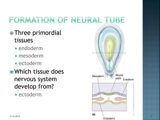
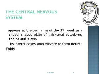
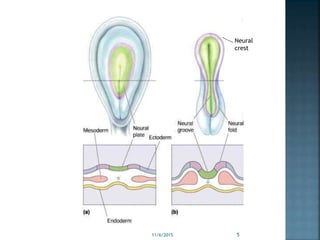
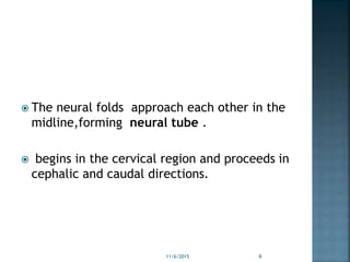
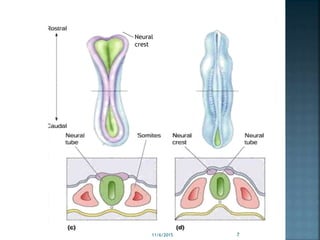
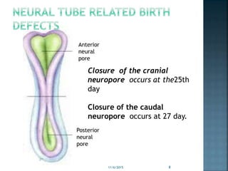
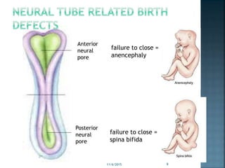
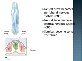
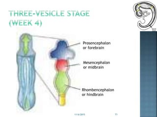
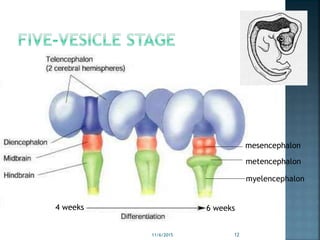
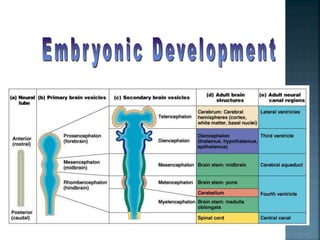
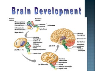
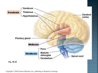
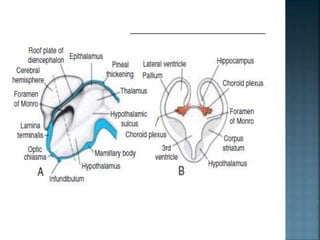
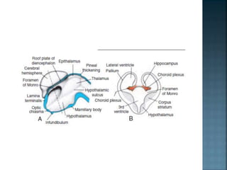
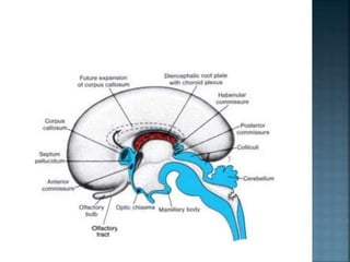
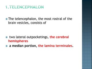
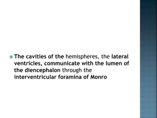
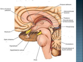
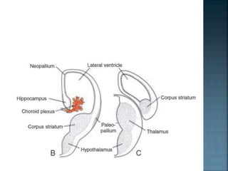
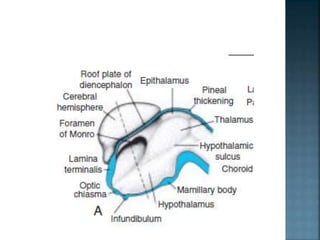
DR SHABANA ALI
 Three primordial
tissues
 endoderm
 mesoderm
 ectoderm
 Which tissue does
nervous system
develop from?
 ectoderm
11/6/2015 3
appears at the beginning of the 3rd week as a
slipper-shaped plate of thickened ectoderm,
the neural plate.
Its lateral edges soon elevate to form neural
Folds.
11/6/2015 4
Neural
crest
11/6/2015 5
 The neural folds approach each other in the
midline,forming neural tube .
 begins in the cervical region and proceeds in
cephalic and caudal directions.
11/6/2015 6
Neural
crest
11/6/2015 7
Anterior
neural
pore
Posterior
neural
pore
11/6/2015 8
Closure of the cranial
neuropore occurs at the25th
day
Closure of the caudal
neuropore occurs at 27 day.
Anterior
neural
pore
Posterior
neural
pore
failure to close =
anencephaly
failure to close =
spina bifida
11/6/2015 9
 Neural crest becomes
peripheral nervous
system (PNS)
 Neural tube becomes
central nervous system
(CNS)
 Somites become spinal
vertebrae.
Somites
11/6/2015 10
Prosencephalon
or forebrain
Mesencephalon
or midbrain
Rhombencephalon
or hindbrain
11/6/2015 11
mesencephalon
metencephalon
myelencephalon
6 weeks4 weeks
11/6/2015 12
Copyright © 2002 Pearson Education, Inc., publishing as Benjamin Cummings
Fig. 48.20
 THE TELENCEPHALON, which forms the
cerebral hemispheres.
 THE DIENCEPHALON, which forms the optic
cup and stalk,pituitary gland , thalamus,
hypothalamus, and epiphysis.
 The diencephalon, consist of a roof plate
and two alar plates but there are no floor
and basal plates
 a single layer of ependymal cells with
vascular mesenchyme give rise to the
choroid plexus of the third ventricle .
 The most caudal part of the roof plate
develops into the pineal body, or epiphysis.
 serves as a channel through which light and
darkness affect endocrine and behavioral
rhythms.
 lateral walls of the diencephalon.
 the hypothalamic sulcus, divides the plate
into a dorsal and a ventral region, the
thalamus and hypothalamus,
 As a result of proliferative activity, the
thalamus gradually projects into the lumen
of the diencephalon,forming the massa
intermedia, or interthalamic connexus.
 The telencephalon, the most rostral of the
brain vesicles, consists of
 two lateral outpocketings, the cerebral
hemispheres
 a median portion, the lamina terminales.
 The cavities of the hemispheres, the lateral
ventricles, communicate with the lumen of
the diencephalon through the
interventricular foramina of Monro
 arise at the beginning of the fifth week of
development as bilateral evaginations of the
lateral wall of the prosencephalon.
 By the middle of the second month the basal
part of the hemispheres begins to grow and
bulges, has a striated appearance and is
known as the corpus striatum .
 Near the diencephlon a single layer of ependymal
cells covered by vascular mesenchyme form the
choroid plexus.
 Immediately above the choroidal fissure, the
wall of the hemisphere thickens, forming the
hippocampus.
 With further expansion, the hemispheres
cover the lateral aspect of the diencephalon,
mesencephalon, and cephalic portion of the
metencephalon

 internal capsule divide nuclear mass of the
corpus striatum by axons passing to and from
the cortex of the hemisphere.
 At the same time, the medial wall of the
area between the frontal and temporal lobes
becomes depressed and is known as the
insula .
 During the final part of fetal life, the surface
of the cerebral hemispheres grows so rapidly
that a great many convolutions (gyri)
separated by fissures and sulci appear on
its surface
 Cortex Development.
 The cerebral cortex develops from the
pallium, which has two regions:
 (a) the paleopallium, or archipallium,
immediately lateral to the corpus striatum,
 (b) the neopallium,between the
hippocampus and the paleopallium
 a number of fiber bundles, cross the
midline, connect the right and left halves of
the hemispheres.
 The most important fiber bundles make use
of the lamina terminalis.
 anterior commissure.
 hippocampal commissure, or
 fornix commissure.
 corpus callosum.
 posterior and habenular commissures,
 the optic chiasma
 Holoprosencephaly
 Exencephaly
 Hydrocephalus
 Microcephaly
1.Mylencephlon ----medulla oblangata
 Basal plate-------- three motor nuclei
 Alar plate----------three sensory nuclei
 Roof plate------ ---ependymal layer(choroid
plexus)
 Floor plate---------region of median sulcus
11/6/2015 38
Metencephlon -----cerebellum,pons .
 Basal plate----------three motor nuclei
 Marginal zone of basal plate---pons
 Alar plate-----------pontine nuclei,three
sensory nuclei
11/6/2015 39
Basal plate ; 2 groups of efferent nuclei
 Marginal layer- --------- crus cerebri
Alar plate---------------superior and inferior
colliculi
11/6/2015 40
 THE TELENCEPHALON, which forms the
cerebral hemispheres.
 THE DIENCEPHALON, which forms the optic
cup and stalk,pituitary gland , thalamus,
hypothalamus, and epiphysis.
Cns development

Cns development

  • 2.
  • 3.
     Three primordial tissues endoderm  mesoderm  ectoderm  Which tissue does nervous system develop from?  ectoderm 11/6/2015 3
  • 4.
    appears at thebeginning of the 3rd week as a slipper-shaped plate of thickened ectoderm, the neural plate. Its lateral edges soon elevate to form neural Folds. 11/6/2015 4
  • 5.
  • 6.
     The neuralfolds approach each other in the midline,forming neural tube .  begins in the cervical region and proceeds in cephalic and caudal directions. 11/6/2015 6
  • 7.
  • 8.
    Anterior neural pore Posterior neural pore 11/6/2015 8 Closure ofthe cranial neuropore occurs at the25th day Closure of the caudal neuropore occurs at 27 day.
  • 9.
    Anterior neural pore Posterior neural pore failure to close= anencephaly failure to close = spina bifida 11/6/2015 9
  • 10.
     Neural crestbecomes peripheral nervous system (PNS)  Neural tube becomes central nervous system (CNS)  Somites become spinal vertebrae. Somites 11/6/2015 10
  • 11.
  • 12.
  • 15.
    Copyright © 2002Pearson Education, Inc., publishing as Benjamin Cummings Fig. 48.20
  • 17.
     THE TELENCEPHALON,which forms the cerebral hemispheres.  THE DIENCEPHALON, which forms the optic cup and stalk,pituitary gland , thalamus, hypothalamus, and epiphysis.
  • 18.
     The diencephalon,consist of a roof plate and two alar plates but there are no floor and basal plates
  • 19.
     a singlelayer of ependymal cells with vascular mesenchyme give rise to the choroid plexus of the third ventricle .  The most caudal part of the roof plate develops into the pineal body, or epiphysis.  serves as a channel through which light and darkness affect endocrine and behavioral rhythms.
  • 20.
     lateral wallsof the diencephalon.  the hypothalamic sulcus, divides the plate into a dorsal and a ventral region, the thalamus and hypothalamus,  As a result of proliferative activity, the thalamus gradually projects into the lumen of the diencephalon,forming the massa intermedia, or interthalamic connexus.
  • 23.
     The telencephalon,the most rostral of the brain vesicles, consists of  two lateral outpocketings, the cerebral hemispheres  a median portion, the lamina terminales.
  • 24.
     The cavitiesof the hemispheres, the lateral ventricles, communicate with the lumen of the diencephalon through the interventricular foramina of Monro
  • 26.
     arise atthe beginning of the fifth week of development as bilateral evaginations of the lateral wall of the prosencephalon.  By the middle of the second month the basal part of the hemispheres begins to grow and bulges, has a striated appearance and is known as the corpus striatum .
  • 27.
     Near thediencephlon a single layer of ependymal cells covered by vascular mesenchyme form the choroid plexus.  Immediately above the choroidal fissure, the wall of the hemisphere thickens, forming the hippocampus.
  • 29.
     With furtherexpansion, the hemispheres cover the lateral aspect of the diencephalon, mesencephalon, and cephalic portion of the metencephalon 
  • 30.
     internal capsuledivide nuclear mass of the corpus striatum by axons passing to and from the cortex of the hemisphere.  At the same time, the medial wall of the area between the frontal and temporal lobes becomes depressed and is known as the insula .
  • 31.
     During thefinal part of fetal life, the surface of the cerebral hemispheres grows so rapidly that a great many convolutions (gyri) separated by fissures and sulci appear on its surface
  • 32.
     Cortex Development. The cerebral cortex develops from the pallium, which has two regions:  (a) the paleopallium, or archipallium, immediately lateral to the corpus striatum,  (b) the neopallium,between the hippocampus and the paleopallium
  • 34.
     a numberof fiber bundles, cross the midline, connect the right and left halves of the hemispheres.  The most important fiber bundles make use of the lamina terminalis.
  • 36.
     anterior commissure. hippocampal commissure, or  fornix commissure.  corpus callosum.  posterior and habenular commissures,  the optic chiasma
  • 37.
     Holoprosencephaly  Exencephaly Hydrocephalus  Microcephaly
  • 38.
    1.Mylencephlon ----medulla oblangata Basal plate-------- three motor nuclei  Alar plate----------three sensory nuclei  Roof plate------ ---ependymal layer(choroid plexus)  Floor plate---------region of median sulcus 11/6/2015 38
  • 39.
    Metencephlon -----cerebellum,pons . Basal plate----------three motor nuclei  Marginal zone of basal plate---pons  Alar plate-----------pontine nuclei,three sensory nuclei 11/6/2015 39
  • 40.
    Basal plate ;2 groups of efferent nuclei  Marginal layer- --------- crus cerebri Alar plate---------------superior and inferior colliculi 11/6/2015 40
  • 41.
     THE TELENCEPHALON,which forms the cerebral hemispheres.  THE DIENCEPHALON, which forms the optic cup and stalk,pituitary gland , thalamus, hypothalamus, and epiphysis.