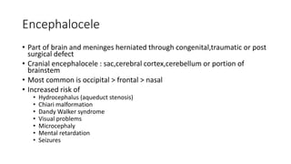
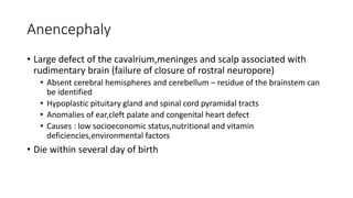
Neural tube defect
Definition
• Dysraphism
• Group of disorder
• Resulting from failure of closure embryonic posterior and anterior
neuropores
• Associated with abnormal development of other part of neural tube and bony
& soft tissue structures
• Most congenital anomalies of the CNS
• Incidence is declining
• Folic acid supplementation
• Earlier prenatal diagnosis
Etiology
• Hyperthermia
• Drugs
• Malnutrition
• Maternal obesity or DM
• Genetic : mutation at the folate responsive and folate dependent
pathway
• Exposure to radiation before conception
Types
• Spina bifida occulta
• Meningocele (cystica)
• Myelomeningocele (cystica)
• Encephalocele
• Anencephaly
• Others
Spina bifida occulta
• Defect in closure of posterior vertebral arches and laminae,usually
occur at L5 and S1,and no protrusion of spinal cord or meninges
• Clinical features
• Asymptomatic and lack of neurological signs
• Cutaneous manifestation : haemangioma,dimple,lump or hairy patch
Meningocele (cystica)
• Meninges herniates through a posterior vertebral arches (normal spinal cord
• Mostly covered with skin : less meningitis
• Anterior meningoceles project into the pelvis through a defect at the sacrum
• Constipation
• Bladder dysfunction
• Investigations
• Plain reontgenogram,US and MRI : extent of neural tissue involvement
• CT scan of the head : any concomitant presence of hydrocephalus
• Management
• Surgery : delayed in normal neurological development,thick skin covering meningocele and
immediate leaking of CSF,thin skin (to prevent meningitis)
• Surgical referral of female : associated with anomalies of genital tract (rectovaginal
fistula,anorectal septa)
Myelomeningocele (cystica)
• Spinal cord and meninges protrude through the vertebral arch (most
severe form of NTD)
• Dysfunction of many organ and structures along the neural axis but
lumbasacral region (75%) affected:
• Peripheral NS
• Skeleton
• Skin
• GIT
• Genitourinary tract
• Hydrocephalus occur in associated with type II Chiari-Malformation
Meningomyelocele
• Low sacral region
• Bowel and bladder incontinence
• Anesthesia of perianal area
• No impairment of motor function
• Mid lumbar region (abnormality and disruption of conus medularis)
• Flaccid paralysis of LL
• Absence deep tendon reflexes
• Lack response to touch and pain
• Deformities:clubfeet,subluxation of the hip
• Constant urinary dribbling and a relaxed anal sphincter
• High pressure bladder and sphincter dyssynergy
Meningomyelocele
• Treatment
• Surgery
• Repair defect,shunt the hydrocephalus,early surgical decompression of medulla and
cervical cord(if sign and symptoms of hindbrain dysfunction occur)
• Regular cathetherized neurogenic bladder
• Bowel trained
• With regimen of timed enema or suppositories
• At a predetermined time
• Once/twice per day
Encephalocele
• Part of brain and meninges herniated through congenital,traumatic or post
surgical defect
• Cranial encephalocele : sac,cerebral cortex,cerebellum or portion of
brainstem
• Most common is occipital > frontal > nasal
• Increased risk of
• Hydrocephalus (aqueduct stenosis)
• Chiari malformation
• Dandy Walker syndrome
• Visual problems
• Microcephaly
• Mental retardation
• Seizures
Anencephaly
• Large defect of the cavalrium,meninges and scalp associated with
rudimentary brain (failure of closure of rostral neuropore)
• Absent cerebral hemispheres and cerebellum – residue of the brainstem can
be identified
• Hypoplastic pituitary gland and spinal cord pyramidal tracts
• Anomalies of ear,cleft palate and congenital heart defect
• Causes : low socioeconomic status,nutritional and vitamin
deficiencies,environmental factors
• Die within several day of birth
Others
• Dermal sinus
• Tethered cord
• Syringomyelia
• Diastematomyelia
• Lipoma of conus medullaris

Neural tube defect

  • 1.
  • 2.
    Definition • Dysraphism • Groupof disorder • Resulting from failure of closure embryonic posterior and anterior neuropores • Associated with abnormal development of other part of neural tube and bony & soft tissue structures • Most congenital anomalies of the CNS • Incidence is declining • Folic acid supplementation • Earlier prenatal diagnosis
  • 3.
    Etiology • Hyperthermia • Drugs •Malnutrition • Maternal obesity or DM • Genetic : mutation at the folate responsive and folate dependent pathway • Exposure to radiation before conception
  • 4.
    Types • Spina bifidaocculta • Meningocele (cystica) • Myelomeningocele (cystica) • Encephalocele • Anencephaly • Others
  • 5.
    Spina bifida occulta •Defect in closure of posterior vertebral arches and laminae,usually occur at L5 and S1,and no protrusion of spinal cord or meninges • Clinical features • Asymptomatic and lack of neurological signs • Cutaneous manifestation : haemangioma,dimple,lump or hairy patch
  • 6.
    Meningocele (cystica) • Meningesherniates through a posterior vertebral arches (normal spinal cord • Mostly covered with skin : less meningitis • Anterior meningoceles project into the pelvis through a defect at the sacrum • Constipation • Bladder dysfunction • Investigations • Plain reontgenogram,US and MRI : extent of neural tissue involvement • CT scan of the head : any concomitant presence of hydrocephalus • Management • Surgery : delayed in normal neurological development,thick skin covering meningocele and immediate leaking of CSF,thin skin (to prevent meningitis) • Surgical referral of female : associated with anomalies of genital tract (rectovaginal fistula,anorectal septa)
  • 7.
    Myelomeningocele (cystica) • Spinalcord and meninges protrude through the vertebral arch (most severe form of NTD) • Dysfunction of many organ and structures along the neural axis but lumbasacral region (75%) affected: • Peripheral NS • Skeleton • Skin • GIT • Genitourinary tract • Hydrocephalus occur in associated with type II Chiari-Malformation
  • 8.
    Meningomyelocele • Low sacralregion • Bowel and bladder incontinence • Anesthesia of perianal area • No impairment of motor function • Mid lumbar region (abnormality and disruption of conus medularis) • Flaccid paralysis of LL • Absence deep tendon reflexes • Lack response to touch and pain • Deformities:clubfeet,subluxation of the hip • Constant urinary dribbling and a relaxed anal sphincter • High pressure bladder and sphincter dyssynergy
  • 9.
    Meningomyelocele • Treatment • Surgery •Repair defect,shunt the hydrocephalus,early surgical decompression of medulla and cervical cord(if sign and symptoms of hindbrain dysfunction occur) • Regular cathetherized neurogenic bladder • Bowel trained • With regimen of timed enema or suppositories • At a predetermined time • Once/twice per day
  • 10.
    Encephalocele • Part ofbrain and meninges herniated through congenital,traumatic or post surgical defect • Cranial encephalocele : sac,cerebral cortex,cerebellum or portion of brainstem • Most common is occipital > frontal > nasal • Increased risk of • Hydrocephalus (aqueduct stenosis) • Chiari malformation • Dandy Walker syndrome • Visual problems • Microcephaly • Mental retardation • Seizures
  • 11.
    Anencephaly • Large defectof the cavalrium,meninges and scalp associated with rudimentary brain (failure of closure of rostral neuropore) • Absent cerebral hemispheres and cerebellum – residue of the brainstem can be identified • Hypoplastic pituitary gland and spinal cord pyramidal tracts • Anomalies of ear,cleft palate and congenital heart defect • Causes : low socioeconomic status,nutritional and vitamin deficiencies,environmental factors • Die within several day of birth
  • 12.
    Others • Dermal sinus •Tethered cord • Syringomyelia • Diastematomyelia • Lipoma of conus medullaris