Downloaded 409 times


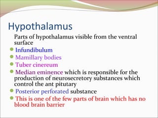


The diencephalon is the area surrounding the third ventricle of the brain. It contains several parts including the thalamus, hypothalamus, epithalamus, and subthalamus. The thalamus relays sensory information to the cortex and is involved in visual and auditory processing. The hypothalamus regulates autonomic functions and homeostasis through connections to the pituitary gland and autonomic nervous system. Lesions in the diencephalon can disrupt temperature regulation, appetite, water balance, and other homeostatic processes.


![The Diencephalon[1] vvtyyyyytttttttvvv.pdf](https://cdn.slidesharecdn.com/ss_thumbnails/thediencephalon1-240606092454-c768ad3c-thumbnail.jpg?width=640&height=640&fit=bounds)