What is Neuraltube defect ?
Failure of normal fusion of the neural plate to
form neural tube during the first 28 days
following conception .
Neural tube defects (NTDs) are one of the most
common birth defects, occurring in
approximately one in 1000 live births in the
United States.
3.
Prevalence
Increasedincidence in families of Celtic and Irish
heritage .
Increased incidence in minorities (genetic or
environmental?)
Increased incidence in families
Neural tube defects (NTDs) are among the most
common birth defects that cause infant mortality
(death) and serious disability .
4.
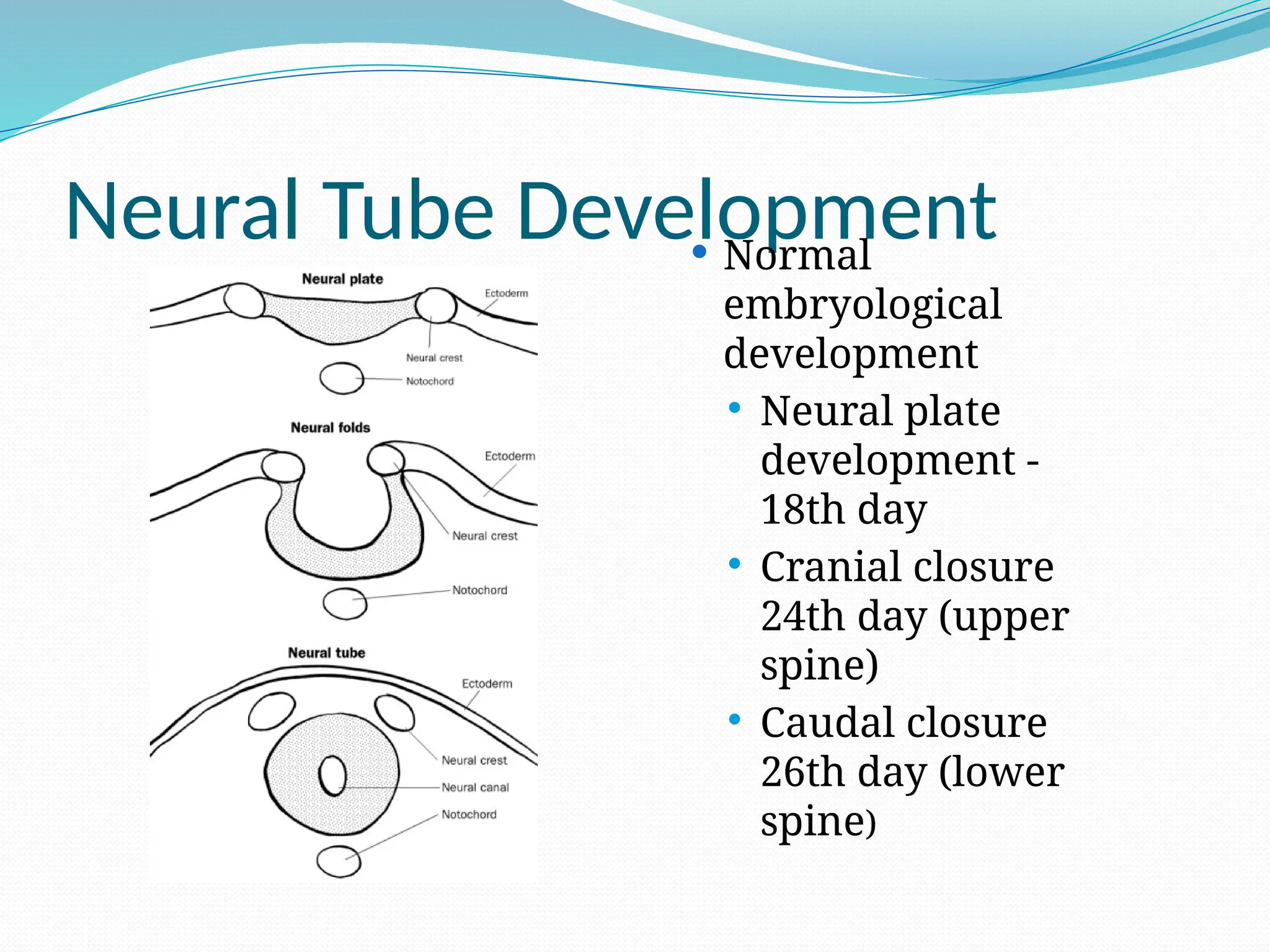
Neural Tube Development
Normal
embryological
development
Neural plate
development -
18th day
Cranial closure
24th day (upper
spine)
Caudal closure
26th day (lower
spine)
5.
Etiology of NTDs
Combination of environmental and genetic
causes .
Teratogens :
- Drugs
-Rdiation
Infection and maternal illnesses.
Nutritional deficiencies . - notably, folic acid
deficiency
6.
RISK FACTOR :
All pregnancies are at risk for an NTD. However,
women with a history of a previous pregnancy
with ( NTD).
women with first degree relative with(NTD)
women with type 1 diabetes mellitus
women with seizure disorders on Na valproic
acid.
women or their partners who themselves have
an NTD.
7.
NTDs :
Twotypes of NTDs:
1- Open NTDs ( most common) :
- occur when the brain and/or spinal cord are
exposed at birth through a defect in the skull or
vertebrae.
Spina bifida
Anencephaly
Encephalocele
8.
2- closed NTDs(Rarer type ):
- occur when the spinal defect is covered by skin.
lipomyelomeningocel
lipomeningocele
tethered spinal cord.
9.
Neural Tube Defects
What are the common Neural Tube Defects
(NTDs) ?
Spina Bifida - 60%
Anencephaly - 30%
Encephalocele - 10%
10.
What is SpinaBifida?
- A midline defect of the :
bone,
skin,
spinal column, &/or
spinal cord.
11.
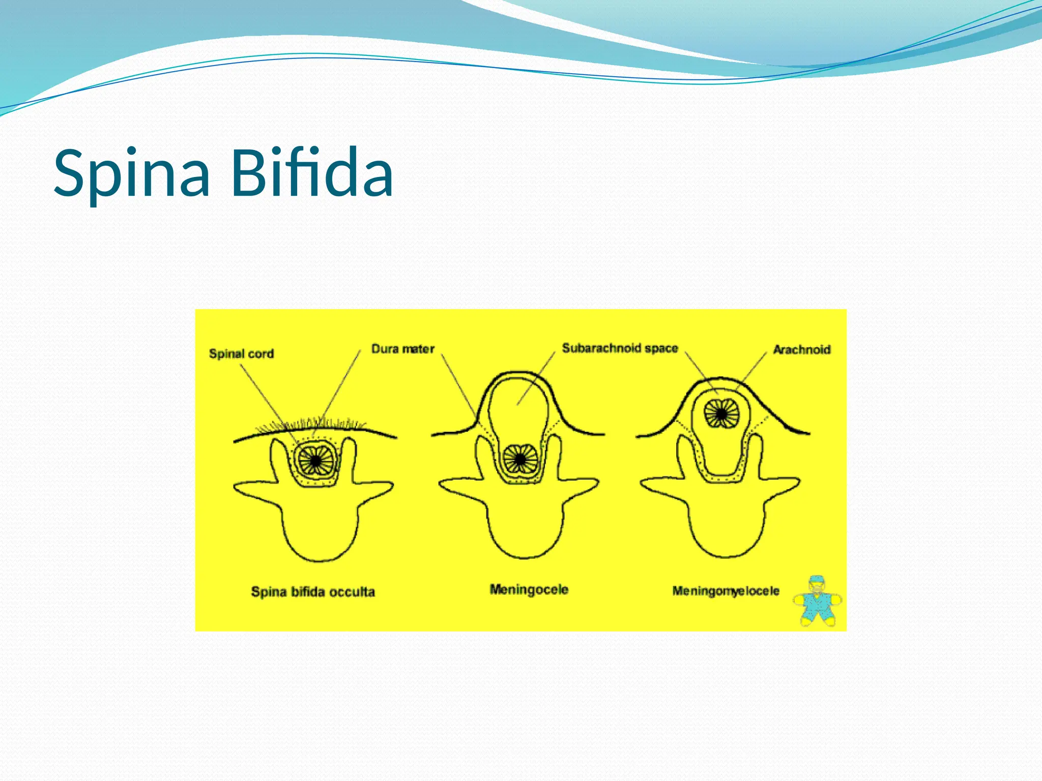
Spina Bifida
SpinaBifida is divided into two subclasses :
1 - Spina Bifida Occulta(closed ) :
- mildest form ( meninges do not herniate
through the opening in the spinal canal )
2 -Spina Bifida Cystic ( open) :
- meningocele and myelomeningocele .
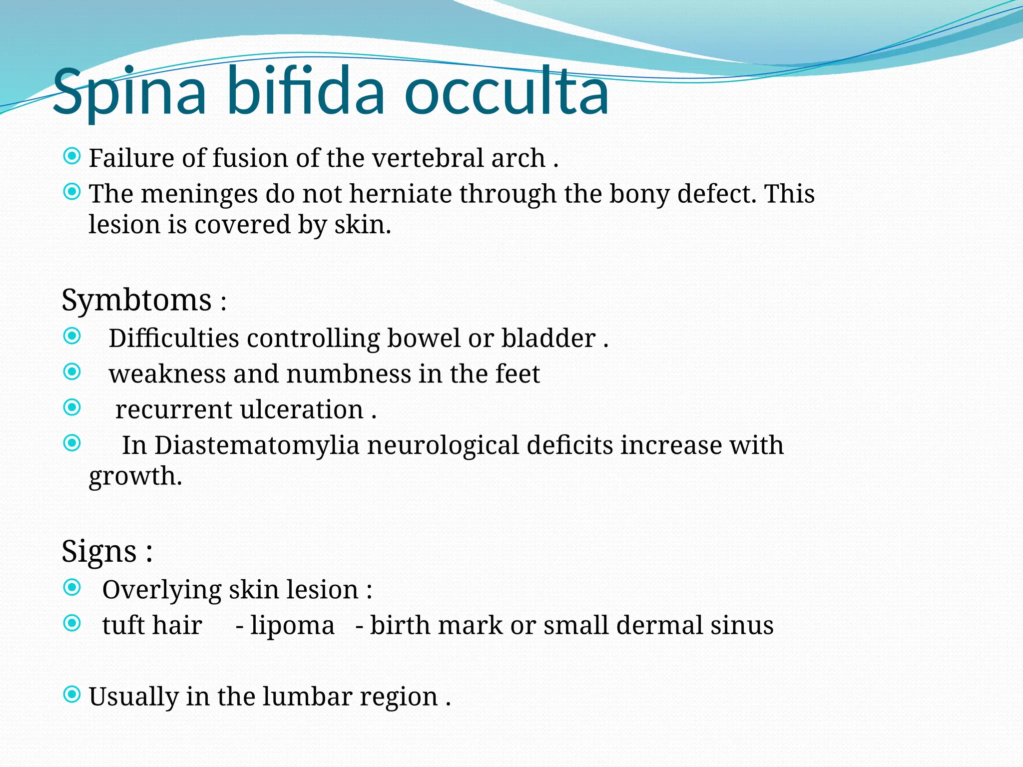
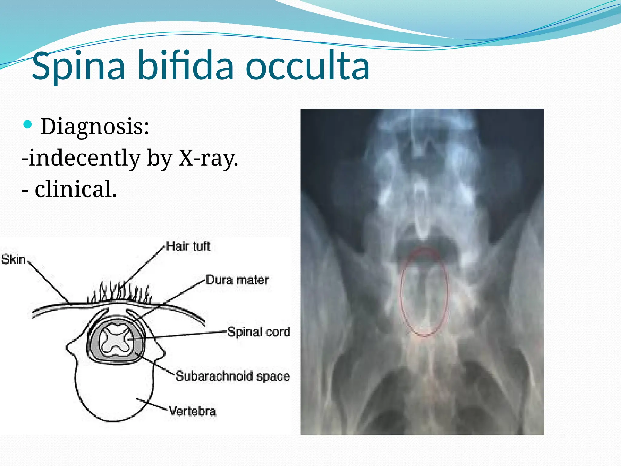
Spina bifida occulta
Failure of fusion of the vertebral arch .
The meninges do not herniate through the bony defect. This
lesion is covered by skin.
Symbtoms :
Difficulties controlling bowel or bladder .
weakness and numbness in the feet
recurrent ulceration .
In Diastematomylia neurological deficits increase with
growth.
Signs :
Overlying skin lesion :
tuft hair - lipoma - birth mark or small dermal sinus
Usually in the lumbar region .
Spina bifida manifesta
The 2 major types of defects seen here are
myelomeningoceles and meningoceles.
lumobosacral regions are the most common sites
for these lesions .
Cervical and thoracic regions are the least
common sites.
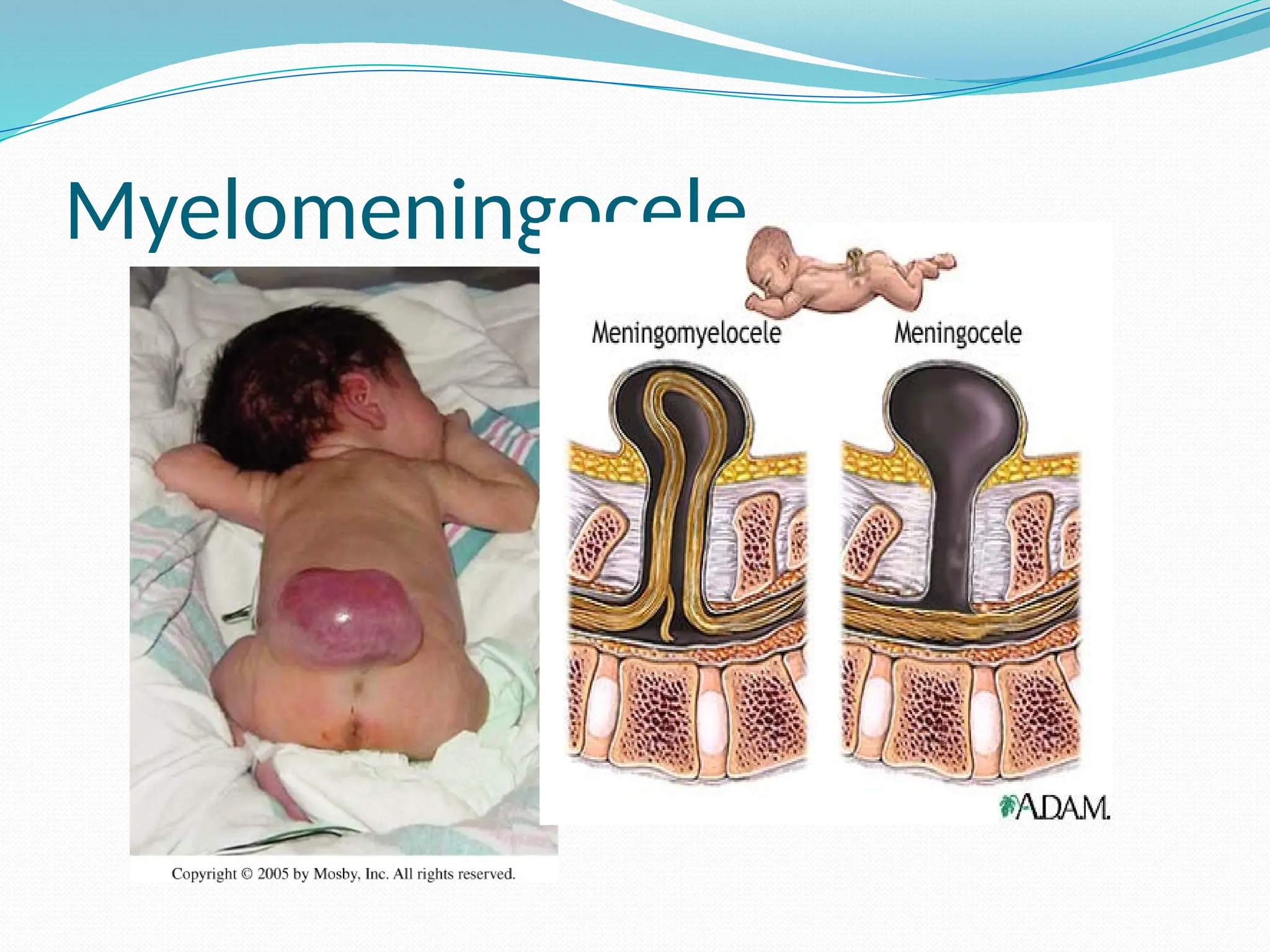
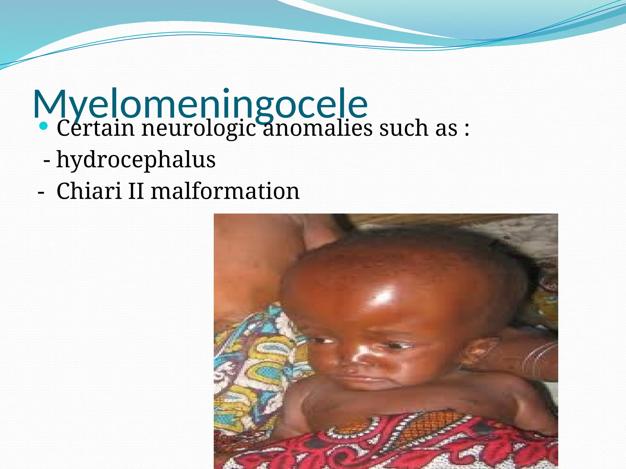
Myelomeningocele
The spinalcord and nerve roots herniate into a
sac comprising the meninges.
This sac protrudes through the bone and
musculocutaneous defect.
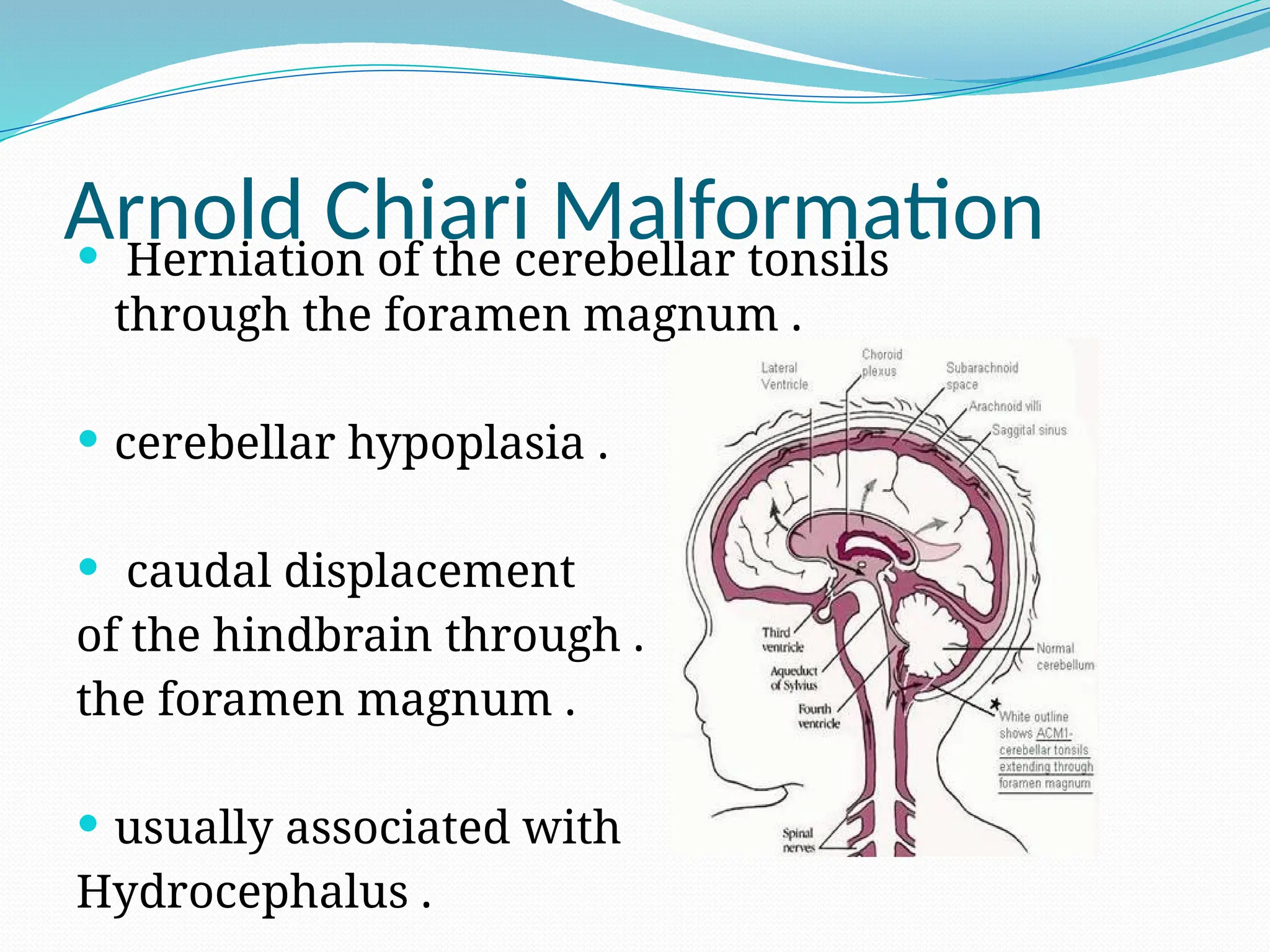
Arnold Chiari Malformation
Herniation of the cerebellar tonsils
through the foramen magnum .
cerebellar hypoplasia .
caudal displacement
of the hindbrain through .
the foramen magnum .
usually associated with
Hydrocephalus .
23.
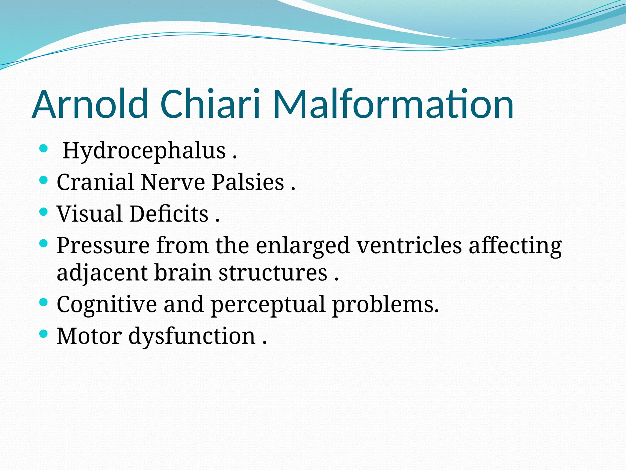
Arnold Chiari Malformation
Hydrocephalus .
Cranial Nerve Palsies .
Visual Deficits .
Pressure from the enlarged ventricles affecting
adjacent brain structures .
Cognitive and perceptual problems.
Motor dysfunction .
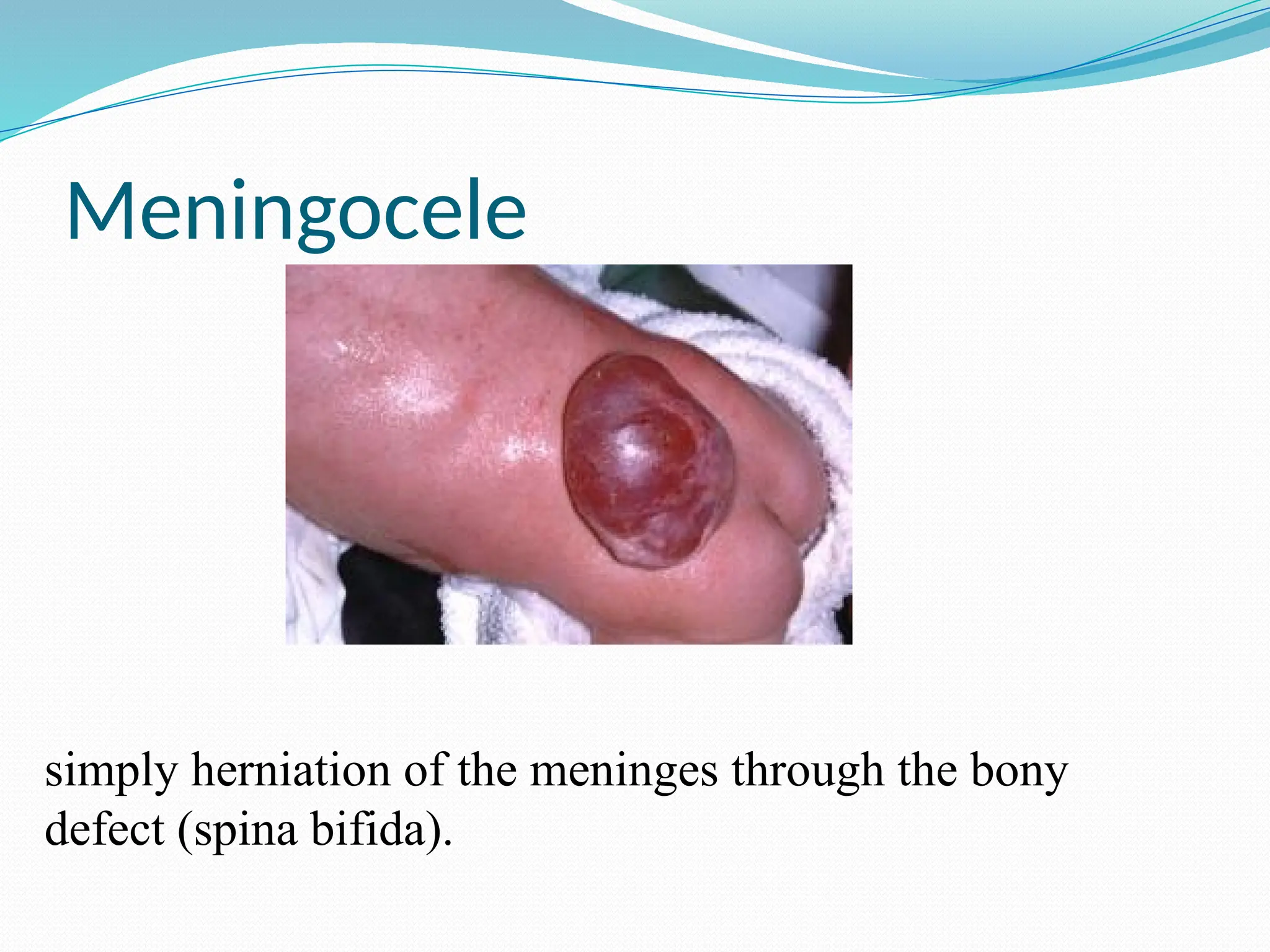
Meningocele
Fluid-filled sacwith meninges involved but
neural tissue unaffected .
The spinal cord and nerve roots do not herniate
into this dorsal dural sac.
The primary problems with this deformity
are cosmetic
26.
Meningocele
Neonates witha meningocele usually have
normal findings upon physical examination and
a covered (closed) dural sac.
Neonates with meningocele do not have
associated neurologic malformations such as
hydrocephalus or Chiari II.
May complicted by CSF infection.
27.
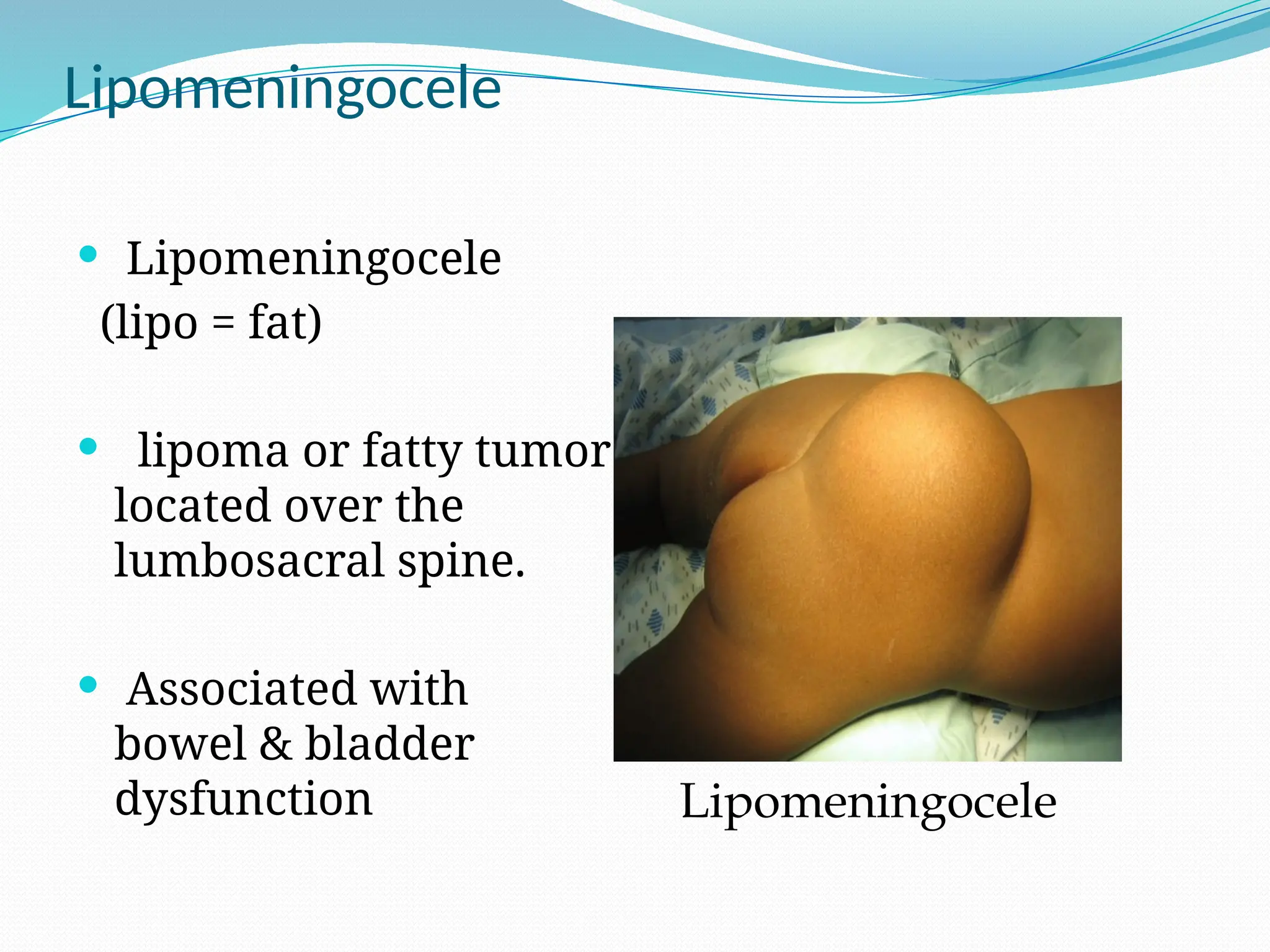
Lipomeningocele
Lipomeningocele
(lipo =fat)
lipoma or fatty tumor
located over the
lumbosacral spine.
Associated with
bowel & bladder
dysfunction Lipomeningocele
28.
Prognosis of Spinabifida
o static
o non-progressive defect
o with worsening from secondary problems.
- The prognosis for a normal life span is generally
good for a child with good health habits and a
supportive family/caregiver.
29.
Impairments associated withSpina Bifida
Abnormal eye movement
Pressure sore and skin irritations.
Latex allergy.
Bladder and bowel control problems
musculoskeletal deformities (scoliosis).
joint and extremity deformities (joint
contractures, club foot, hip subluxations,
diminished growth of non-weight bearing
limbs)
Osteoporosis.
tethered spinal cord after surgery .
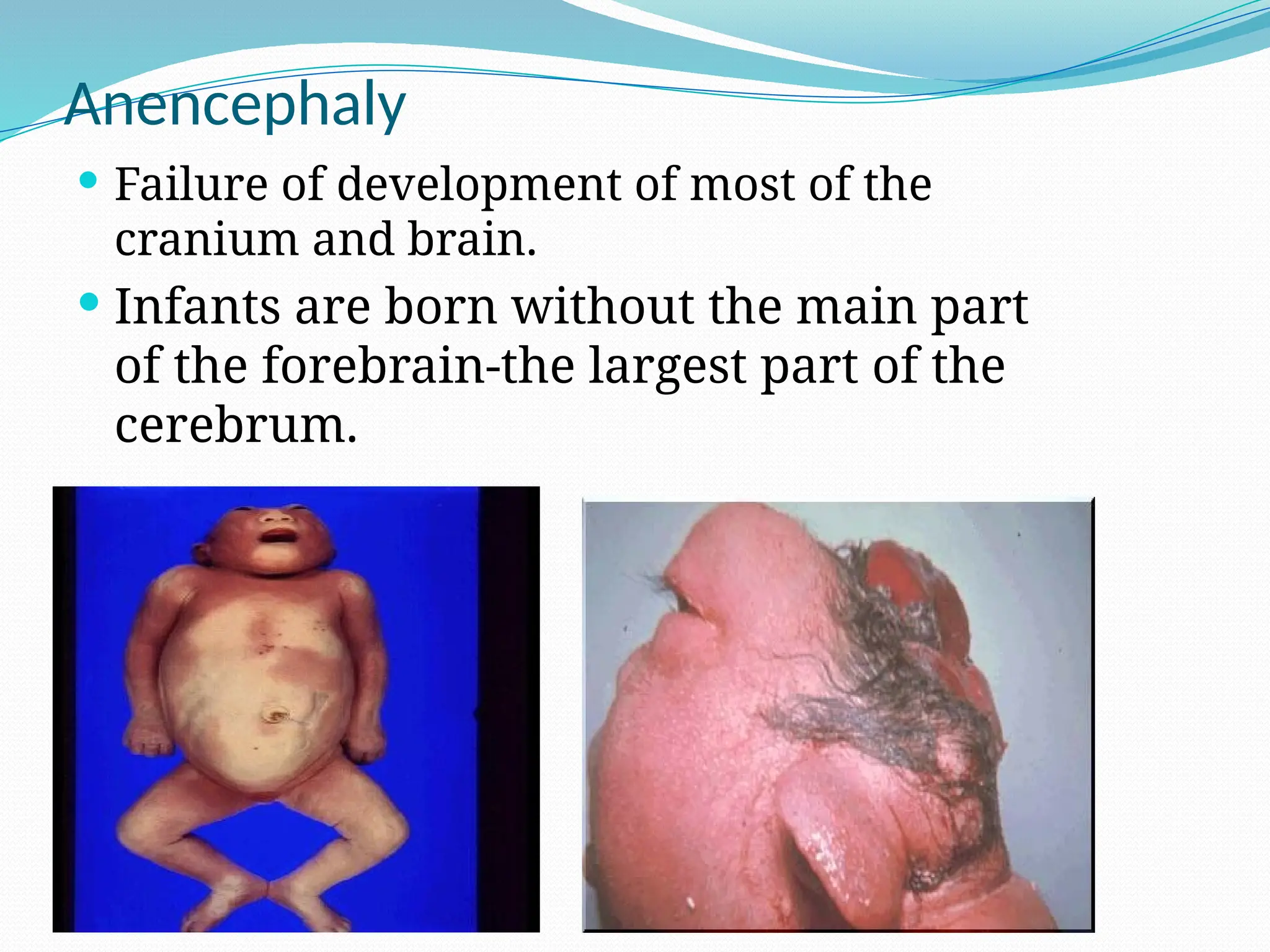
Anencephaly
Failure ofdevelopment of most of the
cranium and brain.
Infants are born without the main part
of the forebrain-the largest part of the
cerebrum.
32.
The fetususually blind, deaf and unconscious
. partially destroyed brain, deformed
forehead, and large ears and eyes with
often relatively normal lower facial
structures.
Both genetic and environmental insults
appear to be responsible for this outcome.
The defect normally occurs after neural
fold development at day 16 of gestation
but before closure of the anterior
neuropore at 24-26 days' gestation.
33.
Anencephaly
Anencephaly isthe most common major CNS
malformation in the Western world,
no neonates survive. It is seen 37 times more in
females than in males.
The recurrence rate in families can be as high as
35%.
34.
Anencephaly
Symptoms
Mom-Polyhydramnios
Baby- absence of brain/skull
Diagnosis
Ultrasound
Treatment
None, incompatible with life
Management
Comfort Measures
Support Parents
35.
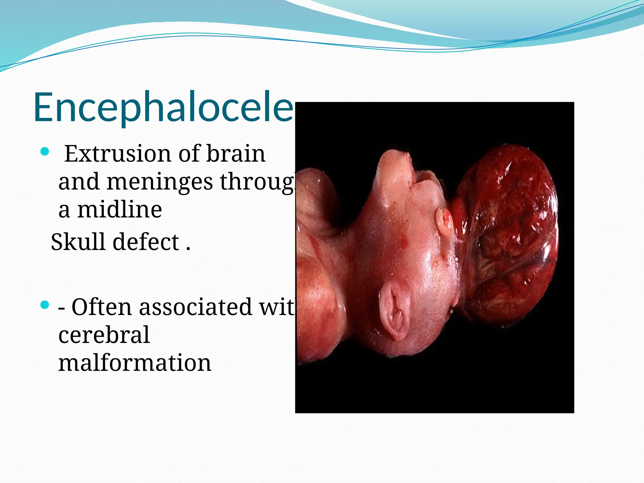
Encephalocele
Extrusion ofbrain
and meninges through
a midline
Skull defect .
- Often associated with
cerebral
malformation
36.
Diagnosis and Detection
Amniocentesis
AFP - indication of abnormal leakage
Blood test
Maternal blood samples of AFP
Ultrasonography
For locating back lesion vs. cranial signs
History
C/C :
Bulging onthe back or other deformity .
HPI :
Onset(at birth).
Size.
Course( progressive or constant)
Associated symptoms .
Past medical hx :
Previous medical problems .
Previous hospitalization.
Previous surgery or shunt .
39.
Pregnancy & neonatalhx :
Follow up during pregnancy or no .
Mother’s illness during pregnancy .
Mother’s medication during pregnancy (anticonvlsion)
Exposure of the mother to radiation.
Exposure to high temperatures early in pregnancy
Taking Folic acid in 1st
trimester.
Gestational age
Type of delivery
Birth weight
Other Congenital anomalies
Apgar scores
Admission to NICU
Developmental hx:
According to age .
40.
Family & socialhx :
Age of parents.
Consanguinity.
History of NTD in family .
History of diabetes of mother.
History of using anti-seizure for mother.
Obesity mother .
History of stillbirth or abortion
History of neonatal death in family.
41.
Physical Examination
General examination:
Childappearance
Vital signs.
Growth parameter ( HC imp)
Examination of the head & neck :
Anterior Fontanel : wide bulging
Separated suture .
Dilated scalp vein .
Setting sun eye sign .
May be neck stiffness .
42.
Examination of cranialnerve .
Examination of the back:
Inspection for deformity , scar, bulging( size, content)
pressure sores and skin irritations
sensation .
Examination of lower limps :
Inspection for deformity, muscle bulk .
Exam for tone and power (maybe paralysis)
Reflex and sensation ,
Gait .
Remember : urinary and bowel sphincters (maybe
affected)
Treatment of mylomenigocele
-Genetic counseling may be recommended. In some cases
where severe defect is detected early in the pregnancy, a
therapeutic abortion may be considered
After birth - surgery to repair the defect is usually
recommended at an early age. Before surgery, the infant
must be handled carefully to reduce damage to the
exposed spinal cord. This may include special care and
positioning, protective devices, and changes in the
methods of handling, feeding, and bathing.
46.
Hydrocephalus:
- Children whoalso have hydrocephalus may need
a ventricular peritoneal shunt
This will help drain the extra fluid
- Antibiotics may be used to treat or prevent
infections such as meningitis or urinary tract
infections
47.
Most children willrequire lifelong treatment for problems that
result from damage to the spinal cord and spinal nerves. This
includes :
- Gentle downward pressure over the bladder may help drain the
bladder. In severe cases, drainage tubes, called catheters, may
be needed. Bowel training programs and a high fiber diet may
improve bowel function
- Orthopedic or physical therapy may be needed to treat
musculoskeletal symptoms. Braces may be needed for muscle
and joint problems
- Neurological losses are treated according to the type and severity
of function loss
48.
- Follow-up examinationsgenerally continue
throughout the child's life. These are done to
check the child's developmental level and to treat
any intellectual, neurological, or physical
problems
49.
Treatment of menigocele
Thekey priorities in the treatment of meningocele
are to prevent infection from developing through
the tissue of the defect on the spine and to
protect the exposed structures from additional
trauma. Most children with meningocele are
treated with surgery (within the first few days of
life) to close the defect and to prevent infection
or further trauma
50.
Management of spinabifida occulta
- can remove fat or fibrous tissues which are
affecting the functioning of the spinal cord
- can drain syrinxes or cysts in the spinal canal to
reduce pressure on the spinal cord and
- can be performed on the legs or feet to improve
their functioning
51.
General management
- braces,supports and corrective casts
- physiotherapy to improve physical strength and coordination
- therapeutic strategies for improving mobility
- surgical care
- medical strategies for improving bladder and bowel functioning :
intermittent catheterization
voiding and cleansing routines
medications
diet with adequate fiber and fluids
possible surgical reconstruction (urinary)
- psychological strategies for personal and social adjustment
medications
52.
SUMMERY :
Prevention
folic acid 0.4 mg daily pre, 1 mg daily preg
Identify
Prenatal
At birth
Protect pre-op and post-op
Skin integrity to prevent infection
Special handling to reduce nerve damage
Support
Parental coping
Pictures of similar defects corrected
Genetic Counseling
For future pregnancy
In early pregnancy, therapeutic abortion
Complications
Permanent disability
Education
Symptoms of hydrocephalus
Symptoms of meningitis
Follow up for monitoring to assess neurologic damage
53.
How Can NTDsbe Prevented?
All women of childbearing age should receive 0.4
mg (400 micrograms) of folic acid daily prior to
conception of planned or unplanned pregnancies
and continue thru 1st trimester
Women with a history of NTD and should receive
daily supplementation of (4000 micrograms) of
folic acid starting three months prior to
conception and continuing thru the 1st trimester