Downloaded 268 times


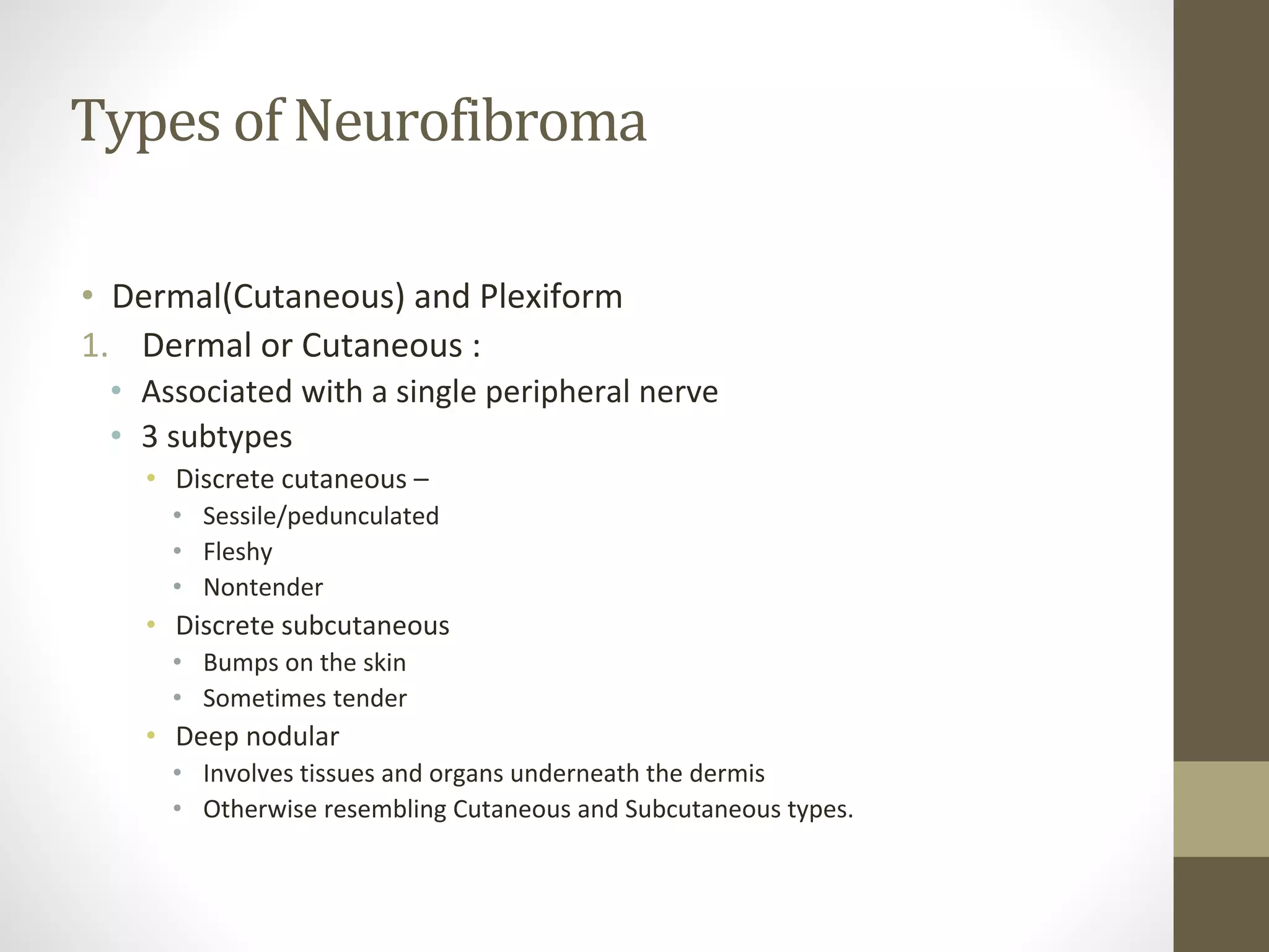


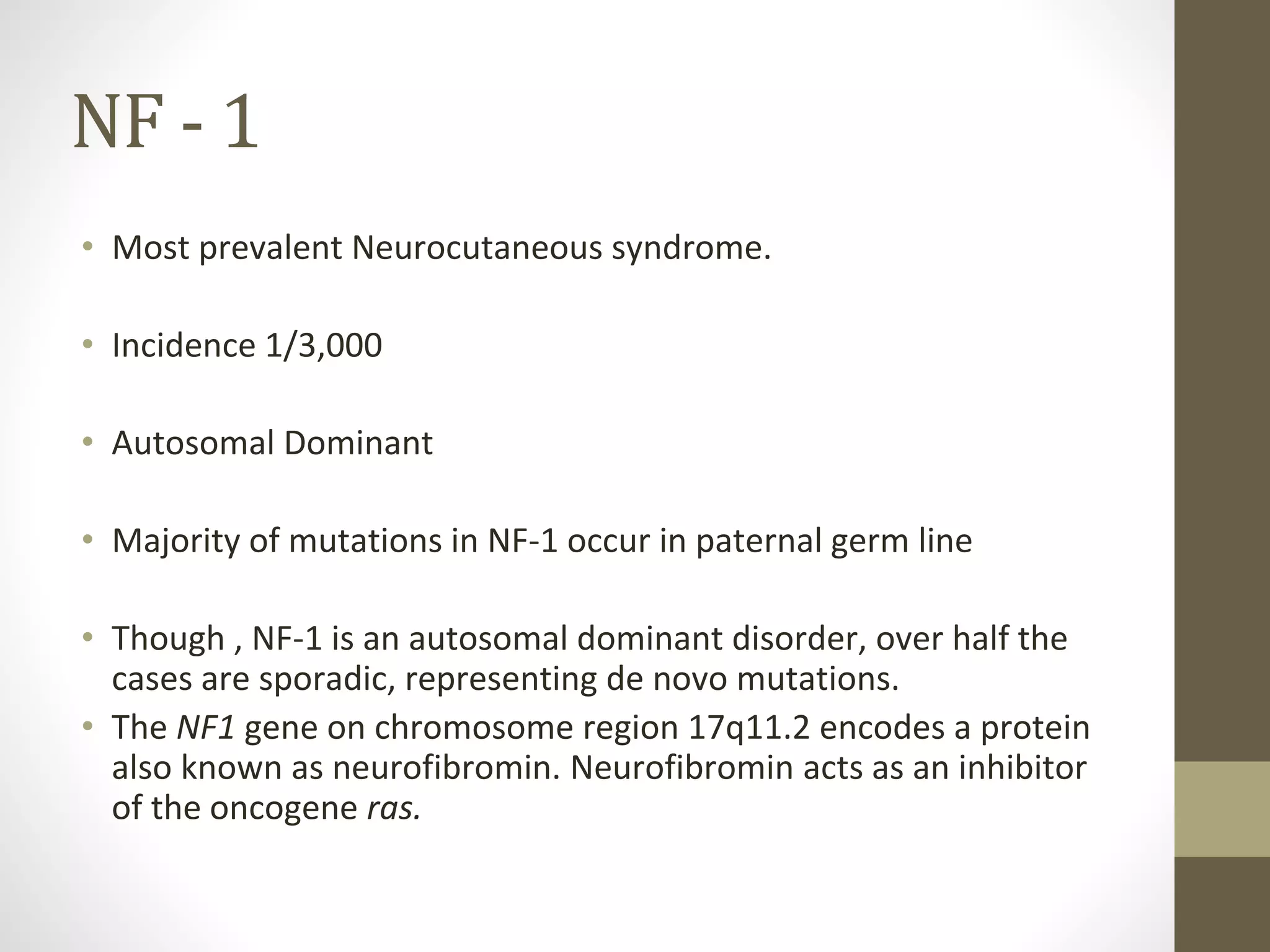


The document provides an overview of several neurocutaneous syndromes from a pediatric perspective. It discusses the defining features and management of neurofibromatosis types 1 and 2, tuberous sclerosis complex, and Sturge-Weber syndrome. Key points include: neurofibromatosis type 1 is characterized by café-au-lait spots and neurofibromas; neurofibromatosis type 2 features tumors of the cranial and spinal nerves; tuberous sclerosis complex causes non-cancerous tumors in many organs and features epilepsy and intellectual disability; and Sturge-Weber syndrome is identified by a port-wine stain on the face and glaucoma of the ipsilateral eye. Close multidisciplinary monitoring


