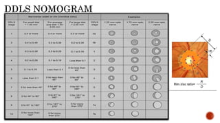
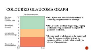
SlideShare a Scribd company logo
1 of 16
Dr. Sumit Kumar
Department of Ophthalmology
Maulana Azad Medical College
and associated hospitals Delhi
• Glaucoma is a multifactorial, chronic optic neuropathy characterized by progressive retinal
ganglion cell death, optic disc cupping, and corresponding visual field loss mostly due to an
abnormally high intraocular pressure, and is a second leading cause of blindness worldwide.1-2
• Stuctural changes in optic disc precedes functional damage so their early assessment is crucial,
structural assessment can detect glaucoma in early stage and diminishes the risk of irreversible
visual imapiarement.
Disc pattern associated with glaucoma are progressive and asymmetric
Glaucomatous optic disc morphology can be divided into 3 categories:-
1.) Disc pattern Focal atrophy
Concentric atrophy
Deepening of cup
Pallor cup dicrepency
Advanced glaucomatous cupping
2.) Vascular sign Optic disc haemorrhage
Tortuous retinal vessels Baring of circumlinear blood vessel
Bayonetting of vessel
3.) Peripapillary changes RNFL Defect
β- zone peripapillary atrophy
FOCALATROPHY
o Selective Neural rim tissue loss primarily in
Inferotemporal region of the Optic Nerve Head
(Polar notching/focal notching)
+
o Lesser extent in the superotemporal sector in
early stage of damage.
Vertical enlargement of cup
o Characteristic of glaucoma especially with
average/normal IOP
CONCENTRIC ATROPHY
o Glaucomatous damage lead to enlargement of
cup in concentric circles.
o Because loss of neural rim tissue begins
temporally and then progress
circumferentially towards superior and
inferior pole this has been called Temporal
unfolding.
o Characteristic of glaucoma with high IOP
Focal notch is characteristic of glaucoma.
NOTCH is characterised by Rim/Disc ratio narrower than 0.1, limited to an area of less than 2
o’clock hour.
Since the late 1960s, the most commonly used quantitative classification of the optic nerve has been
Armaly’s cup/disc ratio. 3
This staging scale describes the disc using cup diameter as a percentage of overall disc diameter. The
cup/disc ratio, especially the vertical cup/disc ratio, represented a significant advance in quantifying
glaucomatous optic neuropathy.
Advatages- Ease of use and lack of magnification artifacts
Limitations-
1.) This system does not account for disc size.
2.) The second issue is that focal changes in the neuroretinal rim that are so
characteristic of glaucoma are not readily detected by the cup/disc ratio (doesn’t
consider the eccentricity of the cup)
 The disc damage likelihood scale (DDLS) was devised by Spaeth et al in 2002 to
incorporate the effect of disc size and focal rim width into a clinical
grading scale.4
 It is highly reproducible and does correlate strongly with the degree of field loss.5
 The system categorises the disc as small (<1.5mm), medium(1.5-2.0mm) or
large(>2.0mm). This ensures that the disc size is measured thereby reducing
misclassification bias based on disc size.
STEP 1. Perform dilation if necessary. The pupils must be sufficiently large to allow a clear view
of the fundus.
STEP 2. Get an idea of both of the patient’s optic nerves via a brief examination at the
biomicroscope with a strong plus lens (eg, +66.00 D). Determine the vertical size of the
discs. Disc size can be measured using a fundus lens at the slit-lamp. A slit beam is
directed onto the disc and the graticule at the top used to reduce the height of the beam
until it corresponds in size to the disc. The lens used will determine the correction
factor. A 66D gives the exact measure from the graticule.
Correction factors for the other lenses are -
Volk
60D-0.88
78D-1.1
90D-1.3
Nikon
60D-1
90D-1.63
Goldmann 3-mirror
1.27
STEP 3. Choose one of the patient’s eyes to concentrate on first. With a direct ophthalmoscope,
examine the optic disc for an area where its outer edge is clearly distinguished from other
ocular tissue such as sclera. Then, determine the full circumference of the outer edge.
STEP 4. Define the inside edge of the neuroretinal rim (outer edge of the cup) by direct
ophthalmoscopy. Estimate the rim-to-disc ratio by comparing the width of the neuroretinal
rim with that of the disc diameter on the same axis. Perform this comparison at several
clock positions. If the rim-to-disc ratio is different at various parts of the rim, note the area
at which the rim is narrowest and calculate the rim-to-disc ratio there.
STEP 5. Draw the shape of the optic disc. When sketching the neuroretinal rim’s inner edge, indicate
clear demarcation with a thick line and less clear demarcation with a thin or hatched line. Note
the course of blood vessels that help determine the rim’s width and any pertinent features of the
disc (eg, notches, pallor, hemorrhage).
STEP 6. Determine the DDLS by using your drawing of the disc, the narrowest rim-to-disc ratio, the size
of the disc, and the DDLS nomogram. If the nerve is smaller or larger than average, you must
adjust the DDLS score appropriately. An easy method is to stage the nerve as if it were of
average size and then increase the stage by one if the nerve is small or decrease the stage by
one if it is large. Note the DDLS score in the patient’s chart.
Repeat steps No. 3 through 6 for the patient’s other eye.
• DDLS provides a quantitative method of
assessing the glaucomatous damage.
• DDLS can be used for diagnosing, staging
and monitoring the rate of change of the
patient’s condition.
• Because each grade is assigned a numerical
value the system can then be used in
research settings to determine severity or
degree of progression.
GREEN ZONE
• All cases with IOP >40 mm Hg.
• Most cases with IOP >30 mmHg, especially where this represents a rise from previously lower pressure
and life expectancy is greater than one year.
• Marked anxiety about possible deterioration of disease.
YELLOW ZONE
• Life expectancy >2 year.
• All cases in which there is reasonable possibility that the glaucomatous disease process is active, that is, the
cause for developing disk damage will cause further disk damage.
RED ZONE
• All cases
• The DDLS cannot be used to evaluate certain types of discs, such as those that are
congenitally anomalous and Myopic discs may also be difficult to grade
• Another problem with the DDLS is that a disc may show progressive damage by having a
continuing generalized narrowing of the neuroretinal rim, but not have an increase in the
circumferential extent of rim absence. In such a situation the disc would unquestionably have
become worse, but the DDLS score will not change.
1.Wang W, He M, Zhou M, Zhang X. Fornix-based versus limbus-based conjunctival flap in
trabeculectomy: a quantitative evaluation of the evidence. PLOS One. 2013;1-8.
2.Delbeke H, Stalmans I, Vandewalle E, Zeyen T. The Effect of Trabeculectomy on
Astigmatism.J Glaucoma. 2015;11;1-5.
3. Armaly M. Genetic determination of cup/disc ratio of the optic nerve. Arch Ophthalmol
1967;78:35-43.
4.Bayer A. Harasymowycz P. Henderer JD, et al. Validity of a new disc grading scale for
estimating glaucomatous damage: correlation with visual field damage. Am J Ophthalmol
2002;133:758-763.
5.Henderer JD, Liu C, Kesen M, et al. Reliability of the disc damage likelihood scale. Am J
Ophthalmol 2003;135:44-8.
It takes some effort to learn it and initially a copy of the table
should always be to hand. However, given practice and used
accordingly the DDLS is an excellent tool for classifying and
monitoring the optic nerve in glaucoma.

More Related Content

What's hot

Glaucoma Guided Progression Analysis - Dr Shylesh Dabke
Glaucoma Guided Progression Analysis - Dr Shylesh DabkeGlaucoma Guided Progression Analysis - Dr Shylesh Dabke
Glaucoma Guided Progression Analysis - Dr Shylesh DabkeShylesh Dabke
 
Yag capsulotomy
Yag capsulotomyYag capsulotomy
Yag capsulotomyRohit Rao
 
Embryology of angle of anterior chamber
Embryology of angle of anterior chamberEmbryology of angle of anterior chamber
Embryology of angle of anterior chamberMahendar Bathina
 
Vitreous substitutes
Vitreous substitutesVitreous substitutes
Vitreous substitutesSSSIHMS-PG
 
Choroidal neovascularisation(cnv)
Choroidal neovascularisation(cnv)Choroidal neovascularisation(cnv)
Choroidal neovascularisation(cnv)Nikhil Rp
 
Diagnosis of pre perimetric glaucoma
Diagnosis of pre perimetric glaucomaDiagnosis of pre perimetric glaucoma
Diagnosis of pre perimetric glaucomaSadhwini Harish
 
Assessment of corneal endothelium
Assessment of corneal endotheliumAssessment of corneal endothelium
Assessment of corneal endotheliumdrvasant162
 
IOL power calculation special situations
IOL power calculation special situations IOL power calculation special situations
IOL power calculation special situations Laxmi Eye Institute
 
Pseudophakic Bullous Keratopathy case
Pseudophakic Bullous Keratopathy case Pseudophakic Bullous Keratopathy case
Pseudophakic Bullous Keratopathy case Noor Munirah Aab
 
Anterior segment OCT & UBM
Anterior segment OCT & UBMAnterior segment OCT & UBM
Anterior segment OCT & UBMDinesh Madduri
 
Choroidal neovascularization
Choroidal neovascularizationChoroidal neovascularization
Choroidal neovascularizationdrkvasantha
 

What's hot (20)

Glaucoma Guided Progression Analysis - Dr Shylesh Dabke
Glaucoma Guided Progression Analysis - Dr Shylesh DabkeGlaucoma Guided Progression Analysis - Dr Shylesh Dabke
Glaucoma Guided Progression Analysis - Dr Shylesh Dabke
 
Nonpenetrating glaucoma surgery
Nonpenetrating glaucoma surgeryNonpenetrating glaucoma surgery
Nonpenetrating glaucoma surgery
 
Yag capsulotomy
Yag capsulotomyYag capsulotomy
Yag capsulotomy
 
Retina drwaing
Retina drwaingRetina drwaing
Retina drwaing
 
Embryology of angle of anterior chamber
Embryology of angle of anterior chamberEmbryology of angle of anterior chamber
Embryology of angle of anterior chamber
 
Toric iol
Toric iolToric iol
Toric iol
 
Macular hole
Macular holeMacular hole
Macular hole
 
Vitreous substitutes
Vitreous substitutesVitreous substitutes
Vitreous substitutes
 
Choroidal neovascularisation(cnv)
Choroidal neovascularisation(cnv)Choroidal neovascularisation(cnv)
Choroidal neovascularisation(cnv)
 
Diagnosis of pre perimetric glaucoma
Diagnosis of pre perimetric glaucomaDiagnosis of pre perimetric glaucoma
Diagnosis of pre perimetric glaucoma
 
Assessment of corneal endothelium
Assessment of corneal endotheliumAssessment of corneal endothelium
Assessment of corneal endothelium
 
IOL power calculation special situations
IOL power calculation special situations IOL power calculation special situations
IOL power calculation special situations
 
Pseudophakic Bullous Keratopathy case
Pseudophakic Bullous Keratopathy case Pseudophakic Bullous Keratopathy case
Pseudophakic Bullous Keratopathy case
 
EDOF IOL
EDOF IOLEDOF IOL
EDOF IOL
 
Oct in glaucoma
Oct in glaucomaOct in glaucoma
Oct in glaucoma
 
Ocular surface analysers
Ocular surface analysersOcular surface analysers
Ocular surface analysers
 
Anterior segment OCT & UBM
Anterior segment OCT & UBMAnterior segment OCT & UBM
Anterior segment OCT & UBM
 
Choroidal neovascularization
Choroidal neovascularizationChoroidal neovascularization
Choroidal neovascularization
 
OCT Angiography
OCT AngiographyOCT Angiography
OCT Angiography
 
anatomy of retina
 anatomy of retina anatomy of retina
anatomy of retina
 

Viewers also liked

Viewers also liked (9)

Optic nerve Clinical significance
Optic nerve Clinical significance Optic nerve Clinical significance
Optic nerve Clinical significance
 
Oftalmoscopia
Oftalmoscopia  Oftalmoscopia
Oftalmoscopia
 
Glaucoma optic disc changes
Glaucoma optic disc changesGlaucoma optic disc changes
Glaucoma optic disc changes
 
Diagnosis of glaucoma
Diagnosis of glaucomaDiagnosis of glaucoma
Diagnosis of glaucoma
 
Revisão OFTALMO
Revisão OFTALMORevisão OFTALMO
Revisão OFTALMO
 
Fundoscopia para iniciantes
Fundoscopia para iniciantesFundoscopia para iniciantes
Fundoscopia para iniciantes
 
Primary open angle glaucoma (poag)
Primary open angle glaucoma (poag)Primary open angle glaucoma (poag)
Primary open angle glaucoma (poag)
 
Optic nerve head evaluation in glaucoma
Optic nerve head evaluation in glaucomaOptic nerve head evaluation in glaucoma
Optic nerve head evaluation in glaucoma
 
Glaucoma
GlaucomaGlaucoma
Glaucoma
 

Similar to Disc Damage Likelihod Scale (DDLS)

Criterios diagnosticos en glaucoma
Criterios diagnosticos en glaucomaCriterios diagnosticos en glaucoma
Criterios diagnosticos en glaucomaMelissa Garcia Felix
 
SUPERPIXEL CLASSIFICATION BASED OPTIC DISC AND OPTIC CUP SEGMENTATION FOR GLA...
SUPERPIXEL CLASSIFICATION BASED OPTIC DISC AND OPTIC CUP SEGMENTATION FOR GLA...SUPERPIXEL CLASSIFICATION BASED OPTIC DISC AND OPTIC CUP SEGMENTATION FOR GLA...
SUPERPIXEL CLASSIFICATION BASED OPTIC DISC AND OPTIC CUP SEGMENTATION FOR GLA...pharmaindexing
 
Evaluating the optic nerve head in glaucoma
Evaluating the optic nerve head in glaucomaEvaluating the optic nerve head in glaucoma
Evaluating the optic nerve head in glaucomaRiyad Banayot
 
22 optic-disc-evaluation-in-glaucoma
22 optic-disc-evaluation-in-glaucoma22 optic-disc-evaluation-in-glaucoma
22 optic-disc-evaluation-in-glaucomaUsamaIqbal18
 
Optical Coherence Tomography in Multiple Sclerosis
Optical Coherence Tomography in Multiple SclerosisOptical Coherence Tomography in Multiple Sclerosis
Optical Coherence Tomography in Multiple Sclerosisneurophq8
 
H0366051058
H0366051058H0366051058
H0366051058theijes
 
Glaucoma Screening Test By Segmentation of Optical Disc& Cup Segmentation Usi...
Glaucoma Screening Test By Segmentation of Optical Disc& Cup Segmentation Usi...Glaucoma Screening Test By Segmentation of Optical Disc& Cup Segmentation Usi...
Glaucoma Screening Test By Segmentation of Optical Disc& Cup Segmentation Usi...IJERA Editor
 
Visual Field presentation.nagla.ppt
Visual Field presentation.nagla.pptVisual Field presentation.nagla.ppt
Visual Field presentation.nagla.pptnanoAly
 
Glucoma detection.pptx
Glucoma detection.pptxGlucoma detection.pptx
Glucoma detection.pptxpriyaghate2
 
Glaucoma and OCT – Are Macula Scans More Valuable than Disc Scans
Glaucoma and OCT – Are Macula Scans More Valuable than Disc ScansGlaucoma and OCT – Are Macula Scans More Valuable than Disc Scans
Glaucoma and OCT – Are Macula Scans More Valuable than Disc ScansJason Higginbotham
 
Retinal Thickness In Macular Region Of High Myopic Eyes Using Sd-Oct
Retinal Thickness In Macular Region Of High Myopic Eyes  Using Sd-OctRetinal Thickness In Macular Region Of High Myopic Eyes  Using Sd-Oct
Retinal Thickness In Macular Region Of High Myopic Eyes Using Sd-OctDr. Jagannath Boramani
 
Optometric examination and management of geriatric problems.pptx
Optometric examination and management of geriatric problems.pptxOptometric examination and management of geriatric problems.pptx
Optometric examination and management of geriatric problems.pptxAnisha Heka
 
22-Optic-Disc-Evaluation-IN-Glaucoma.ppt
22-Optic-Disc-Evaluation-IN-Glaucoma.ppt22-Optic-Disc-Evaluation-IN-Glaucoma.ppt
22-Optic-Disc-Evaluation-IN-Glaucoma.pptudayasree30
 
Optic Nerve Head Evaluation.pptx
Optic Nerve Head Evaluation.pptxOptic Nerve Head Evaluation.pptx
Optic Nerve Head Evaluation.pptxGariyashee Lahkar
 
Optical coherence tomography in glaucoma - Dr Shylesh Dabke
Optical coherence tomography in glaucoma - Dr Shylesh DabkeOptical coherence tomography in glaucoma - Dr Shylesh Dabke
Optical coherence tomography in glaucoma - Dr Shylesh DabkeShylesh Dabke
 

Similar to Disc Damage Likelihod Scale (DDLS) (20)

Criterios diagnosticos en glaucoma
Criterios diagnosticos en glaucomaCriterios diagnosticos en glaucoma
Criterios diagnosticos en glaucoma
 
Oct in glaucoma
Oct in glaucomaOct in glaucoma
Oct in glaucoma
 
28. ONH evaluation in glaucoma
28.  ONH evaluation in glaucoma 28.  ONH evaluation in glaucoma
28. ONH evaluation in glaucoma
 
Fundus in Glaucoma
Fundus in GlaucomaFundus in Glaucoma
Fundus in Glaucoma
 
SUPERPIXEL CLASSIFICATION BASED OPTIC DISC AND OPTIC CUP SEGMENTATION FOR GLA...
SUPERPIXEL CLASSIFICATION BASED OPTIC DISC AND OPTIC CUP SEGMENTATION FOR GLA...SUPERPIXEL CLASSIFICATION BASED OPTIC DISC AND OPTIC CUP SEGMENTATION FOR GLA...
SUPERPIXEL CLASSIFICATION BASED OPTIC DISC AND OPTIC CUP SEGMENTATION FOR GLA...
 
Evaluating the optic nerve head in glaucoma
Evaluating the optic nerve head in glaucomaEvaluating the optic nerve head in glaucoma
Evaluating the optic nerve head in glaucoma
 
22 optic-disc-evaluation-in-glaucoma
22 optic-disc-evaluation-in-glaucoma22 optic-disc-evaluation-in-glaucoma
22 optic-disc-evaluation-in-glaucoma
 
AMA series design
AMA series designAMA series design
AMA series design
 
Optical Coherence Tomography in Multiple Sclerosis
Optical Coherence Tomography in Multiple SclerosisOptical Coherence Tomography in Multiple Sclerosis
Optical Coherence Tomography in Multiple Sclerosis
 
H0366051058
H0366051058H0366051058
H0366051058
 
Glaucoma Screening Test By Segmentation of Optical Disc& Cup Segmentation Usi...
Glaucoma Screening Test By Segmentation of Optical Disc& Cup Segmentation Usi...Glaucoma Screening Test By Segmentation of Optical Disc& Cup Segmentation Usi...
Glaucoma Screening Test By Segmentation of Optical Disc& Cup Segmentation Usi...
 
Glaucoma
GlaucomaGlaucoma
Glaucoma
 
Visual Field presentation.nagla.ppt
Visual Field presentation.nagla.pptVisual Field presentation.nagla.ppt
Visual Field presentation.nagla.ppt
 
Glucoma detection.pptx
Glucoma detection.pptxGlucoma detection.pptx
Glucoma detection.pptx
 
Glaucoma and OCT – Are Macula Scans More Valuable than Disc Scans
Glaucoma and OCT – Are Macula Scans More Valuable than Disc ScansGlaucoma and OCT – Are Macula Scans More Valuable than Disc Scans
Glaucoma and OCT – Are Macula Scans More Valuable than Disc Scans
 
Retinal Thickness In Macular Region Of High Myopic Eyes Using Sd-Oct
Retinal Thickness In Macular Region Of High Myopic Eyes  Using Sd-OctRetinal Thickness In Macular Region Of High Myopic Eyes  Using Sd-Oct
Retinal Thickness In Macular Region Of High Myopic Eyes Using Sd-Oct
 
Optometric examination and management of geriatric problems.pptx
Optometric examination and management of geriatric problems.pptxOptometric examination and management of geriatric problems.pptx
Optometric examination and management of geriatric problems.pptx
 
22-Optic-Disc-Evaluation-IN-Glaucoma.ppt
22-Optic-Disc-Evaluation-IN-Glaucoma.ppt22-Optic-Disc-Evaluation-IN-Glaucoma.ppt
22-Optic-Disc-Evaluation-IN-Glaucoma.ppt
 
Optic Nerve Head Evaluation.pptx
Optic Nerve Head Evaluation.pptxOptic Nerve Head Evaluation.pptx
Optic Nerve Head Evaluation.pptx
 
Optical coherence tomography in glaucoma - Dr Shylesh Dabke
Optical coherence tomography in glaucoma - Dr Shylesh DabkeOptical coherence tomography in glaucoma - Dr Shylesh Dabke
Optical coherence tomography in glaucoma - Dr Shylesh Dabke
 

Recently uploaded

Interdisciplinary_Insights_Data_Collection_Methods.pptx
Interdisciplinary_Insights_Data_Collection_Methods.pptxInterdisciplinary_Insights_Data_Collection_Methods.pptx
Interdisciplinary_Insights_Data_Collection_Methods.pptxPooja Bhuva
 
Introduction to TechSoup’s Digital Marketing Services and Use Cases
Introduction to TechSoup’s Digital Marketing  Services and Use CasesIntroduction to TechSoup’s Digital Marketing  Services and Use Cases
Introduction to TechSoup’s Digital Marketing Services and Use CasesTechSoup
 
On National Teacher Day, meet the 2024-25 Kenan Fellows
On National Teacher Day, meet the 2024-25 Kenan FellowsOn National Teacher Day, meet the 2024-25 Kenan Fellows
On National Teacher Day, meet the 2024-25 Kenan FellowsMebane Rash
 
QUATER-1-PE-HEALTH-LC2- this is just a sample of unpacked lesson
QUATER-1-PE-HEALTH-LC2- this is just a sample of unpacked lessonQUATER-1-PE-HEALTH-LC2- this is just a sample of unpacked lesson
QUATER-1-PE-HEALTH-LC2- this is just a sample of unpacked lessonhttgc7rh9c
 
Accessible Digital Futures project (20/03/2024)
Accessible Digital Futures project (20/03/2024)Accessible Digital Futures project (20/03/2024)
Accessible Digital Futures project (20/03/2024)Jisc
 
Economic Importance Of Fungi In Food Additives
Economic Importance Of Fungi In Food AdditivesEconomic Importance Of Fungi In Food Additives
Economic Importance Of Fungi In Food AdditivesSHIVANANDaRV
 
UGC NET Paper 1 Unit 7 DATA INTERPRETATION.pdf
UGC NET Paper 1 Unit 7 DATA INTERPRETATION.pdfUGC NET Paper 1 Unit 7 DATA INTERPRETATION.pdf
UGC NET Paper 1 Unit 7 DATA INTERPRETATION.pdfNirmal Dwivedi
 
How to Manage Global Discount in Odoo 17 POS
How to Manage Global Discount in Odoo 17 POSHow to Manage Global Discount in Odoo 17 POS
How to Manage Global Discount in Odoo 17 POSCeline George
 
What is 3 Way Matching Process in Odoo 17.pptx
What is 3 Way Matching Process in Odoo 17.pptxWhat is 3 Way Matching Process in Odoo 17.pptx
What is 3 Way Matching Process in Odoo 17.pptxCeline George
 
Play hard learn harder: The Serious Business of Play
Play hard learn harder:  The Serious Business of PlayPlay hard learn harder:  The Serious Business of Play
Play hard learn harder: The Serious Business of PlayPooky Knightsmith
 
How to Create and Manage Wizard in Odoo 17
How to Create and Manage Wizard in Odoo 17How to Create and Manage Wizard in Odoo 17
How to Create and Manage Wizard in Odoo 17Celine George
 
Exploring_the_Narrative_Style_of_Amitav_Ghoshs_Gun_Island.pptx
Exploring_the_Narrative_Style_of_Amitav_Ghoshs_Gun_Island.pptxExploring_the_Narrative_Style_of_Amitav_Ghoshs_Gun_Island.pptx
Exploring_the_Narrative_Style_of_Amitav_Ghoshs_Gun_Island.pptxPooja Bhuva
 
On_Translating_a_Tamil_Poem_by_A_K_Ramanujan.pptx
On_Translating_a_Tamil_Poem_by_A_K_Ramanujan.pptxOn_Translating_a_Tamil_Poem_by_A_K_Ramanujan.pptx
On_Translating_a_Tamil_Poem_by_A_K_Ramanujan.pptxPooja Bhuva
 
OSCM Unit 2_Operations Processes & Systems
OSCM Unit 2_Operations Processes & SystemsOSCM Unit 2_Operations Processes & Systems
OSCM Unit 2_Operations Processes & SystemsSandeep D Chaudhary
 
Understanding Accommodations and Modifications
Understanding  Accommodations and ModificationsUnderstanding  Accommodations and Modifications
Understanding Accommodations and ModificationsMJDuyan
 
dusjagr & nano talk on open tools for agriculture research and learning
dusjagr & nano talk on open tools for agriculture research and learningdusjagr & nano talk on open tools for agriculture research and learning
dusjagr & nano talk on open tools for agriculture research and learningMarc Dusseiller Dusjagr
 
PANDITA RAMABAI- Indian political thought GENDER.pptx
PANDITA RAMABAI- Indian political thought GENDER.pptxPANDITA RAMABAI- Indian political thought GENDER.pptx
PANDITA RAMABAI- Indian political thought GENDER.pptxakanksha16arora
 
Model Attribute _rec_name in the Odoo 17
Model Attribute _rec_name in the Odoo 17Model Attribute _rec_name in the Odoo 17
Model Attribute _rec_name in the Odoo 17Celine George
 

Recently uploaded (20)

OS-operating systems- ch05 (CPU Scheduling) ...
OS-operating systems- ch05 (CPU Scheduling) ...OS-operating systems- ch05 (CPU Scheduling) ...
OS-operating systems- ch05 (CPU Scheduling) ...
 
Interdisciplinary_Insights_Data_Collection_Methods.pptx
Interdisciplinary_Insights_Data_Collection_Methods.pptxInterdisciplinary_Insights_Data_Collection_Methods.pptx
Interdisciplinary_Insights_Data_Collection_Methods.pptx
 
Introduction to TechSoup’s Digital Marketing Services and Use Cases
Introduction to TechSoup’s Digital Marketing  Services and Use CasesIntroduction to TechSoup’s Digital Marketing  Services and Use Cases
Introduction to TechSoup’s Digital Marketing Services and Use Cases
 
On National Teacher Day, meet the 2024-25 Kenan Fellows
On National Teacher Day, meet the 2024-25 Kenan FellowsOn National Teacher Day, meet the 2024-25 Kenan Fellows
On National Teacher Day, meet the 2024-25 Kenan Fellows
 
QUATER-1-PE-HEALTH-LC2- this is just a sample of unpacked lesson
QUATER-1-PE-HEALTH-LC2- this is just a sample of unpacked lessonQUATER-1-PE-HEALTH-LC2- this is just a sample of unpacked lesson
QUATER-1-PE-HEALTH-LC2- this is just a sample of unpacked lesson
 
Accessible Digital Futures project (20/03/2024)
Accessible Digital Futures project (20/03/2024)Accessible Digital Futures project (20/03/2024)
Accessible Digital Futures project (20/03/2024)
 
Economic Importance Of Fungi In Food Additives
Economic Importance Of Fungi In Food AdditivesEconomic Importance Of Fungi In Food Additives
Economic Importance Of Fungi In Food Additives
 
UGC NET Paper 1 Unit 7 DATA INTERPRETATION.pdf
UGC NET Paper 1 Unit 7 DATA INTERPRETATION.pdfUGC NET Paper 1 Unit 7 DATA INTERPRETATION.pdf
UGC NET Paper 1 Unit 7 DATA INTERPRETATION.pdf
 
How to Manage Global Discount in Odoo 17 POS
How to Manage Global Discount in Odoo 17 POSHow to Manage Global Discount in Odoo 17 POS
How to Manage Global Discount in Odoo 17 POS
 
What is 3 Way Matching Process in Odoo 17.pptx
What is 3 Way Matching Process in Odoo 17.pptxWhat is 3 Way Matching Process in Odoo 17.pptx
What is 3 Way Matching Process in Odoo 17.pptx
 
Play hard learn harder: The Serious Business of Play
Play hard learn harder:  The Serious Business of PlayPlay hard learn harder:  The Serious Business of Play
Play hard learn harder: The Serious Business of Play
 
How to Create and Manage Wizard in Odoo 17
How to Create and Manage Wizard in Odoo 17How to Create and Manage Wizard in Odoo 17
How to Create and Manage Wizard in Odoo 17
 
Exploring_the_Narrative_Style_of_Amitav_Ghoshs_Gun_Island.pptx
Exploring_the_Narrative_Style_of_Amitav_Ghoshs_Gun_Island.pptxExploring_the_Narrative_Style_of_Amitav_Ghoshs_Gun_Island.pptx
Exploring_the_Narrative_Style_of_Amitav_Ghoshs_Gun_Island.pptx
 
On_Translating_a_Tamil_Poem_by_A_K_Ramanujan.pptx
On_Translating_a_Tamil_Poem_by_A_K_Ramanujan.pptxOn_Translating_a_Tamil_Poem_by_A_K_Ramanujan.pptx
On_Translating_a_Tamil_Poem_by_A_K_Ramanujan.pptx
 
VAMOS CUIDAR DO NOSSO PLANETA! .
VAMOS CUIDAR DO NOSSO PLANETA!                    .VAMOS CUIDAR DO NOSSO PLANETA!                    .
VAMOS CUIDAR DO NOSSO PLANETA! .
 
OSCM Unit 2_Operations Processes & Systems
OSCM Unit 2_Operations Processes & SystemsOSCM Unit 2_Operations Processes & Systems
OSCM Unit 2_Operations Processes & Systems
 
Understanding Accommodations and Modifications
Understanding  Accommodations and ModificationsUnderstanding  Accommodations and Modifications
Understanding Accommodations and Modifications
 
dusjagr & nano talk on open tools for agriculture research and learning
dusjagr & nano talk on open tools for agriculture research and learningdusjagr & nano talk on open tools for agriculture research and learning
dusjagr & nano talk on open tools for agriculture research and learning
 
PANDITA RAMABAI- Indian political thought GENDER.pptx
PANDITA RAMABAI- Indian political thought GENDER.pptxPANDITA RAMABAI- Indian political thought GENDER.pptx
PANDITA RAMABAI- Indian political thought GENDER.pptx
 
Model Attribute _rec_name in the Odoo 17
Model Attribute _rec_name in the Odoo 17Model Attribute _rec_name in the Odoo 17
Model Attribute _rec_name in the Odoo 17
 

Disc Damage Likelihod Scale (DDLS)

  • 1. Dr. Sumit Kumar Department of Ophthalmology Maulana Azad Medical College and associated hospitals Delhi
  • 2. • Glaucoma is a multifactorial, chronic optic neuropathy characterized by progressive retinal ganglion cell death, optic disc cupping, and corresponding visual field loss mostly due to an abnormally high intraocular pressure, and is a second leading cause of blindness worldwide.1-2 • Stuctural changes in optic disc precedes functional damage so their early assessment is crucial, structural assessment can detect glaucoma in early stage and diminishes the risk of irreversible visual imapiarement.
  • 3. Disc pattern associated with glaucoma are progressive and asymmetric Glaucomatous optic disc morphology can be divided into 3 categories:- 1.) Disc pattern Focal atrophy Concentric atrophy Deepening of cup Pallor cup dicrepency Advanced glaucomatous cupping 2.) Vascular sign Optic disc haemorrhage Tortuous retinal vessels Baring of circumlinear blood vessel Bayonetting of vessel 3.) Peripapillary changes RNFL Defect β- zone peripapillary atrophy
  • 4. FOCALATROPHY o Selective Neural rim tissue loss primarily in Inferotemporal region of the Optic Nerve Head (Polar notching/focal notching) + o Lesser extent in the superotemporal sector in early stage of damage. Vertical enlargement of cup o Characteristic of glaucoma especially with average/normal IOP CONCENTRIC ATROPHY o Glaucomatous damage lead to enlargement of cup in concentric circles. o Because loss of neural rim tissue begins temporally and then progress circumferentially towards superior and inferior pole this has been called Temporal unfolding. o Characteristic of glaucoma with high IOP
  • 5. Focal notch is characteristic of glaucoma. NOTCH is characterised by Rim/Disc ratio narrower than 0.1, limited to an area of less than 2 o’clock hour.
  • 6. Since the late 1960s, the most commonly used quantitative classification of the optic nerve has been Armaly’s cup/disc ratio. 3 This staging scale describes the disc using cup diameter as a percentage of overall disc diameter. The cup/disc ratio, especially the vertical cup/disc ratio, represented a significant advance in quantifying glaucomatous optic neuropathy. Advatages- Ease of use and lack of magnification artifacts Limitations- 1.) This system does not account for disc size. 2.) The second issue is that focal changes in the neuroretinal rim that are so characteristic of glaucoma are not readily detected by the cup/disc ratio (doesn’t consider the eccentricity of the cup)
  • 7.  The disc damage likelihood scale (DDLS) was devised by Spaeth et al in 2002 to incorporate the effect of disc size and focal rim width into a clinical grading scale.4  It is highly reproducible and does correlate strongly with the degree of field loss.5  The system categorises the disc as small (<1.5mm), medium(1.5-2.0mm) or large(>2.0mm). This ensures that the disc size is measured thereby reducing misclassification bias based on disc size.
  • 8. STEP 1. Perform dilation if necessary. The pupils must be sufficiently large to allow a clear view of the fundus. STEP 2. Get an idea of both of the patient’s optic nerves via a brief examination at the biomicroscope with a strong plus lens (eg, +66.00 D). Determine the vertical size of the discs. Disc size can be measured using a fundus lens at the slit-lamp. A slit beam is directed onto the disc and the graticule at the top used to reduce the height of the beam until it corresponds in size to the disc. The lens used will determine the correction factor. A 66D gives the exact measure from the graticule.
  • 9. Correction factors for the other lenses are - Volk 60D-0.88 78D-1.1 90D-1.3 Nikon 60D-1 90D-1.63 Goldmann 3-mirror 1.27 STEP 3. Choose one of the patient’s eyes to concentrate on first. With a direct ophthalmoscope, examine the optic disc for an area where its outer edge is clearly distinguished from other ocular tissue such as sclera. Then, determine the full circumference of the outer edge. STEP 4. Define the inside edge of the neuroretinal rim (outer edge of the cup) by direct ophthalmoscopy. Estimate the rim-to-disc ratio by comparing the width of the neuroretinal rim with that of the disc diameter on the same axis. Perform this comparison at several clock positions. If the rim-to-disc ratio is different at various parts of the rim, note the area at which the rim is narrowest and calculate the rim-to-disc ratio there.
  • 10. STEP 5. Draw the shape of the optic disc. When sketching the neuroretinal rim’s inner edge, indicate clear demarcation with a thick line and less clear demarcation with a thin or hatched line. Note the course of blood vessels that help determine the rim’s width and any pertinent features of the disc (eg, notches, pallor, hemorrhage). STEP 6. Determine the DDLS by using your drawing of the disc, the narrowest rim-to-disc ratio, the size of the disc, and the DDLS nomogram. If the nerve is smaller or larger than average, you must adjust the DDLS score appropriately. An easy method is to stage the nerve as if it were of average size and then increase the stage by one if the nerve is small or decrease the stage by one if it is large. Note the DDLS score in the patient’s chart. Repeat steps No. 3 through 6 for the patient’s other eye.
  • 11.
  • 12. • DDLS provides a quantitative method of assessing the glaucomatous damage. • DDLS can be used for diagnosing, staging and monitoring the rate of change of the patient’s condition. • Because each grade is assigned a numerical value the system can then be used in research settings to determine severity or degree of progression.
  • 13. GREEN ZONE • All cases with IOP >40 mm Hg. • Most cases with IOP >30 mmHg, especially where this represents a rise from previously lower pressure and life expectancy is greater than one year. • Marked anxiety about possible deterioration of disease. YELLOW ZONE • Life expectancy >2 year. • All cases in which there is reasonable possibility that the glaucomatous disease process is active, that is, the cause for developing disk damage will cause further disk damage. RED ZONE • All cases
  • 14. • The DDLS cannot be used to evaluate certain types of discs, such as those that are congenitally anomalous and Myopic discs may also be difficult to grade • Another problem with the DDLS is that a disc may show progressive damage by having a continuing generalized narrowing of the neuroretinal rim, but not have an increase in the circumferential extent of rim absence. In such a situation the disc would unquestionably have become worse, but the DDLS score will not change.
  • 15. 1.Wang W, He M, Zhou M, Zhang X. Fornix-based versus limbus-based conjunctival flap in trabeculectomy: a quantitative evaluation of the evidence. PLOS One. 2013;1-8. 2.Delbeke H, Stalmans I, Vandewalle E, Zeyen T. The Effect of Trabeculectomy on Astigmatism.J Glaucoma. 2015;11;1-5. 3. Armaly M. Genetic determination of cup/disc ratio of the optic nerve. Arch Ophthalmol 1967;78:35-43. 4.Bayer A. Harasymowycz P. Henderer JD, et al. Validity of a new disc grading scale for estimating glaucomatous damage: correlation with visual field damage. Am J Ophthalmol 2002;133:758-763. 5.Henderer JD, Liu C, Kesen M, et al. Reliability of the disc damage likelihood scale. Am J Ophthalmol 2003;135:44-8.
  • 16. It takes some effort to learn it and initially a copy of the table should always be to hand. However, given practice and used accordingly the DDLS is an excellent tool for classifying and monitoring the optic nerve in glaucoma.