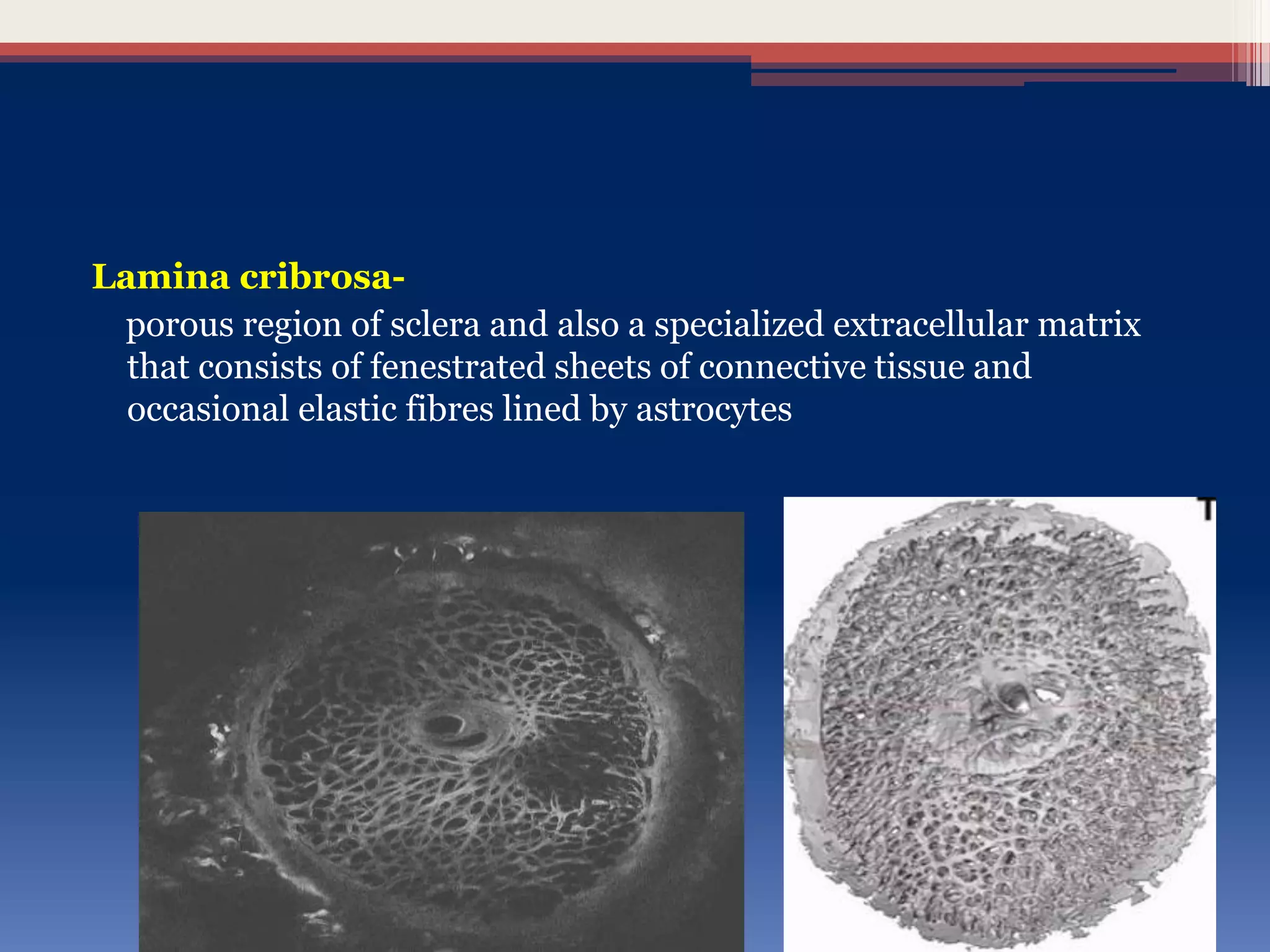
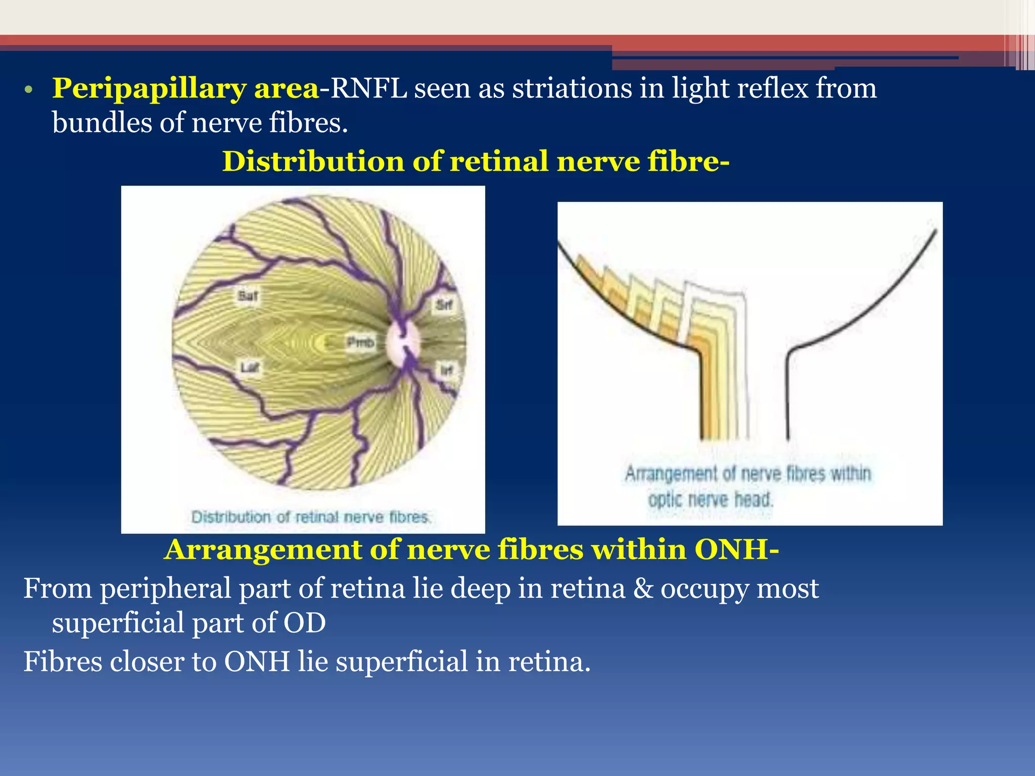
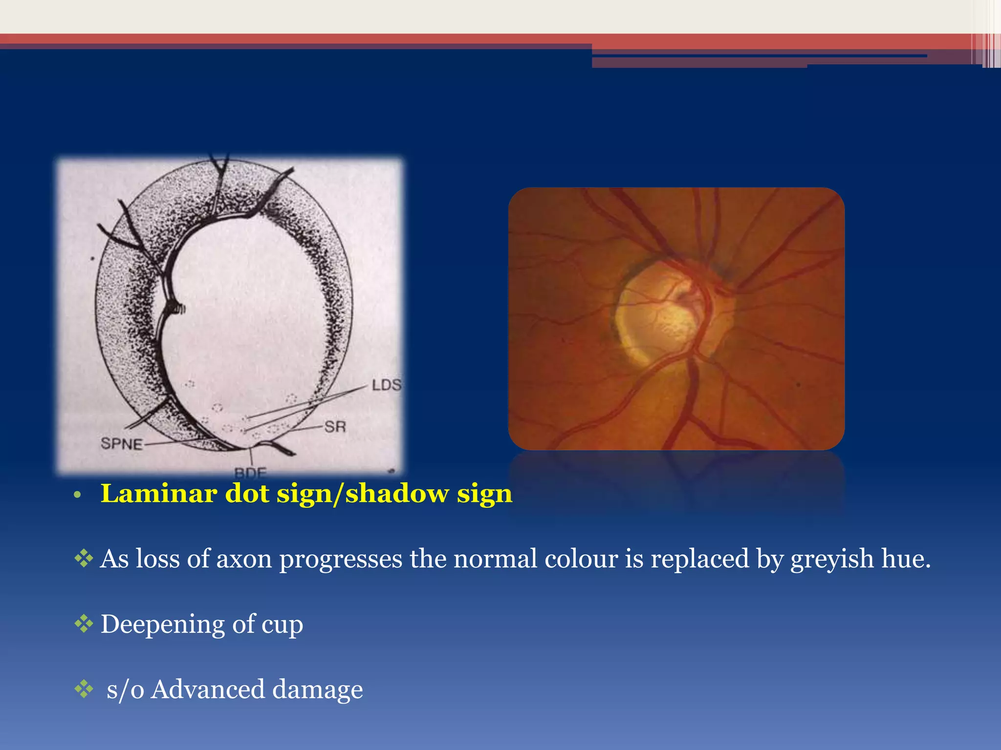
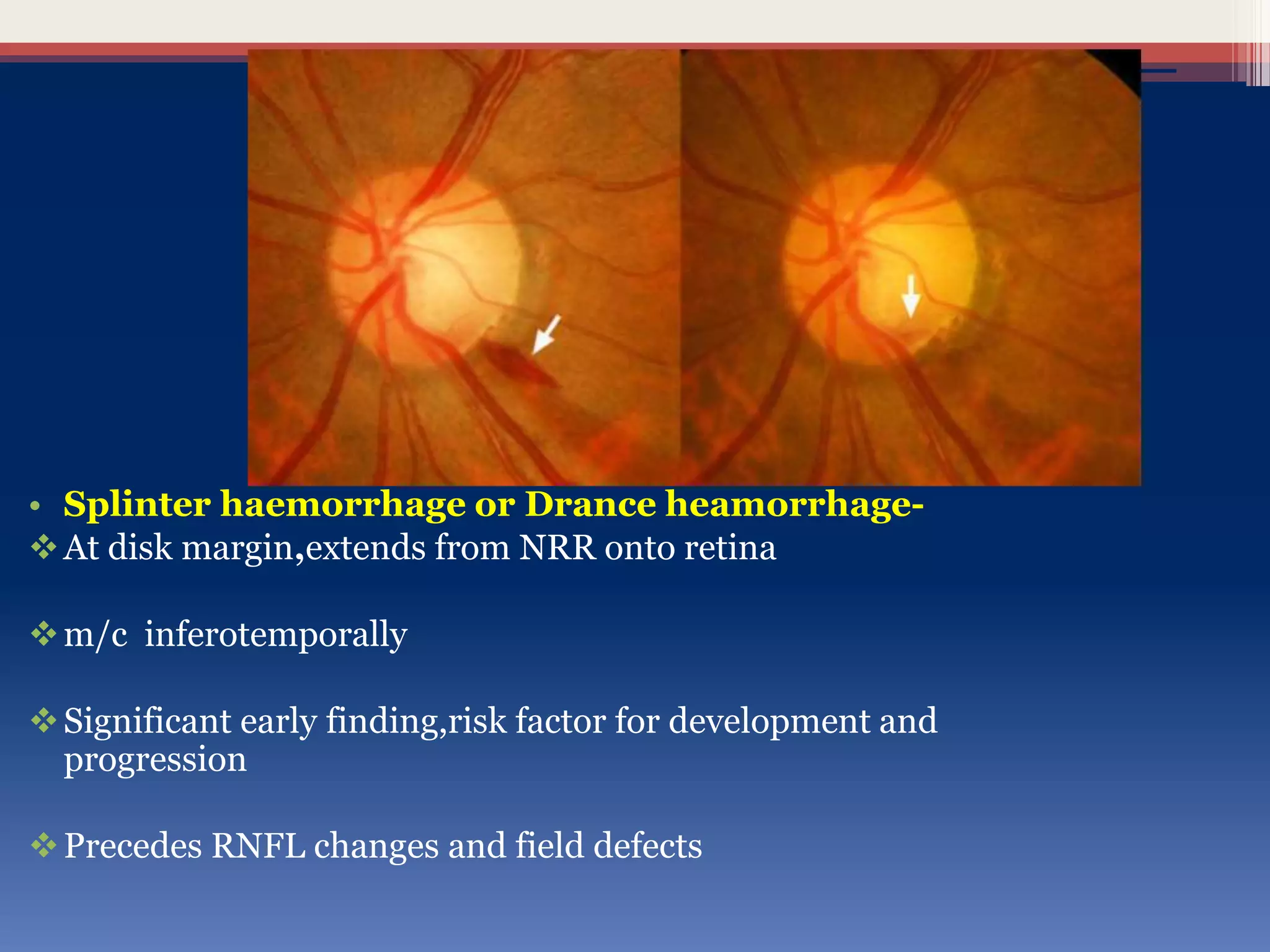
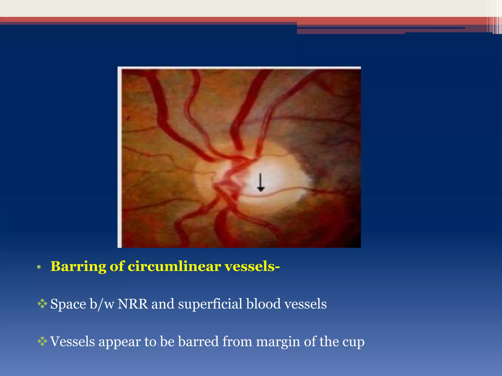
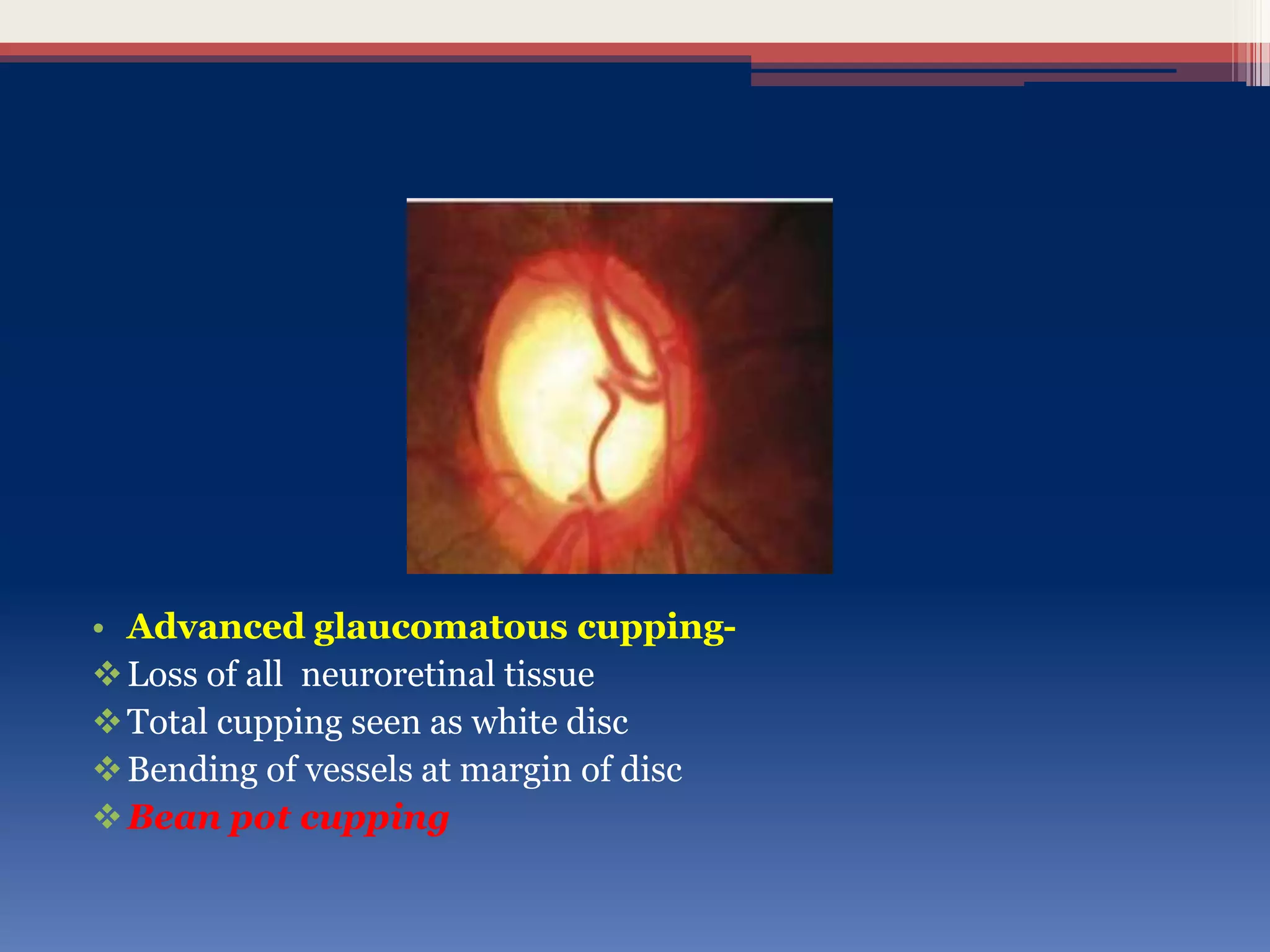
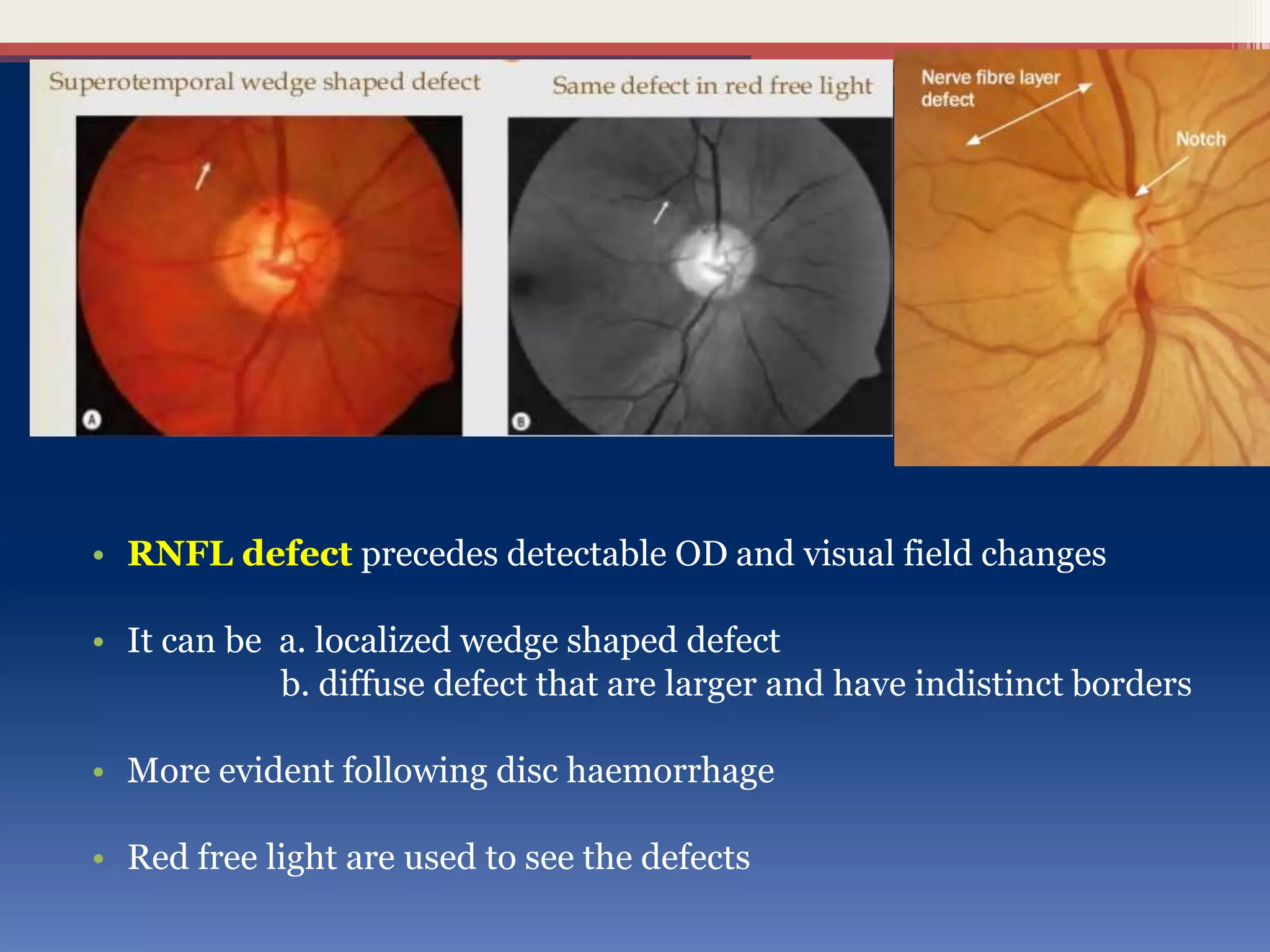
SlideShare a Scribd company logo
GLAUCOMA OPTIC DISC CHANGES
PRESENTED BY-
PRAGATI JAIN
• Chronic progressive optic neuropathy caused by group of ocular
conditions which lead to damage of optic nerve with loss of visual
function.
Definitions
• ONH-distal portion of optic nerve,extends from retinal surface to
myelinated portion of optic nerve.
• Composed of the nerve fibres which originates in the Ganglionic cell
layer of retina & converge upon the nerve head from all points in the
fundus.
• Relation between ONH and glaucoma was first described by von
Graefe in 1857
• Physiological cup-represents partial or complete absence of
axons
Pallor of cup results from exposure of lamina cribrosa and lack of
glial tissue in the centre
• NRR-tissue b/w cup and disc margin
I>S>N>T
• Normal CDR is 0.3 :1
2% have CDR 0.7:1
Lamina cribrosa-
porous region of sclera and also a specialized extracellular matrix
that consists of fenestrated sheets of connective tissue and
occasional elastic fibres lined by astrocytes
• Peripapillary area-RNFL seen as striations in light reflex from
bundles of nerve fibres.
Distribution of retinal nerve fibre-
Arrangement of nerve fibres within ONH-
From peripheral part of retina lie deep in retina & occupy most
superficial part of OD
Fibres closer to ONH lie superficial in retina.
Pathogenesis of ONH changes in glaucoma
• Mechanical effect-raised IOP forces lamina cribrosa backwards
and squeezes nerve fibres within ,disturb axoplasmic flow
• Vascular effect-ischaemic atrophy of nerve fibre
ONH Signs Suggestive Of Glaucoma
• Size of cup in relation to size of ONH-
Small disc with deep cup
Medium disc with deep cup
Meduim disc with large cup
Large disc with large cup
• Asymmetry of cups-
0.2 in both eyes suspicion.
• Vertical enlargement of cup-
Vertical CDR > horizontal CDR
Loss of axons occur more in superior and inferior regions so NRR
thinning at vertical poles
• Focal atrophy and polar notching
Inferotemporal region,sometimes superotemporally
Small discrete defect due to loss of neural rim tissue
• Sharpened nasal margins-
As focal defect enlarges and deepens it may develop sharp nasal
margins adjacent to major blood vessels.
• Sharpened rim-
NRR is lost adjacent to edge of the disc
No visible neural rim is seen
• `
• Bayonetting sign-
double angulation of blood vessel
When retinal vessels cross the sharpened rim,it will bend sharply at
the edge of the disc
• Laminar dot sign/shadow sign
 As loss of axon progresses the normal colour is replaced by greyish hue.
 Deepening of cup
 s/o Advanced damage
• Overpass cupping-
Vessels initially bridge the deepened cup and later collapse into it
• Pallor areas on the disc-
 Kinking of vessels at cup margin
 Colour cup-
extent of central pallor
 Contour cup-
site at which vessel change their contour
 Enlargement of cup may progress ahead of pallor
 Pathognomic of glaucomatous optic atrophy
• Saucerization-
Diffuse shallow cupping extends disc margin with retention of central pale
cup
Focal saucerization –more localized shallow sloping cup
Early s/o glaucoma
• Tinted hollow-
Retention of normal NRR colour in area of focal saucerization
• Splinter haemorrhage or Drance heamorrhage-
At disk margin,extends from NRR onto retina
m/c inferotemporally
Significant early finding,risk factor for development and
progression
Precedes RNFL changes and field defects
• Barring of circumlinear vessels-
Space b/w NRR and superficial blood vessels
Vessels appear to be barred from margin of the cup
• Tortousity of retinal vessels-
As response to chronic ischaemia
• Collaterals b/w 2 veins at the disc may also be seen
• Advanced glaucomatous cupping-
Loss of all neuroretinal tissue
Total cupping seen as white disc
Bending of vessels at margin of disc
Bean pot cupping
• RNFL defect precedes detectable OD and visual field changes
• It can be a. localized wedge shaped defect
b. diffuse defect that are larger and have indistinct borders
• More evident following disc haemorrhage
• Red free light are used to see the defects
• Peripappillary pigmentary disturbances
• Scleral lip/peripappillary halo-even white rim that marks disc
margin,anterior extension of sclera b/w choroid and optic nerve
• Zone beta
• Zone alpha
• Alpha zone-irregular hypo or hyper pigmented zone associated
with chorioretinal thinning.
• Beta zone-represents loss of RPE and choriocapilaries leaving
intact choroidal vasculature.
THANK YOU

More Related Content

What's hot

Anatomy of anterior chamber
Anatomy of anterior chamberAnatomy of anterior chamber
Anatomy of anterior chamber
Dr.Prathibha S
 
Coloboma
ColobomaColoboma
Gonioscopy presentation
Gonioscopy presentationGonioscopy presentation
Gonioscopy presentation
Hira Dahal
 
Anatomy and physiology of cornea
Anatomy and physiology of corneaAnatomy and physiology of cornea
Anatomy and physiology of cornea
SSSIHMS-PG
 
Lens induced glaucoma - DR ARNAV
Lens induced glaucoma - DR ARNAVLens induced glaucoma - DR ARNAV
Lens induced glaucoma - DR ARNAV
DrArnavSaroya
 
Corneal transparency
Corneal transparencyCorneal transparency
Corneal transparency
Hira Dahal
 
Clinical evaluation of optic disc changes
Clinical evaluation of optic disc changesClinical evaluation of optic disc changes
Clinical evaluation of optic disc changes
Dr Saurabh Kushwaha
 
Angle of anterior chamber
Angle of anterior chamberAngle of anterior chamber
Angle of anterior chamber
Kalluri Sahithi Reddy
 
Optic atrophy (b)
Optic atrophy (b)Optic atrophy (b)
Optic atrophy (b)
Muhammad AbdulWahidKarim
 
Rapd
Rapd Rapd
Hfa
HfaHfa
Pediatric cataract
Pediatric cataractPediatric cataract
Pediatric cataract
Dr. Ajay Kumar Singh
 
Amblyopia
AmblyopiaAmblyopia
Amblyopia
Nedhina
 
Keratoplasty
KeratoplastyKeratoplasty
Keratoplasty
Karan Bhatia
 
Blood supply of the optic nerve
Blood supply of the optic nerveBlood supply of the optic nerve
Blood supply of the optic nerve
Dhwanit Khetwani
 
Lenses in ophthalmology
Lenses in ophthalmologyLenses in ophthalmology
Lenses in ophthalmology
Reshma Peter
 
Proptosis
ProptosisProptosis
Proptosis
SSSIHMS-PG
 
Primary open angle glaucoma(POAG)
Primary open angle glaucoma(POAG)Primary open angle glaucoma(POAG)
Primary open angle glaucoma(POAG)
rakshyabasnet1
 

What's hot (20)

Anatomy of anterior chamber
Anatomy of anterior chamberAnatomy of anterior chamber
Anatomy of anterior chamber
 
Coloboma
ColobomaColoboma
Coloboma
 
Gonioscopy presentation
Gonioscopy presentationGonioscopy presentation
Gonioscopy presentation
 
Anatomy and physiology of cornea
Anatomy and physiology of corneaAnatomy and physiology of cornea
Anatomy and physiology of cornea
 
Lens induced glaucoma - DR ARNAV
Lens induced glaucoma - DR ARNAVLens induced glaucoma - DR ARNAV
Lens induced glaucoma - DR ARNAV
 
Corneal transparency
Corneal transparencyCorneal transparency
Corneal transparency
 
Clinical evaluation of optic disc changes
Clinical evaluation of optic disc changesClinical evaluation of optic disc changes
Clinical evaluation of optic disc changes
 
Angle of anterior chamber
Angle of anterior chamberAngle of anterior chamber
Angle of anterior chamber
 
Tear film test
Tear film testTear film test
Tear film test
 
Optic atrophy (b)
Optic atrophy (b)Optic atrophy (b)
Optic atrophy (b)
 
Rapd
Rapd Rapd
Rapd
 
Hfa
HfaHfa
Hfa
 
Pediatric cataract
Pediatric cataractPediatric cataract
Pediatric cataract
 
Amblyopia
AmblyopiaAmblyopia
Amblyopia
 
Keratoplasty
KeratoplastyKeratoplasty
Keratoplasty
 
Anterior uveitis
Anterior uveitisAnterior uveitis
Anterior uveitis
 
Blood supply of the optic nerve
Blood supply of the optic nerveBlood supply of the optic nerve
Blood supply of the optic nerve
 
Lenses in ophthalmology
Lenses in ophthalmologyLenses in ophthalmology
Lenses in ophthalmology
 
Proptosis
ProptosisProptosis
Proptosis
 
Primary open angle glaucoma(POAG)
Primary open angle glaucoma(POAG)Primary open angle glaucoma(POAG)
Primary open angle glaucoma(POAG)
 

Viewers also liked

Optic nerve head evaluation in glaucoma
Optic nerve head evaluation in glaucomaOptic nerve head evaluation in glaucoma
Optic nerve head evaluation in glaucomaDr Laltanpuia Chhangte
 
Primary open angle glaucoma (poag)
Primary open angle glaucoma (poag)Primary open angle glaucoma (poag)
Primary open angle glaucoma (poag)
Om Patel
 
POAG
POAGPOAG
POAG
Lhacha
 
Primary open angle glaucoma
Primary open angle glaucomaPrimary open angle glaucoma
Primary open angle glaucoma
ketan bhardwaj
 
Oct in glaucoma
Oct in glaucomaOct in glaucoma
Oct in glaucoma
Jagdish Dukre
 
IMAGING TECHNIQUES IN GLAUCOMA
IMAGING TECHNIQUES IN GLAUCOMAIMAGING TECHNIQUES IN GLAUCOMA
IMAGING TECHNIQUES IN GLAUCOMA
Laxmi Eye Institute
 
Anatomy of optic nerve and its clinical significance
Anatomy of optic nerve and its clinical significanceAnatomy of optic nerve and its clinical significance
Anatomy of optic nerve and its clinical significance
Pabita Dhungel
 
Disc Damage Likelihod Scale (DDLS)
Disc Damage Likelihod Scale (DDLS)Disc Damage Likelihod Scale (DDLS)
Disc Damage Likelihod Scale (DDLS)
Sumit Kumar
 
Diagnosis of glaucoma
Diagnosis of glaucomaDiagnosis of glaucoma
Diagnosis of glaucoma
Sue Ting Lim
 
Optic nerve
Optic nerveOptic nerve
Optic nerve
Green Green
 
optic nerve head swelling
optic nerve head swellingoptic nerve head swelling
optic nerve head swelling
Jehad Hassan
 
Gonioscopy and optic nerve head evaluation
Gonioscopy and optic nerve head evaluationGonioscopy and optic nerve head evaluation
Gonioscopy and optic nerve head evaluation
Ahmedfaik
 
Revisão OFTALMO
Revisão OFTALMORevisão OFTALMO
Revisão OFTALMO
Pietro de Azevedo
 
Fundoscopia para iniciantes
Fundoscopia para iniciantesFundoscopia para iniciantes
Fundoscopia para iniciantes
Pietro de Azevedo
 
Retina and layers
Retina and layersRetina and layers
Retina and layers
Lhacha
 
Glaucoma
GlaucomaGlaucoma
Glaucoma
enarm
 
Anatomy of optic nerve (Optic Nerve Anatomy), Blood Supply & Clinical Signifi...
Anatomy of optic nerve (Optic Nerve Anatomy), Blood Supply & Clinical Signifi...Anatomy of optic nerve (Optic Nerve Anatomy), Blood Supply & Clinical Signifi...
Anatomy of optic nerve (Optic Nerve Anatomy), Blood Supply & Clinical Signifi...
Bikash Sapkota
 

Viewers also liked (20)

Optic nerve head evaluation in glaucoma
Optic nerve head evaluation in glaucomaOptic nerve head evaluation in glaucoma
Optic nerve head evaluation in glaucoma
 
Primary open angle glaucoma (poag)
Primary open angle glaucoma (poag)Primary open angle glaucoma (poag)
Primary open angle glaucoma (poag)
 
Glaucoma
GlaucomaGlaucoma
Glaucoma
 
POAG
POAGPOAG
POAG
 
Primary open angle glaucoma
Primary open angle glaucomaPrimary open angle glaucoma
Primary open angle glaucoma
 
Oct in glaucoma
Oct in glaucomaOct in glaucoma
Oct in glaucoma
 
IMAGING TECHNIQUES IN GLAUCOMA
IMAGING TECHNIQUES IN GLAUCOMAIMAGING TECHNIQUES IN GLAUCOMA
IMAGING TECHNIQUES IN GLAUCOMA
 
POAG
POAGPOAG
POAG
 
Anatomy of optic nerve and its clinical significance
Anatomy of optic nerve and its clinical significanceAnatomy of optic nerve and its clinical significance
Anatomy of optic nerve and its clinical significance
 
Disc Damage Likelihod Scale (DDLS)
Disc Damage Likelihod Scale (DDLS)Disc Damage Likelihod Scale (DDLS)
Disc Damage Likelihod Scale (DDLS)
 
Oftalmoscopia
Oftalmoscopia  Oftalmoscopia
Oftalmoscopia
 
Diagnosis of glaucoma
Diagnosis of glaucomaDiagnosis of glaucoma
Diagnosis of glaucoma
 
Optic nerve
Optic nerveOptic nerve
Optic nerve
 
optic nerve head swelling
optic nerve head swellingoptic nerve head swelling
optic nerve head swelling
 
Gonioscopy and optic nerve head evaluation
Gonioscopy and optic nerve head evaluationGonioscopy and optic nerve head evaluation
Gonioscopy and optic nerve head evaluation
 
Revisão OFTALMO
Revisão OFTALMORevisão OFTALMO
Revisão OFTALMO
 
Fundoscopia para iniciantes
Fundoscopia para iniciantesFundoscopia para iniciantes
Fundoscopia para iniciantes
 
Retina and layers
Retina and layersRetina and layers
Retina and layers
 
Glaucoma
GlaucomaGlaucoma
Glaucoma
 
Anatomy of optic nerve (Optic Nerve Anatomy), Blood Supply & Clinical Signifi...
Anatomy of optic nerve (Optic Nerve Anatomy), Blood Supply & Clinical Signifi...Anatomy of optic nerve (Optic Nerve Anatomy), Blood Supply & Clinical Signifi...
Anatomy of optic nerve (Optic Nerve Anatomy), Blood Supply & Clinical Signifi...
 

Similar to Glaucoma optic disc changes

Funduscopy evaluation in glaucomatous optic
Funduscopy evaluation in glaucomatous opticFunduscopy evaluation in glaucomatous optic
Funduscopy evaluation in glaucomatous optic
rikhaerina
 
Anatomy of optic nerve
Anatomy of optic nerveAnatomy of optic nerve
Anatomy of optic nerve
Sudheer Kumar
 
Lesions of retina
Lesions of retina Lesions of retina
Lesions of retina
Shruti Laddha
 
22-Optic-Disc-Evaluation-IN-Glaucoma.ppt
22-Optic-Disc-Evaluation-IN-Glaucoma.ppt22-Optic-Disc-Evaluation-IN-Glaucoma.ppt
22-Optic-Disc-Evaluation-IN-Glaucoma.ppt
udayasree30
 
Vascular disorders of retina
Vascular disorders of retinaVascular disorders of retina
Vascular disorders of retina
Haris Khan
 
22 optic-disc-evaluation-in-glaucoma
22 optic-disc-evaluation-in-glaucoma22 optic-disc-evaluation-in-glaucoma
22 optic-disc-evaluation-in-glaucoma
UsamaIqbal18
 
Lesions of retina
Lesions of retinaLesions of retina
Lesions of retina
abhishek ghelani
 
Optic Nerve Head Evaluation.pptx
Optic Nerve Head Evaluation.pptxOptic Nerve Head Evaluation.pptx
Optic Nerve Head Evaluation.pptx
Gariyashee Lahkar
 
Papilledema
Papilledema Papilledema
Papilledema
Ishta Thakur
 
Fundus in Glaucoma
Fundus in GlaucomaFundus in Glaucoma
Fundus in Glaucoma
Bakkiyalakshmi K
 
3 mirror, retinal break.pptx
3 mirror, retinal break.pptx3 mirror, retinal break.pptx
3 mirror, retinal break.pptx
TimothyLiew3
 
Examination of the optic nerve
Examination of the optic nerveExamination of the optic nerve
Examination of the optic nerve
Akshay Nayak
 
Retinal detachment
Retinal detachment Retinal detachment
Retinal detachment
AyushiPatel59
 
Retina for undergraduate students
Retina for undergraduate studentsRetina for undergraduate students
Retina for undergraduate students
faculty of medicine -benha university
 
OCT
OCTOCT
VF in glaucoma.pptx
VF in glaucoma.pptxVF in glaucoma.pptx
VF in glaucoma.pptx
Sheim Elteb
 
Management of retinal detachment
Management of retinal detachmentManagement of retinal detachment
Management of retinal detachment
Amreen Deshmukh
 
Traumatic chorioretinopathies
Traumatic chorioretinopathiesTraumatic chorioretinopathies
Traumatic chorioretinopathies
Shruti Laddha
 
PAPILLEDEMA
PAPILLEDEMAPAPILLEDEMA
PAPILLEDEMA
Nikitha Crasta
 

Similar to Glaucoma optic disc changes (20)

Funduscopy evaluation in glaucomatous optic
Funduscopy evaluation in glaucomatous opticFunduscopy evaluation in glaucomatous optic
Funduscopy evaluation in glaucomatous optic
 
Anatomy of optic nerve
Anatomy of optic nerveAnatomy of optic nerve
Anatomy of optic nerve
 
Lesions of retina
Lesions of retina Lesions of retina
Lesions of retina
 
22-Optic-Disc-Evaluation-IN-Glaucoma.ppt
22-Optic-Disc-Evaluation-IN-Glaucoma.ppt22-Optic-Disc-Evaluation-IN-Glaucoma.ppt
22-Optic-Disc-Evaluation-IN-Glaucoma.ppt
 
Vascular disorders of retina
Vascular disorders of retinaVascular disorders of retina
Vascular disorders of retina
 
22 optic-disc-evaluation-in-glaucoma
22 optic-disc-evaluation-in-glaucoma22 optic-disc-evaluation-in-glaucoma
22 optic-disc-evaluation-in-glaucoma
 
Lesions of retina
Lesions of retinaLesions of retina
Lesions of retina
 
Optic Nerve Head Evaluation.pptx
Optic Nerve Head Evaluation.pptxOptic Nerve Head Evaluation.pptx
Optic Nerve Head Evaluation.pptx
 
Papilledema
Papilledema Papilledema
Papilledema
 
Fundus in Glaucoma
Fundus in GlaucomaFundus in Glaucoma
Fundus in Glaucoma
 
Gluacoma clinical evaluation
Gluacoma clinical evaluationGluacoma clinical evaluation
Gluacoma clinical evaluation
 
3 mirror, retinal break.pptx
3 mirror, retinal break.pptx3 mirror, retinal break.pptx
3 mirror, retinal break.pptx
 
Examination of the optic nerve
Examination of the optic nerveExamination of the optic nerve
Examination of the optic nerve
 
Retinal detachment
Retinal detachment Retinal detachment
Retinal detachment
 
Retina for undergraduate students
Retina for undergraduate studentsRetina for undergraduate students
Retina for undergraduate students
 
OCT
OCTOCT
OCT
 
VF in glaucoma.pptx
VF in glaucoma.pptxVF in glaucoma.pptx
VF in glaucoma.pptx
 
Management of retinal detachment
Management of retinal detachmentManagement of retinal detachment
Management of retinal detachment
 
Traumatic chorioretinopathies
Traumatic chorioretinopathiesTraumatic chorioretinopathies
Traumatic chorioretinopathies
 
PAPILLEDEMA
PAPILLEDEMAPAPILLEDEMA
PAPILLEDEMA
 

Recently uploaded

ANATOMY AND PHYSIOLOGY OF URINARY SYSTEM.pptx
ANATOMY AND PHYSIOLOGY OF URINARY SYSTEM.pptxANATOMY AND PHYSIOLOGY OF URINARY SYSTEM.pptx
ANATOMY AND PHYSIOLOGY OF URINARY SYSTEM.pptx
Swetaba Besh
 
BRACHYTHERAPY OVERVIEW AND APPLICATORS
BRACHYTHERAPY OVERVIEW  AND  APPLICATORSBRACHYTHERAPY OVERVIEW  AND  APPLICATORS
BRACHYTHERAPY OVERVIEW AND APPLICATORS
Krishan Murari
 
MANAGEMENT OF ATRIOVENTRICULAR CONDUCTION BLOCK.pdf
MANAGEMENT OF ATRIOVENTRICULAR CONDUCTION BLOCK.pdfMANAGEMENT OF ATRIOVENTRICULAR CONDUCTION BLOCK.pdf
MANAGEMENT OF ATRIOVENTRICULAR CONDUCTION BLOCK.pdf
Jim Jacob Roy
 
Report Back from SGO 2024: What’s the Latest in Cervical Cancer?
Report Back from SGO 2024: What’s the Latest in Cervical Cancer?Report Back from SGO 2024: What’s the Latest in Cervical Cancer?
Report Back from SGO 2024: What’s the Latest in Cervical Cancer?
bkling
 
Prix Galien International 2024 Forum Program
Prix Galien International 2024 Forum ProgramPrix Galien International 2024 Forum Program
Prix Galien International 2024 Forum Program
Levi Shapiro
 
Antiulcer drugs Advance Pharmacology .pptx
Antiulcer drugs Advance Pharmacology .pptxAntiulcer drugs Advance Pharmacology .pptx
Antiulcer drugs Advance Pharmacology .pptx
Rohit chaurpagar
 
The Normal Electrocardiogram - Part I of II
The Normal Electrocardiogram - Part I of IIThe Normal Electrocardiogram - Part I of II
The Normal Electrocardiogram - Part I of II
MedicoseAcademics
 
Triangles of Neck and Clinical Correlation by Dr. RIG.pptx
Triangles of Neck and Clinical Correlation by Dr. RIG.pptxTriangles of Neck and Clinical Correlation by Dr. RIG.pptx
Triangles of Neck and Clinical Correlation by Dr. RIG.pptx
Dr. Rabia Inam Gandapore
 
Are There Any Natural Remedies To Treat Syphilis.pdf
Are There Any Natural Remedies To Treat Syphilis.pdfAre There Any Natural Remedies To Treat Syphilis.pdf
Are There Any Natural Remedies To Treat Syphilis.pdf
Little Cross Family Clinic
 
New Directions in Targeted Therapeutic Approaches for Older Adults With Mantl...
New Directions in Targeted Therapeutic Approaches for Older Adults With Mantl...New Directions in Targeted Therapeutic Approaches for Older Adults With Mantl...
New Directions in Targeted Therapeutic Approaches for Older Adults With Mantl...
i3 Health
 
Surat @ℂall @Girls ꧁❤8527049040❤꧂@ℂall @Girls Service Vip Top Model Safe
Surat @ℂall @Girls ꧁❤8527049040❤꧂@ℂall @Girls Service Vip Top Model SafeSurat @ℂall @Girls ꧁❤8527049040❤꧂@ℂall @Girls Service Vip Top Model Safe
Surat @ℂall @Girls ꧁❤8527049040❤꧂@ℂall @Girls Service Vip Top Model Safe
Savita Shen $i11
 
24 Upakrama.pptx class ppt useful in all
24 Upakrama.pptx class ppt useful in all24 Upakrama.pptx class ppt useful in all
24 Upakrama.pptx class ppt useful in all
DrSathishMS1
 
KDIGO 2024 guidelines for diabetologists
KDIGO 2024 guidelines for diabetologistsKDIGO 2024 guidelines for diabetologists
KDIGO 2024 guidelines for diabetologists
د.محمود نجيب
 
Couples presenting to the infertility clinic- Do they really have infertility...
Couples presenting to the infertility clinic- Do they really have infertility...Couples presenting to the infertility clinic- Do they really have infertility...
Couples presenting to the infertility clinic- Do they really have infertility...
Sujoy Dasgupta
 
Maxilla, Mandible & Hyoid Bone & Clinical Correlations by Dr. RIG.pptx
Maxilla, Mandible & Hyoid Bone & Clinical Correlations by Dr. RIG.pptxMaxilla, Mandible & Hyoid Bone & Clinical Correlations by Dr. RIG.pptx
Maxilla, Mandible & Hyoid Bone & Clinical Correlations by Dr. RIG.pptx
Dr. Rabia Inam Gandapore
 
For Better Surat #ℂall #Girl Service ❤85270-49040❤ Surat #ℂall #Girls
For Better Surat #ℂall #Girl Service ❤85270-49040❤ Surat #ℂall #GirlsFor Better Surat #ℂall #Girl Service ❤85270-49040❤ Surat #ℂall #Girls
For Better Surat #ℂall #Girl Service ❤85270-49040❤ Surat #ℂall #Girls
Savita Shen $i11
 
Alcohol_Dr. Jeenal Mistry MD Pharmacology.pdf
Alcohol_Dr. Jeenal Mistry MD Pharmacology.pdfAlcohol_Dr. Jeenal Mistry MD Pharmacology.pdf
Alcohol_Dr. Jeenal Mistry MD Pharmacology.pdf
Dr Jeenal Mistry
 
The POPPY STUDY (Preconception to post-partum cardiovascular function in prim...
The POPPY STUDY (Preconception to post-partum cardiovascular function in prim...The POPPY STUDY (Preconception to post-partum cardiovascular function in prim...
The POPPY STUDY (Preconception to post-partum cardiovascular function in prim...
Catherine Liao
 
Ocular injury ppt Upendra pal optometrist upums saifai etawah
Ocular injury  ppt  Upendra pal  optometrist upums saifai etawahOcular injury  ppt  Upendra pal  optometrist upums saifai etawah
Ocular injury ppt Upendra pal optometrist upums saifai etawah
pal078100
 
HOT NEW PRODUCT! BIG SALES FAST SHIPPING NOW FROM CHINA!! EU KU DB BK substit...
HOT NEW PRODUCT! BIG SALES FAST SHIPPING NOW FROM CHINA!! EU KU DB BK substit...HOT NEW PRODUCT! BIG SALES FAST SHIPPING NOW FROM CHINA!! EU KU DB BK substit...
HOT NEW PRODUCT! BIG SALES FAST SHIPPING NOW FROM CHINA!! EU KU DB BK substit...
GL Anaacs
 

Recently uploaded (20)

ANATOMY AND PHYSIOLOGY OF URINARY SYSTEM.pptx
ANATOMY AND PHYSIOLOGY OF URINARY SYSTEM.pptxANATOMY AND PHYSIOLOGY OF URINARY SYSTEM.pptx
ANATOMY AND PHYSIOLOGY OF URINARY SYSTEM.pptx
 
BRACHYTHERAPY OVERVIEW AND APPLICATORS
BRACHYTHERAPY OVERVIEW  AND  APPLICATORSBRACHYTHERAPY OVERVIEW  AND  APPLICATORS
BRACHYTHERAPY OVERVIEW AND APPLICATORS
 
MANAGEMENT OF ATRIOVENTRICULAR CONDUCTION BLOCK.pdf
MANAGEMENT OF ATRIOVENTRICULAR CONDUCTION BLOCK.pdfMANAGEMENT OF ATRIOVENTRICULAR CONDUCTION BLOCK.pdf
MANAGEMENT OF ATRIOVENTRICULAR CONDUCTION BLOCK.pdf
 
Report Back from SGO 2024: What’s the Latest in Cervical Cancer?
Report Back from SGO 2024: What’s the Latest in Cervical Cancer?Report Back from SGO 2024: What’s the Latest in Cervical Cancer?
Report Back from SGO 2024: What’s the Latest in Cervical Cancer?
 
Prix Galien International 2024 Forum Program
Prix Galien International 2024 Forum ProgramPrix Galien International 2024 Forum Program
Prix Galien International 2024 Forum Program
 
Antiulcer drugs Advance Pharmacology .pptx
Antiulcer drugs Advance Pharmacology .pptxAntiulcer drugs Advance Pharmacology .pptx
Antiulcer drugs Advance Pharmacology .pptx
 
The Normal Electrocardiogram - Part I of II
The Normal Electrocardiogram - Part I of IIThe Normal Electrocardiogram - Part I of II
The Normal Electrocardiogram - Part I of II
 
Triangles of Neck and Clinical Correlation by Dr. RIG.pptx
Triangles of Neck and Clinical Correlation by Dr. RIG.pptxTriangles of Neck and Clinical Correlation by Dr. RIG.pptx
Triangles of Neck and Clinical Correlation by Dr. RIG.pptx
 
Are There Any Natural Remedies To Treat Syphilis.pdf
Are There Any Natural Remedies To Treat Syphilis.pdfAre There Any Natural Remedies To Treat Syphilis.pdf
Are There Any Natural Remedies To Treat Syphilis.pdf
 
New Directions in Targeted Therapeutic Approaches for Older Adults With Mantl...
New Directions in Targeted Therapeutic Approaches for Older Adults With Mantl...New Directions in Targeted Therapeutic Approaches for Older Adults With Mantl...
New Directions in Targeted Therapeutic Approaches for Older Adults With Mantl...
 
Surat @ℂall @Girls ꧁❤8527049040❤꧂@ℂall @Girls Service Vip Top Model Safe
Surat @ℂall @Girls ꧁❤8527049040❤꧂@ℂall @Girls Service Vip Top Model SafeSurat @ℂall @Girls ꧁❤8527049040❤꧂@ℂall @Girls Service Vip Top Model Safe
Surat @ℂall @Girls ꧁❤8527049040❤꧂@ℂall @Girls Service Vip Top Model Safe
 
24 Upakrama.pptx class ppt useful in all
24 Upakrama.pptx class ppt useful in all24 Upakrama.pptx class ppt useful in all
24 Upakrama.pptx class ppt useful in all
 
KDIGO 2024 guidelines for diabetologists
KDIGO 2024 guidelines for diabetologistsKDIGO 2024 guidelines for diabetologists
KDIGO 2024 guidelines for diabetologists
 
Couples presenting to the infertility clinic- Do they really have infertility...
Couples presenting to the infertility clinic- Do they really have infertility...Couples presenting to the infertility clinic- Do they really have infertility...
Couples presenting to the infertility clinic- Do they really have infertility...
 
Maxilla, Mandible & Hyoid Bone & Clinical Correlations by Dr. RIG.pptx
Maxilla, Mandible & Hyoid Bone & Clinical Correlations by Dr. RIG.pptxMaxilla, Mandible & Hyoid Bone & Clinical Correlations by Dr. RIG.pptx
Maxilla, Mandible & Hyoid Bone & Clinical Correlations by Dr. RIG.pptx
 
For Better Surat #ℂall #Girl Service ❤85270-49040❤ Surat #ℂall #Girls
For Better Surat #ℂall #Girl Service ❤85270-49040❤ Surat #ℂall #GirlsFor Better Surat #ℂall #Girl Service ❤85270-49040❤ Surat #ℂall #Girls
For Better Surat #ℂall #Girl Service ❤85270-49040❤ Surat #ℂall #Girls
 
Alcohol_Dr. Jeenal Mistry MD Pharmacology.pdf
Alcohol_Dr. Jeenal Mistry MD Pharmacology.pdfAlcohol_Dr. Jeenal Mistry MD Pharmacology.pdf
Alcohol_Dr. Jeenal Mistry MD Pharmacology.pdf
 
The POPPY STUDY (Preconception to post-partum cardiovascular function in prim...
The POPPY STUDY (Preconception to post-partum cardiovascular function in prim...The POPPY STUDY (Preconception to post-partum cardiovascular function in prim...
The POPPY STUDY (Preconception to post-partum cardiovascular function in prim...
 
Ocular injury ppt Upendra pal optometrist upums saifai etawah
Ocular injury  ppt  Upendra pal  optometrist upums saifai etawahOcular injury  ppt  Upendra pal  optometrist upums saifai etawah
Ocular injury ppt Upendra pal optometrist upums saifai etawah
 
HOT NEW PRODUCT! BIG SALES FAST SHIPPING NOW FROM CHINA!! EU KU DB BK substit...
HOT NEW PRODUCT! BIG SALES FAST SHIPPING NOW FROM CHINA!! EU KU DB BK substit...HOT NEW PRODUCT! BIG SALES FAST SHIPPING NOW FROM CHINA!! EU KU DB BK substit...
HOT NEW PRODUCT! BIG SALES FAST SHIPPING NOW FROM CHINA!! EU KU DB BK substit...
 

Glaucoma optic disc changes

  • 1. GLAUCOMA OPTIC DISC CHANGES PRESENTED BY- PRAGATI JAIN
  • 2. • Chronic progressive optic neuropathy caused by group of ocular conditions which lead to damage of optic nerve with loss of visual function.
  • 3. Definitions • ONH-distal portion of optic nerve,extends from retinal surface to myelinated portion of optic nerve. • Composed of the nerve fibres which originates in the Ganglionic cell layer of retina & converge upon the nerve head from all points in the fundus. • Relation between ONH and glaucoma was first described by von Graefe in 1857
  • 4. • Physiological cup-represents partial or complete absence of axons Pallor of cup results from exposure of lamina cribrosa and lack of glial tissue in the centre • NRR-tissue b/w cup and disc margin I>S>N>T • Normal CDR is 0.3 :1 2% have CDR 0.7:1
  • 5. Lamina cribrosa- porous region of sclera and also a specialized extracellular matrix that consists of fenestrated sheets of connective tissue and occasional elastic fibres lined by astrocytes
  • 6. • Peripapillary area-RNFL seen as striations in light reflex from bundles of nerve fibres. Distribution of retinal nerve fibre- Arrangement of nerve fibres within ONH- From peripheral part of retina lie deep in retina & occupy most superficial part of OD Fibres closer to ONH lie superficial in retina.
  • 7. Pathogenesis of ONH changes in glaucoma • Mechanical effect-raised IOP forces lamina cribrosa backwards and squeezes nerve fibres within ,disturb axoplasmic flow • Vascular effect-ischaemic atrophy of nerve fibre
  • 8. ONH Signs Suggestive Of Glaucoma • Size of cup in relation to size of ONH- Small disc with deep cup Medium disc with deep cup Meduim disc with large cup Large disc with large cup
  • 9. • Asymmetry of cups- 0.2 in both eyes suspicion.
  • 10. • Vertical enlargement of cup- Vertical CDR > horizontal CDR Loss of axons occur more in superior and inferior regions so NRR thinning at vertical poles
  • 11. • Focal atrophy and polar notching Inferotemporal region,sometimes superotemporally Small discrete defect due to loss of neural rim tissue
  • 12. • Sharpened nasal margins- As focal defect enlarges and deepens it may develop sharp nasal margins adjacent to major blood vessels.
  • 13. • Sharpened rim- NRR is lost adjacent to edge of the disc No visible neural rim is seen
  • 14. • ` • Bayonetting sign- double angulation of blood vessel When retinal vessels cross the sharpened rim,it will bend sharply at the edge of the disc
  • 15. • Laminar dot sign/shadow sign  As loss of axon progresses the normal colour is replaced by greyish hue.  Deepening of cup  s/o Advanced damage
  • 16. • Overpass cupping- Vessels initially bridge the deepened cup and later collapse into it
  • 17. • Pallor areas on the disc-  Kinking of vessels at cup margin  Colour cup- extent of central pallor  Contour cup- site at which vessel change their contour  Enlargement of cup may progress ahead of pallor  Pathognomic of glaucomatous optic atrophy
  • 18. • Saucerization- Diffuse shallow cupping extends disc margin with retention of central pale cup Focal saucerization –more localized shallow sloping cup Early s/o glaucoma • Tinted hollow- Retention of normal NRR colour in area of focal saucerization
  • 19. • Splinter haemorrhage or Drance heamorrhage- At disk margin,extends from NRR onto retina m/c inferotemporally Significant early finding,risk factor for development and progression Precedes RNFL changes and field defects
  • 20. • Barring of circumlinear vessels- Space b/w NRR and superficial blood vessels Vessels appear to be barred from margin of the cup
  • 21. • Tortousity of retinal vessels- As response to chronic ischaemia • Collaterals b/w 2 veins at the disc may also be seen
  • 22. • Advanced glaucomatous cupping- Loss of all neuroretinal tissue Total cupping seen as white disc Bending of vessels at margin of disc Bean pot cupping
  • 23. • RNFL defect precedes detectable OD and visual field changes • It can be a. localized wedge shaped defect b. diffuse defect that are larger and have indistinct borders • More evident following disc haemorrhage • Red free light are used to see the defects
  • 24. • Peripappillary pigmentary disturbances • Scleral lip/peripappillary halo-even white rim that marks disc margin,anterior extension of sclera b/w choroid and optic nerve • Zone beta • Zone alpha
  • 25. • Alpha zone-irregular hypo or hyper pigmented zone associated with chorioretinal thinning. • Beta zone-represents loss of RPE and choriocapilaries leaving intact choroidal vasculature.