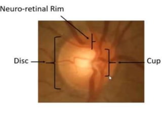
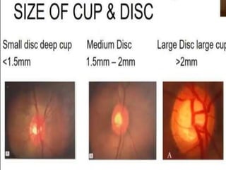
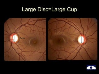
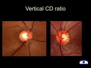
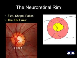
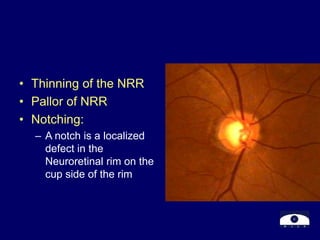
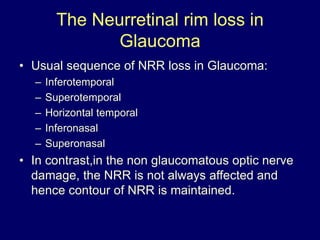
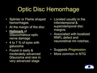
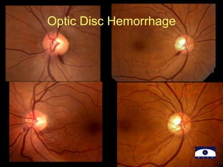
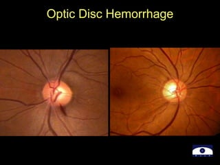
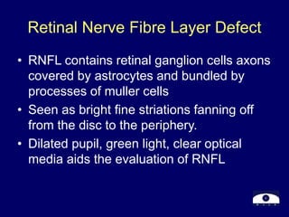
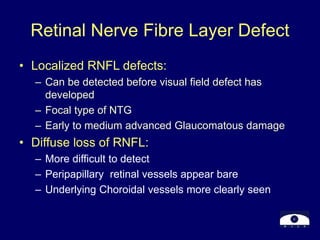
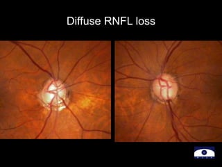
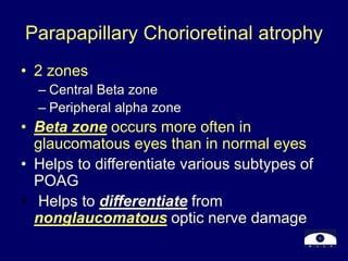
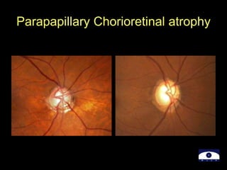
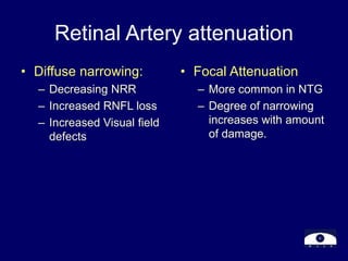
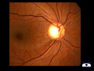
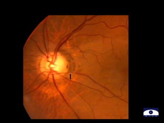
Optic Disc Evaluation
By
Dr.Nirupama
Glaucoma =Optic neuropathy
• The evaluation of the appearance of the
optic disc is central to the diagnosis and
management of Glaucoma
• Optic Disc Evaluation:
– Why..?
– How..?
– What to look for..?
The 4 goals of optic disc evaluation
• Distinguishing between the healthy and
the sick -Diagnosing.
• Quantifying the amount of damage:
Healthy,Mild,Moderate,Advanced Disease
• Monitoring Change,for better or for worse
• Quantitating the rate of change
Optic Disc Evaluation ..
How?
• Slit lamp biomicroscopy : Ideal
– Stereoscopic View –Cupping
– Measuring the optic disc size
• Direct Ophthalmoscopy
– Good Magnification
• Indirect Ophthalmoscopy
– Overall View
• Optic disc Photoghraphy.
– Documentation,Monitoring for progression.
The 7 parameters to look for…
what ?
1)Disc: Size and Shape
2)Neuroretinal Rim (NRR):
 Size,Shape,Pallor
 ISNT rule
3)Cup: Size and Shape in relation to the optic disc
size,
-Vertical C/ D Ratio, Cup depth / Excavation
4) Optic Disc Hemorrhage: Presence & Location
The 7 parameters to look for…
5) Nerve Fibre Layer Defect:
 focal & diffuse
6) Para Papillary Atrophy;
 Size,location & Configuration
7) Retinal Arterial Attenuation:
 focal & diffuse
1)Optic Disc: Size & Shape
• Determining the size of the disc =Crucial
– Helps to differentiate Physiological cupping from
Pathological.
• Large discs have big physiological cups.
• Small Discs have small cups or no cups
• Measurement of Vertical Disc diameter :
– Length of the vertical beam of slit lamp light
– Multiplied by correction factor of the condensing lens
– Volk 60 D= X 1
– Volk 90D=X 1.3
– Volk 78D= X1.1
Cup: Size, Shape, location in
relation to the disc size
• Optic Cup= Excavation in the optic nerve head
– Stereoscopic evaluation
• In normal eyes= Areas of optic disc & Optic cup
are corelated
• Large optic discs=Large cup
• Small optic disc =Small cup or no cup
• Early & moderate glaucomatous damage in
small disc may be missed because of the
erroneously low cup disc ratios
Large Disc=Large Cup
3mm
Early & moderate glaucomatous damage in small disc may
be missed because of the erroneously low cup disc ratios
Vertical Cup Disc Ratio
• Vertically oval optic disc
• Horizontally oval optic cup
• In normal eyes: Horizontal CD ratio > than
vertical CD ratio
• In Glaucomatous eyes: Vertical CD ratio >
than the horizontal CD ratio
Vertical CD ratio
The Neuroretinal Rim
• Size, Shape, Pallor.
• The ISNT rule:
I>S>N>T
• Thinning of the NRR
• Pallor of NRR
• Notching:
– A notch is a localized
defect in the
Neuroretinal rim on the
cup side of the rim
The Neurretinal rim loss in
Glaucoma
• Usual sequence of NRR loss in Glaucoma:
– Inferotemporal
– Superotemporal
– Horizontal temporal
– Inferonasal
– Superonasal
• In contrast,in the non glaucomatous optic nerve
damage, the NRR is not always affected and
hence contour of NRR is maintained.
NRR , the “ISNT Rule”
I>S>N>T
I>S>N>T
Optic Disc Hemorrhage
• Splinter or Flame shaped
hemorrhages
• At the margin of the disc
• Hallmark of
Glaucomatous optic
nerve damage
• 4 to 7 % of eyes with
galucoma
• Found in early &
moderately advanced
Glaucoma and rare in
very advanced stage
• Located usually in the
inferotemporal &
superotemporal disc
margins
• Associated with localized
RNFL defect and
neuroretinal rim notches .
• Suggests Progression.
• More common in NTG
Optic Disc Hemorrhage
Retinal Nerve Fibre Layer Defect
• RNFL contains retinal ganglion cells axons
covered by astrocytes and bundled by
processes of muller cells
• Seen as bright fine striations fanning off
from the disc to the periphery.
• Dilated pupil, green light, clear optical
media aids the evaluation of RNFL
Retinal Nerve Fibre Layer Defect
• Localized RNFL defects:
– Can be detected before visual field defect has
developed
– Focal type of NTG
– Early to medium advanced Glaucomatous damage
• Diffuse loss of RNFL:
– More difficult to detect
– Peripapillary retinal vessels appear bare
– Underlying Choroidal vessels more clearly seen
.
Diffuse RNFL loss
Parapapillary Chorioretinal atrophy
• 2 zones
– Central Beta zone
– Peripheral alpha zone
• Beta zone occurs more often in
glaucomatous eyes than in normal eyes.
• Helps to differentiate various subtypes of
POAG
• Helps to differentiate from
nonglaucomatous optic nerve damage
Parapapillary Chorioretinal atrophy
Retinal Artery attenuation
• Diffuse narrowing:
– Decreasing NRR
– Increased RNFL loss
– Increased Visual field
defects
• Focal Attenuation
– More common in NTG
– Degree of narrowing
increases with amount
of damage.
Diffuse Retinal arterial attenuation
Disc Damage Likelihood Scale
Optic Disc Photographs
Optic Disc Drawings
Documentation of disc
damage:
Monitoring change for
progression
Rate of change
Thank You

22-Optic-Disc-Evaluation-IN-Glaucoma.ppt

  • 1.
  • 2.
    Glaucoma =Optic neuropathy •The evaluation of the appearance of the optic disc is central to the diagnosis and management of Glaucoma • Optic Disc Evaluation: – Why..? – How..? – What to look for..?
  • 4.
    The 4 goalsof optic disc evaluation • Distinguishing between the healthy and the sick -Diagnosing. • Quantifying the amount of damage: Healthy,Mild,Moderate,Advanced Disease • Monitoring Change,for better or for worse • Quantitating the rate of change
  • 5.
    Optic Disc Evaluation.. How? • Slit lamp biomicroscopy : Ideal – Stereoscopic View –Cupping – Measuring the optic disc size • Direct Ophthalmoscopy – Good Magnification • Indirect Ophthalmoscopy – Overall View • Optic disc Photoghraphy. – Documentation,Monitoring for progression.
  • 6.
    The 7 parametersto look for… what ? 1)Disc: Size and Shape 2)Neuroretinal Rim (NRR):  Size,Shape,Pallor  ISNT rule 3)Cup: Size and Shape in relation to the optic disc size, -Vertical C/ D Ratio, Cup depth / Excavation 4) Optic Disc Hemorrhage: Presence & Location
  • 7.
    The 7 parametersto look for… 5) Nerve Fibre Layer Defect:  focal & diffuse 6) Para Papillary Atrophy;  Size,location & Configuration 7) Retinal Arterial Attenuation:  focal & diffuse
  • 8.
    1)Optic Disc: Size& Shape • Determining the size of the disc =Crucial – Helps to differentiate Physiological cupping from Pathological. • Large discs have big physiological cups. • Small Discs have small cups or no cups • Measurement of Vertical Disc diameter : – Length of the vertical beam of slit lamp light – Multiplied by correction factor of the condensing lens – Volk 60 D= X 1 – Volk 90D=X 1.3 – Volk 78D= X1.1
  • 10.
    Cup: Size, Shape,location in relation to the disc size • Optic Cup= Excavation in the optic nerve head – Stereoscopic evaluation • In normal eyes= Areas of optic disc & Optic cup are corelated • Large optic discs=Large cup • Small optic disc =Small cup or no cup • Early & moderate glaucomatous damage in small disc may be missed because of the erroneously low cup disc ratios
  • 12.
  • 13.
    Early & moderateglaucomatous damage in small disc may be missed because of the erroneously low cup disc ratios
  • 14.
    Vertical Cup DiscRatio • Vertically oval optic disc • Horizontally oval optic cup • In normal eyes: Horizontal CD ratio > than vertical CD ratio • In Glaucomatous eyes: Vertical CD ratio > than the horizontal CD ratio
  • 15.
  • 16.
    The Neuroretinal Rim •Size, Shape, Pallor. • The ISNT rule: I>S>N>T
  • 17.
    • Thinning ofthe NRR • Pallor of NRR • Notching: – A notch is a localized defect in the Neuroretinal rim on the cup side of the rim
  • 18.
    The Neurretinal rimloss in Glaucoma • Usual sequence of NRR loss in Glaucoma: – Inferotemporal – Superotemporal – Horizontal temporal – Inferonasal – Superonasal • In contrast,in the non glaucomatous optic nerve damage, the NRR is not always affected and hence contour of NRR is maintained.
  • 19.
    NRR , the“ISNT Rule” I>S>N>T I>S>N>T
  • 20.
    Optic Disc Hemorrhage •Splinter or Flame shaped hemorrhages • At the margin of the disc • Hallmark of Glaucomatous optic nerve damage • 4 to 7 % of eyes with galucoma • Found in early & moderately advanced Glaucoma and rare in very advanced stage • Located usually in the inferotemporal & superotemporal disc margins • Associated with localized RNFL defect and neuroretinal rim notches . • Suggests Progression. • More common in NTG
  • 21.
  • 22.
  • 23.
    Retinal Nerve FibreLayer Defect • RNFL contains retinal ganglion cells axons covered by astrocytes and bundled by processes of muller cells • Seen as bright fine striations fanning off from the disc to the periphery. • Dilated pupil, green light, clear optical media aids the evaluation of RNFL
  • 24.
    Retinal Nerve FibreLayer Defect • Localized RNFL defects: – Can be detected before visual field defect has developed – Focal type of NTG – Early to medium advanced Glaucomatous damage • Diffuse loss of RNFL: – More difficult to detect – Peripapillary retinal vessels appear bare – Underlying Choroidal vessels more clearly seen .
  • 26.
  • 27.
    Parapapillary Chorioretinal atrophy •2 zones – Central Beta zone – Peripheral alpha zone • Beta zone occurs more often in glaucomatous eyes than in normal eyes. • Helps to differentiate various subtypes of POAG • Helps to differentiate from nonglaucomatous optic nerve damage
  • 28.
  • 30.
    Retinal Artery attenuation •Diffuse narrowing: – Decreasing NRR – Increased RNFL loss – Increased Visual field defects • Focal Attenuation – More common in NTG – Degree of narrowing increases with amount of damage.
  • 33.
  • 34.
  • 35.
    Optic Disc Photographs OpticDisc Drawings Documentation of disc damage: Monitoring change for progression Rate of change
  • 36.