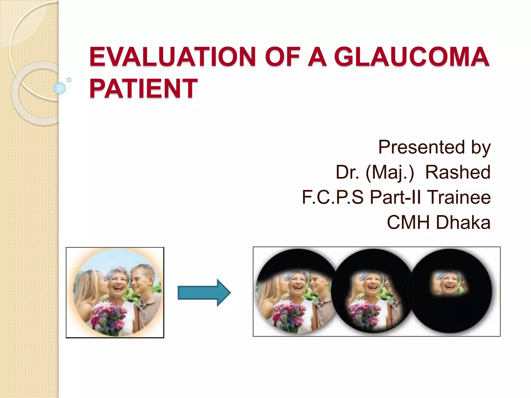
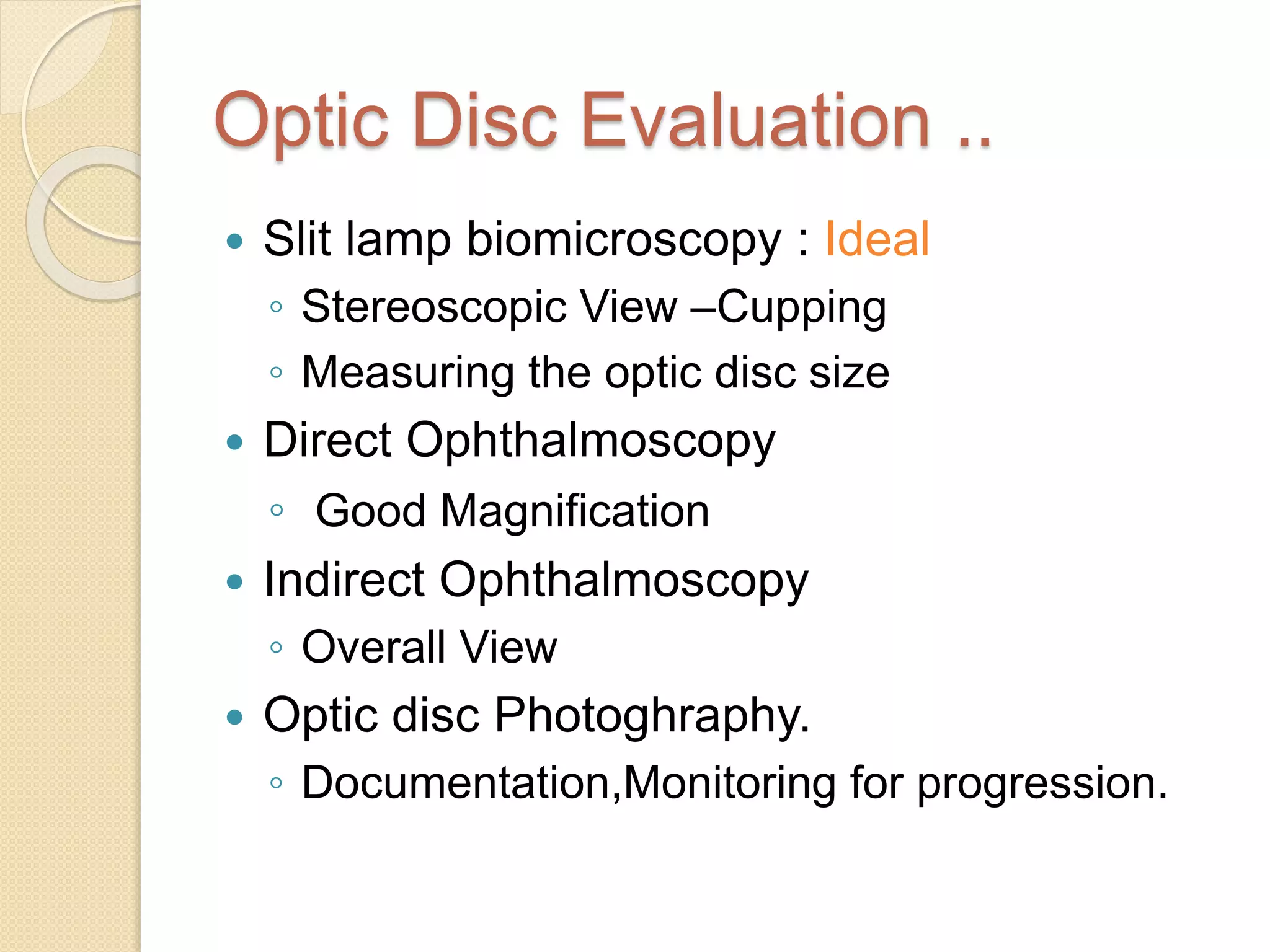
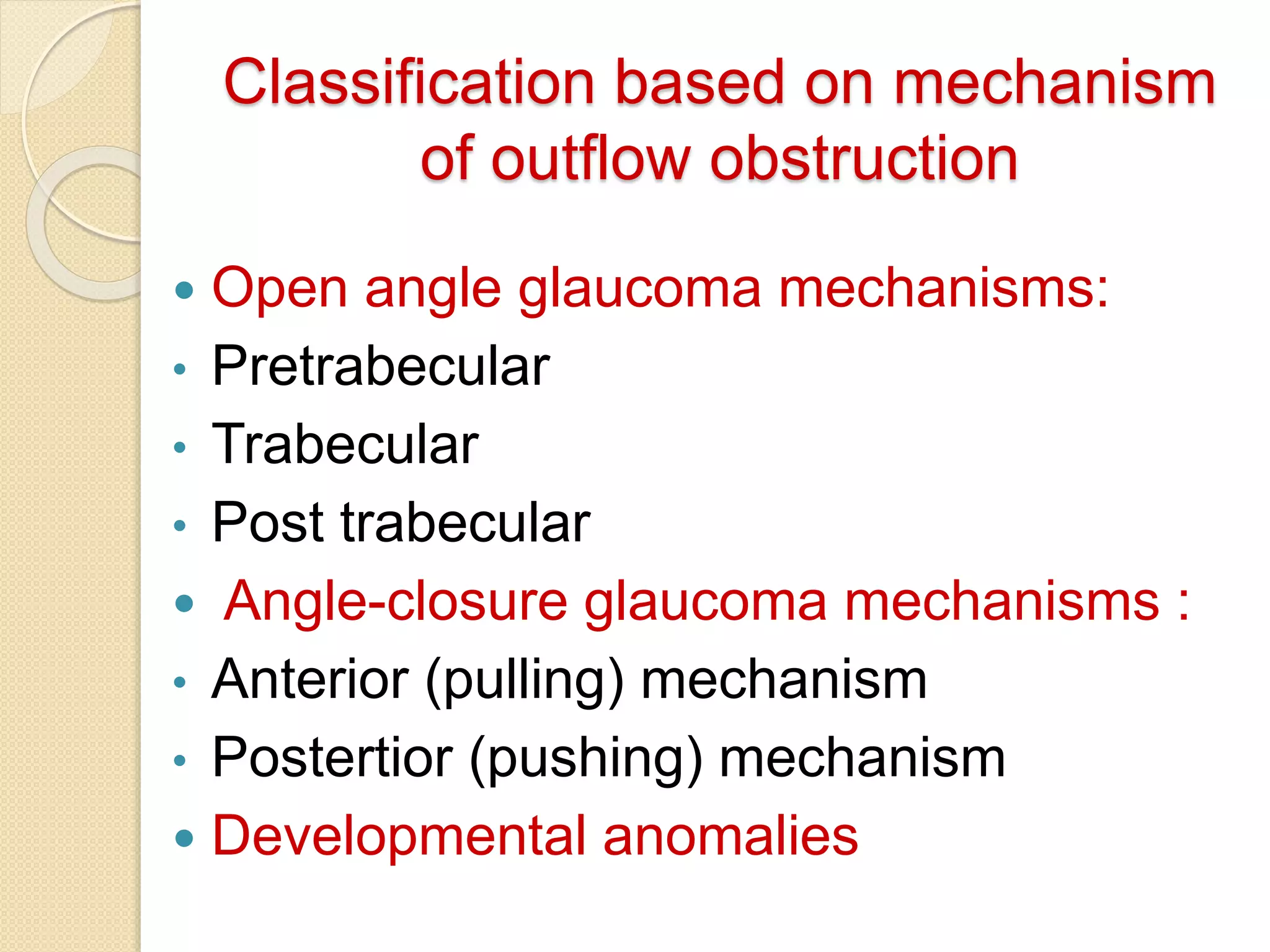
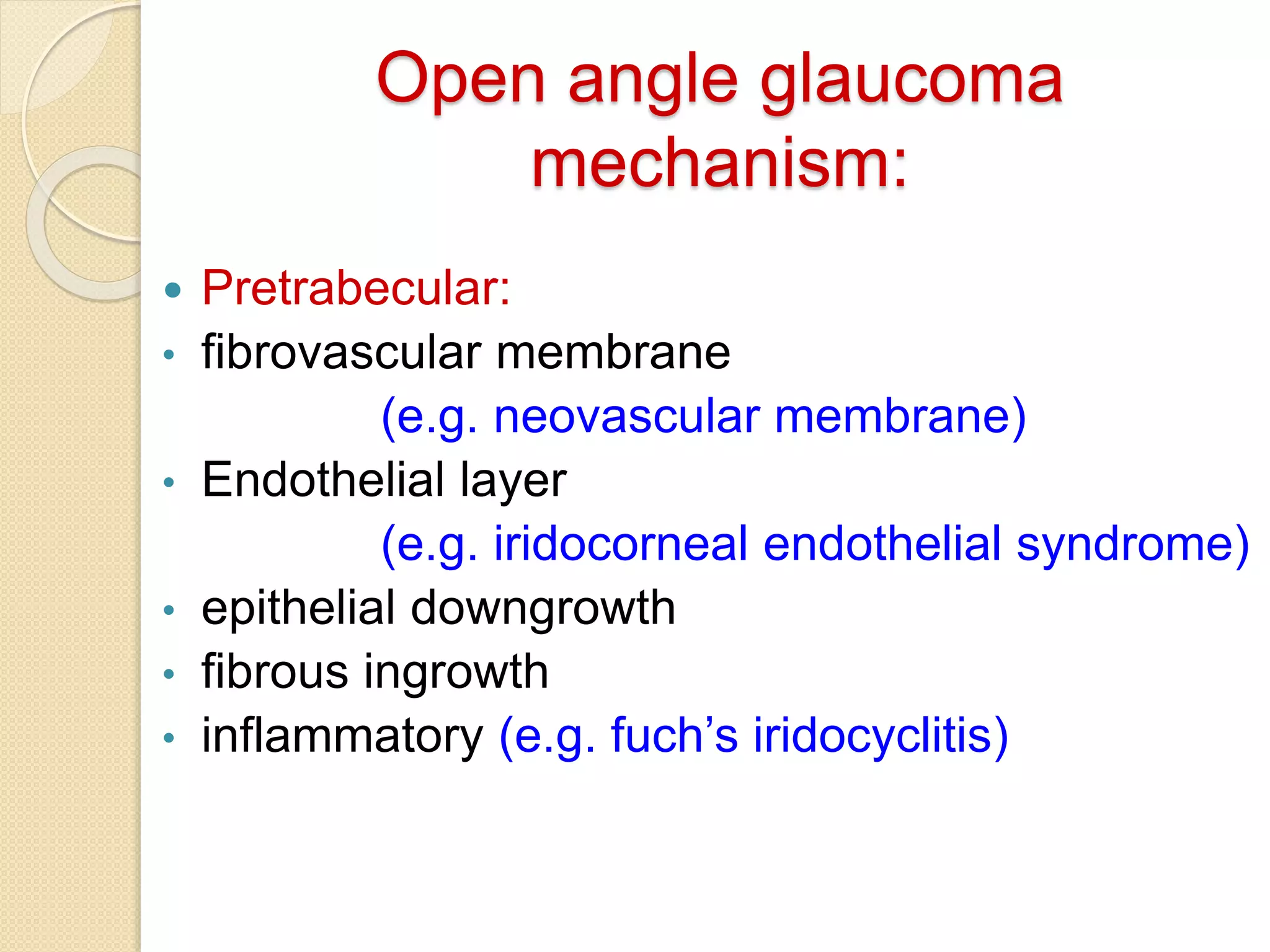
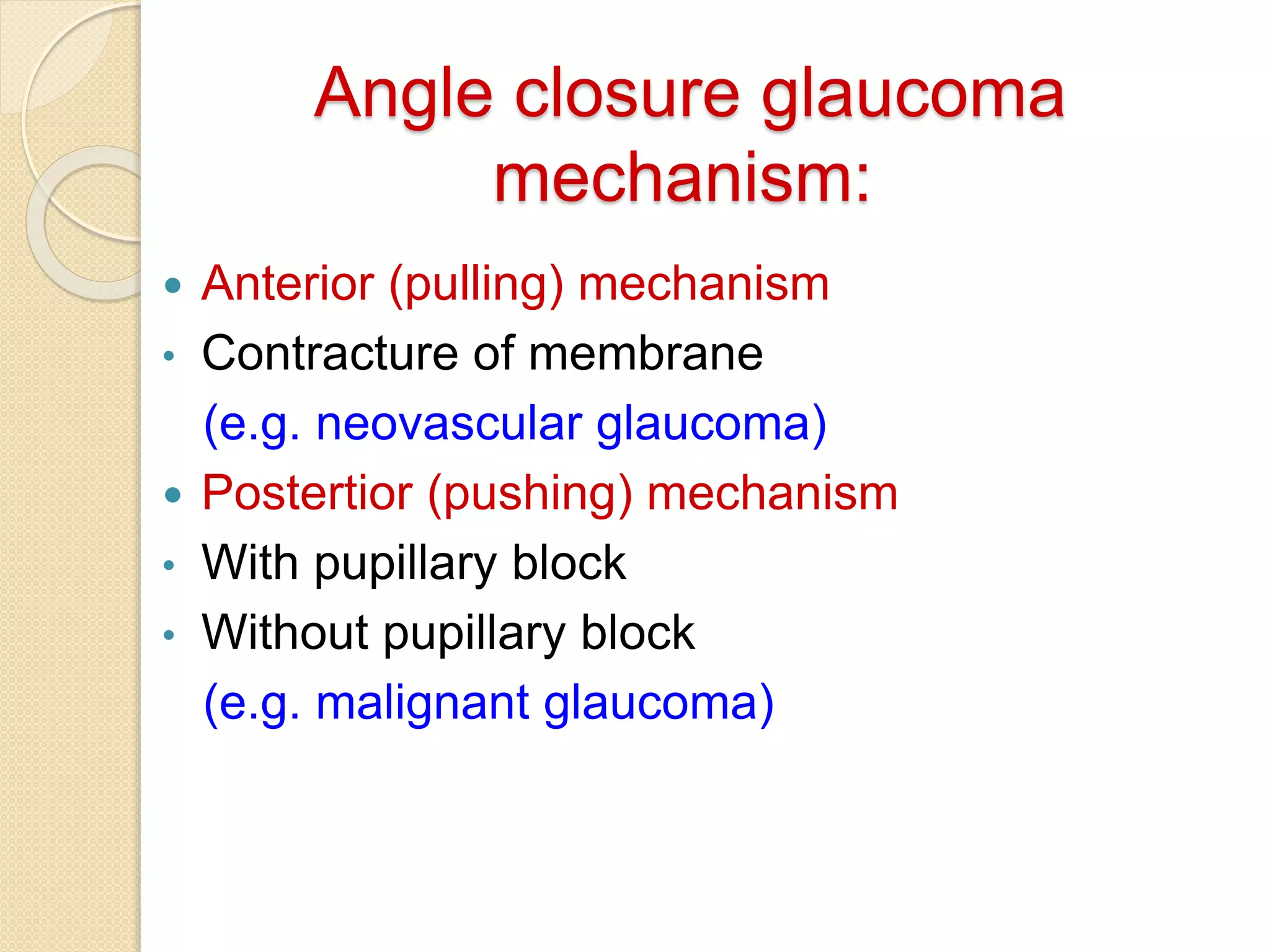
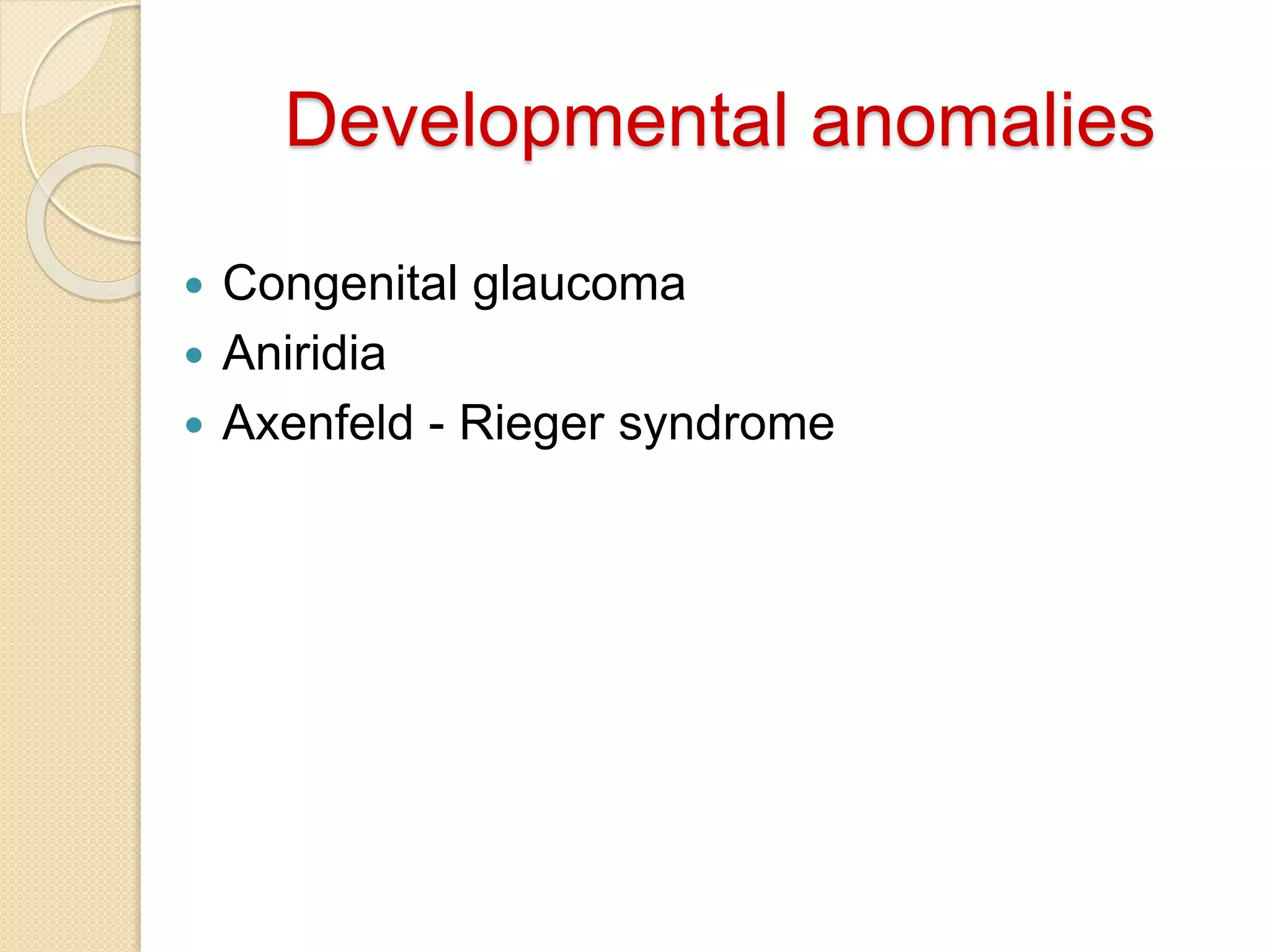
EVALUATION OF A GLAUCOMA
PATIENT
Presented by
Dr. (Maj.) Rashed
F.C.P.S Part-II Trainee
CMH Dhaka
Introduction
 Glaucoma refers to a group of diseases
having common characteristic of progressive
optic neuropathy with associated visual field
loss as progresses in which elevated
intraocular pressure (IOP) is one of the key
modifieable factors.
 Normal IOP: 10-22 mmHg.
 Three risk factor determine the IOP:
◦ rate of aqueous humor production by ciliary body
◦ resistance to aqueous outflow across the trabecular
meshwork-Schlemm’s canal system
◦ The level of epischeral venous pressure
Classification
 Congenital & Acquired
 Primary & Secondary
 Open angle & Angle-closure
Clinical Evaluation
 Appropriate management of glaucoma
depends on the clinician's ability to
diagnose the
◦ specific form of glaucoma in a given
patient,
◦ severity of the condition,
◦ progression in that patient's disease
status.
History
 Patient's current complaint
 History of present illness
 Past ocular, medical, and surgical
history
 Refraction history
 Past medications history
 Social history
 History of alcohol and tobacco use
 Occupation
 Family history
Clinical examination and
investigations
• BCVA distant & near
• Pupillary reaction, relative afferent pupillary
defect
• Slit lamp examination of lids, lid margins,
conjunctiva, cornea, anterior chamber & lens
• IOP and time of measurement
• CCT
• Gonioscopy
• Detailed examination of:
Lens
Biomicroscopy of ONH and RNFL
Fundus
• Automated perimetry
• OCT of ONH & RNFL
Clinical examination
 External Adnexa
◦ Variety of conditions associated with
secondary glaucomas
 Neurofibromatosis type- I
 Oculodermal melanoeytosis (nevus of Ota)
 Axenfeld-Rieger syndrome
 Orbital varices
 Thyroid associated orbitopathy
◦ External ocular manifestations of
glaucoma therapy.
 Prostaglandin analogue
Slit lamp biomicroscopy
 Conjunctiva
◦ Acute IOP
◦ Chronic IOP
◦ Long term use of sympathomimetics
and prostaglandin analogoues
◦ Long term use of epinephrine
derivatives
◦ Filtering bleb +/-
 Episclera & Sclera
◦ Dilation of the episcleral vessels
◦ Sentinal vessels
 Cornea
◦ Developmental glaucoma
◦ Acute IOP
◦ Medication toxicity
◦ Corneal endothelial abnormality
◦ Scar
◦ CCT
 A/C
◦ Uniformity of depth of the anterior chamber
◦ Width of the chamber angle
** Before dilation of pupil; pupillary light
reaction, iris & A/C should be examined **
 Iris
◦ Heterochromia, iris atrophy, transillumination
defects, ectropion uveae. corectopia, nevi,
nodules, exfoliative material & NVI
◦ Trauma
 Lens
◦ Size, shape, clarity & stability of the lens
 Fundus
IOP
 Schiotz tonometry: indentation procedure, now
obsolete.
 Goldmann applanation tonometer : most
appropriate
 Noncontact tonometer (NCT): screening
purpose
 Tonopen: very small area, useful in corneal scar or
edema
 Digital pressure
Possible source of error in
Tonometry
 Squeezing of the eyelids
 Breath holding or Valsalva maneuver
 Pressure on the globe
 Extra-ocular muscle force applied to a restricted
globe
 Tight collar or tight necktie
 Obesity or straining to sit on slit lamp
 An inaccurately calibrated tonometer
 Excessive or inadequate amount of fluorescein
 High corneal astigmatism
 Corneal thickness greater or less than normal
 Corneal scarring or band keratopathy
 Dynamic contour tonometry
The tip of the device exactly matches the contour of
the cornea, the pressure measured by a transducer
placed on its tip.
Measure true IOP irrespective of CCT
Capable of measuring the ocular pulse amplitude
Gonioscopy
CLASSIFICATION
INDIRECT GONIOSCOPY
 Non-Indentation
 Indentatioin
DIRECT GONIOSCOPY
Gonioscopy
 Angle
 Blood vessles
 PAS
 Pigmentation
 Sign of trauma
VAN HERICK METHOD
Peripheral
Chamber
Depth
In Corneal
Thickness
Angle
Grade
Comment
≥ Cornea 4 Wide open
1/4 to 1/2 3 Incapable of closure
1/4 2
Should be
gonioscoped
<1/4 1
Dangerously narrowed
angle
Shaffer system
Optic Disc Evaluation ..
 Slit lamp biomicroscopy : Ideal
◦ Stereoscopic View –Cupping
◦ Measuring the optic disc size
 Direct Ophthalmoscopy
◦ Good Magnification
 Indirect Ophthalmoscopy
◦ Overall View
 Optic disc Photoghraphy.
◦ Documentation,Monitoring for progression.
The 7 parameters to look for…
1)Disc size
2)Neuroretinal Rim (NRR):
- ISNT rule
3)Cup: Disc ratio
- Vertical C/ D Ratio.
4) Optic Disc Hemorrhage
5) Nerve Fiber Layer Defect:
- focal & diffuse
6) Para Papillary Atrophy:
- Size, location & Configuration
7) Retinal Arterial Attenuation:
- focal & diffuse
Ophthalmoscopic sign of glaucoma
Generalized Focal Less specific
o Large optic cup
o Asymetrical of the
cup
o Progressive
enlargement of
cup
o Notching of the rim
o Vertical elongation
of the cup
o Region pallor
o Splinter
hemorrhage
o Nerve fiber layer
loss
o Exposed lamina
cribrosa
o Nasal
displacement of
the vessels
o Baring of
circumlinear
vessels
o Peripapillary
crescent
I>S>N>T I<S<N>T
The visual field
An island hill of vision in a sea of darkness
Perimentry
 Kinetic
◦ Two dimensional
◦ Moving stimulus
◦ Known Luminance
◦ From non-seeing area
to seeing area until it is
perceived.
◦ e.g. confrontation
Tangent screen
Lister perimeter
Goldmann
perimeter
 Static
◦ Three dimensional
◦ Static stimulus
◦ varying luminance in
the same position
◦ e.g. Octopus
Humphrey
Hensen
Glaucomatous visual field defects
Generalized depression
Paracentral scotoma
Arcuate scotoma
Nasal step
Altitudinal defect
Temporal wedge
Paracentral scotoma
Arcuate scotoma
Nasal step
Gross cupping with
ring scotoma Tubular
field
Progression of glaucomatous damage
OCT
Normal Glaucoma
patient
Classification based on mechanism
of outflow obstruction
 Open angle glaucoma mechanisms:
• Pretrabecular
• Trabecular
• Post trabecular
 Angle-closure glaucoma mechanisms :
• Anterior (pulling) mechanism
• Postertior (pushing) mechanism
 Developmental anomalies
Open angle glaucoma
mechanism:
 Pretrabecular:
• fibrovascular membrane
(e.g. neovascular membrane)
• Endothelial layer
(e.g. iridocorneal endothelial syndrome)
• epithelial downgrowth
• fibrous ingrowth
• inflammatory (e.g. fuch’s iridocyclitis)
Open angle glaucoma
mechanism:
 Trabecular:
• Idiopathic (e.g. chronic & juvenile open angle)
• ‘Clogging’ of trabecular meshwork
(e.g. pigmentary, red cell, ghost cell glaucoma)
• Alteration of trabecular meshwork
(e.g. steroid induced glaucoma)
• Trauma (e.g. angle recession)
• Intra ocular foreign bodies
Open angle glaucoma
mechanism:
 Post trabecular:
• Obstruction of Schlemm’s canal
• Elevated intra ocular pressure
(e.g. sturge weber syndrome
thyroid ophthalmopathy
retrobulbar tumours)
Angle closure glaucoma
mechanism:
 Anterior (pulling) mechanism
• Contracture of membrane
(e.g. neovascular glaucoma)
 Postertior (pushing) mechanism
• With pupillary block
• Without pupillary block
(e.g. malignant glaucoma)
Developmental anomalies
 Congenital glaucoma
 Aniridia
 Axenfeld - Rieger syndrome
In-direct
gonioscopy
 Non-Indentation
◦ Goldmann three
mirrors
 Indentatioin
◦ Zeiss
◦ Posner
◦ Sussman
Direct gonioscopy
 Diagnostic – Koeppe
 Surgical –
Medical workshop
Barken
Swan- Jacob

Evaluation of a glaucoma patient

  • 1.
    EVALUATION OF AGLAUCOMA PATIENT Presented by Dr. (Maj.) Rashed F.C.P.S Part-II Trainee CMH Dhaka
  • 2.
    Introduction  Glaucoma refersto a group of diseases having common characteristic of progressive optic neuropathy with associated visual field loss as progresses in which elevated intraocular pressure (IOP) is one of the key modifieable factors.  Normal IOP: 10-22 mmHg.  Three risk factor determine the IOP: ◦ rate of aqueous humor production by ciliary body ◦ resistance to aqueous outflow across the trabecular meshwork-Schlemm’s canal system ◦ The level of epischeral venous pressure
  • 4.
    Classification  Congenital &Acquired  Primary & Secondary  Open angle & Angle-closure
  • 5.
    Clinical Evaluation  Appropriatemanagement of glaucoma depends on the clinician's ability to diagnose the ◦ specific form of glaucoma in a given patient, ◦ severity of the condition, ◦ progression in that patient's disease status.
  • 6.
    History  Patient's currentcomplaint  History of present illness  Past ocular, medical, and surgical history  Refraction history  Past medications history  Social history  History of alcohol and tobacco use  Occupation  Family history
  • 7.
    Clinical examination and investigations •BCVA distant & near • Pupillary reaction, relative afferent pupillary defect • Slit lamp examination of lids, lid margins, conjunctiva, cornea, anterior chamber & lens • IOP and time of measurement • CCT • Gonioscopy • Detailed examination of: Lens Biomicroscopy of ONH and RNFL Fundus • Automated perimetry • OCT of ONH & RNFL
  • 8.
    Clinical examination  ExternalAdnexa ◦ Variety of conditions associated with secondary glaucomas  Neurofibromatosis type- I  Oculodermal melanoeytosis (nevus of Ota)  Axenfeld-Rieger syndrome  Orbital varices  Thyroid associated orbitopathy ◦ External ocular manifestations of glaucoma therapy.  Prostaglandin analogue
  • 9.
    Slit lamp biomicroscopy Conjunctiva ◦ Acute IOP ◦ Chronic IOP ◦ Long term use of sympathomimetics and prostaglandin analogoues ◦ Long term use of epinephrine derivatives ◦ Filtering bleb +/-  Episclera & Sclera ◦ Dilation of the episcleral vessels ◦ Sentinal vessels
  • 10.
     Cornea ◦ Developmentalglaucoma ◦ Acute IOP ◦ Medication toxicity ◦ Corneal endothelial abnormality ◦ Scar ◦ CCT  A/C ◦ Uniformity of depth of the anterior chamber ◦ Width of the chamber angle ** Before dilation of pupil; pupillary light reaction, iris & A/C should be examined **
  • 11.
     Iris ◦ Heterochromia,iris atrophy, transillumination defects, ectropion uveae. corectopia, nevi, nodules, exfoliative material & NVI ◦ Trauma  Lens ◦ Size, shape, clarity & stability of the lens  Fundus
  • 12.
    IOP  Schiotz tonometry:indentation procedure, now obsolete.  Goldmann applanation tonometer : most appropriate  Noncontact tonometer (NCT): screening purpose  Tonopen: very small area, useful in corneal scar or edema  Digital pressure
  • 14.
    Possible source oferror in Tonometry  Squeezing of the eyelids  Breath holding or Valsalva maneuver  Pressure on the globe  Extra-ocular muscle force applied to a restricted globe  Tight collar or tight necktie  Obesity or straining to sit on slit lamp  An inaccurately calibrated tonometer  Excessive or inadequate amount of fluorescein  High corneal astigmatism  Corneal thickness greater or less than normal  Corneal scarring or band keratopathy
  • 15.
     Dynamic contourtonometry The tip of the device exactly matches the contour of the cornea, the pressure measured by a transducer placed on its tip. Measure true IOP irrespective of CCT Capable of measuring the ocular pulse amplitude
  • 16.
  • 17.
  • 18.
    Gonioscopy  Angle  Bloodvessles  PAS  Pigmentation  Sign of trauma
  • 19.
    VAN HERICK METHOD Peripheral Chamber Depth InCorneal Thickness Angle Grade Comment ≥ Cornea 4 Wide open 1/4 to 1/2 3 Incapable of closure 1/4 2 Should be gonioscoped <1/4 1 Dangerously narrowed angle
  • 20.
  • 21.
    Optic Disc Evaluation..  Slit lamp biomicroscopy : Ideal ◦ Stereoscopic View –Cupping ◦ Measuring the optic disc size  Direct Ophthalmoscopy ◦ Good Magnification  Indirect Ophthalmoscopy ◦ Overall View  Optic disc Photoghraphy. ◦ Documentation,Monitoring for progression.
  • 22.
    The 7 parametersto look for… 1)Disc size 2)Neuroretinal Rim (NRR): - ISNT rule 3)Cup: Disc ratio - Vertical C/ D Ratio. 4) Optic Disc Hemorrhage 5) Nerve Fiber Layer Defect: - focal & diffuse 6) Para Papillary Atrophy: - Size, location & Configuration 7) Retinal Arterial Attenuation: - focal & diffuse
  • 23.
    Ophthalmoscopic sign ofglaucoma Generalized Focal Less specific o Large optic cup o Asymetrical of the cup o Progressive enlargement of cup o Notching of the rim o Vertical elongation of the cup o Region pallor o Splinter hemorrhage o Nerve fiber layer loss o Exposed lamina cribrosa o Nasal displacement of the vessels o Baring of circumlinear vessels o Peripapillary crescent
  • 24.
  • 27.
    The visual field Anisland hill of vision in a sea of darkness
  • 28.
    Perimentry  Kinetic ◦ Twodimensional ◦ Moving stimulus ◦ Known Luminance ◦ From non-seeing area to seeing area until it is perceived. ◦ e.g. confrontation Tangent screen Lister perimeter Goldmann perimeter  Static ◦ Three dimensional ◦ Static stimulus ◦ varying luminance in the same position ◦ e.g. Octopus Humphrey Hensen
  • 29.
    Glaucomatous visual fielddefects Generalized depression Paracentral scotoma Arcuate scotoma Nasal step Altitudinal defect Temporal wedge
  • 30.
  • 31.
  • 32.
  • 33.
    Gross cupping with ringscotoma Tubular field
  • 34.
  • 35.
  • 37.
    Classification based onmechanism of outflow obstruction  Open angle glaucoma mechanisms: • Pretrabecular • Trabecular • Post trabecular  Angle-closure glaucoma mechanisms : • Anterior (pulling) mechanism • Postertior (pushing) mechanism  Developmental anomalies
  • 38.
    Open angle glaucoma mechanism: Pretrabecular: • fibrovascular membrane (e.g. neovascular membrane) • Endothelial layer (e.g. iridocorneal endothelial syndrome) • epithelial downgrowth • fibrous ingrowth • inflammatory (e.g. fuch’s iridocyclitis)
  • 39.
    Open angle glaucoma mechanism: Trabecular: • Idiopathic (e.g. chronic & juvenile open angle) • ‘Clogging’ of trabecular meshwork (e.g. pigmentary, red cell, ghost cell glaucoma) • Alteration of trabecular meshwork (e.g. steroid induced glaucoma) • Trauma (e.g. angle recession) • Intra ocular foreign bodies
  • 40.
    Open angle glaucoma mechanism: Post trabecular: • Obstruction of Schlemm’s canal • Elevated intra ocular pressure (e.g. sturge weber syndrome thyroid ophthalmopathy retrobulbar tumours)
  • 41.
    Angle closure glaucoma mechanism: Anterior (pulling) mechanism • Contracture of membrane (e.g. neovascular glaucoma)  Postertior (pushing) mechanism • With pupillary block • Without pupillary block (e.g. malignant glaucoma)
  • 42.
    Developmental anomalies  Congenitalglaucoma  Aniridia  Axenfeld - Rieger syndrome
  • 43.
    In-direct gonioscopy  Non-Indentation ◦ Goldmannthree mirrors  Indentatioin ◦ Zeiss ◦ Posner ◦ Sussman Direct gonioscopy  Diagnostic – Koeppe  Surgical – Medical workshop Barken Swan- Jacob

Editor's Notes