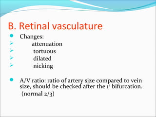
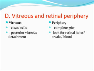
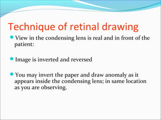
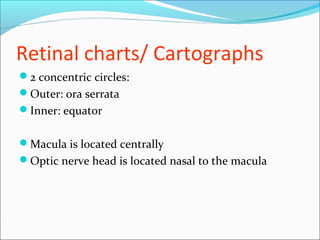
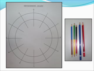
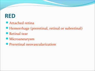
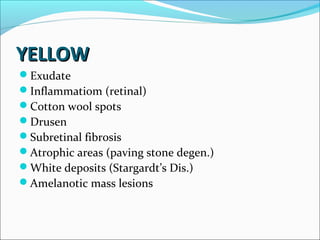
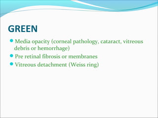
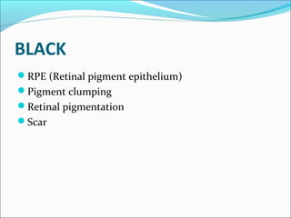
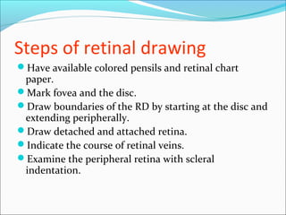
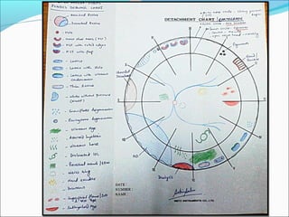
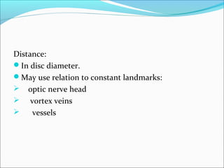
Dr Sabin Sahu
M.S. Ophthalmology
SCEH, Lahan
Retinal diagrams
Most retinal surgeons are trained to create formal
retinal drawings of the fundus.
Retinal drawings are useful to document pathology,
although more and more people now prefer fundus
photographs.
Can be used for serial follow up of patients to
document changes in the pathology.
Fundus evaluation
A. Optic Disc evaluation
Size, shape, colour of the disc
Vertical cup-to-disc ratio (CDR)
Neuroretinal rim
Disc margins: distinct/ blurred
Peripapillary changes
B. Retinal vasculature
 Changes:
 attenuation
 tortuous
 dilated
 nicking
 A/V ratio: ratio of artery size compared to vein
size, should be checked after the 1st
bifurcation.
(normal 2/3)
C. Macula
Flat/intact and uniformly pigmented
Yellowish foveal reflex
Look for any abnormal pigment/ blood or fluid
D. Vitreous and retinal periphery
Vitreous:
 clear/ cells
 posterior vitreous
detachment
Periphery
 complete 3600
 look for retinal holes/
breaks/ blood
Technique of retinal drawing
View in the condensing lens is real and in front of the
patient:
Image is inverted and reversed
You may invert the paper and draw anomaly as it
appears inside the condensing lens; in same location
as you are observing.
Retinal charts/ Cartographs
2 concentric circles:
Outer: ora serrata
Inner: equator
Macula is located centrally
Optic nerve head is located nasal to the macula
BLUE
Retinal vessels
Sub retinal fluid
Detached Retina
Edema
RED
Attached retina
Hemorrhage (preretinal, retinal or subretinal)
Retinal tear
Microaneurysm
Preretinal neovascularization
YELLOWYELLOW
Exudate
Inflammatiom (retinal)
Cotton wool spots
Drusen
Subretinal fibrosis
Atrophic areas (paving stone degen.)
White deposits (Stargardt’s Dis.)
Amelanotic mass lesions
GREEN
Media opacity (corneal pathology, cataract, vitreous
debris or hemorrhage)
Pre retinal fibrosis or membranes
Vitreous detachment (Weiss ring)
BROWN
Melanocytic lesions
Uveal tissue
Malignant choroidal melanomas
Edge of buckle beneath detached retina
Choroidal detachment
BLACK
RPE (Retinal pigment epithelium)
Pigment clumping
Retinal pigmentation
Scar
Steps of retinal drawing
Have available colored pensils and retinal chart
paper.
Mark fovea and the disc.
Draw boundaries of the RD by starting at the disc and
extending peripherally.
Draw detached and attached retina.
Indicate the course of retinal veins.
Examine the peripheral retina with scleral
indentation.
How big is the lesion?
Size in disc diameters (DD)
compare lesion to optic nerve head size.
Where is the lesion:
Location:
Clock-dial
superior, supero-nasal, nasal, infero-nasal, inferior,
infero-temporal, temporal, supero-temporal.
Anterior/ posterior to the equator/ oral lesion.
Distance:
In disc diameter.
May use relation to constant landmarks:
 optic nerve head
 vortex veins
 vessels
Retinal diagram dr sabin sahu

Retinal diagram dr sabin sahu

  • 1.
    Dr Sabin Sahu M.S.Ophthalmology SCEH, Lahan
  • 2.
    Retinal diagrams Most retinalsurgeons are trained to create formal retinal drawings of the fundus. Retinal drawings are useful to document pathology, although more and more people now prefer fundus photographs. Can be used for serial follow up of patients to document changes in the pathology.
  • 3.
    Fundus evaluation A. OpticDisc evaluation Size, shape, colour of the disc Vertical cup-to-disc ratio (CDR) Neuroretinal rim Disc margins: distinct/ blurred Peripapillary changes
  • 4.
    B. Retinal vasculature Changes:  attenuation  tortuous  dilated  nicking  A/V ratio: ratio of artery size compared to vein size, should be checked after the 1st bifurcation. (normal 2/3)
  • 5.
    C. Macula Flat/intact anduniformly pigmented Yellowish foveal reflex Look for any abnormal pigment/ blood or fluid
  • 6.
    D. Vitreous andretinal periphery Vitreous:  clear/ cells  posterior vitreous detachment Periphery  complete 3600  look for retinal holes/ breaks/ blood
  • 7.
    Technique of retinaldrawing View in the condensing lens is real and in front of the patient: Image is inverted and reversed You may invert the paper and draw anomaly as it appears inside the condensing lens; in same location as you are observing.
  • 8.
    Retinal charts/ Cartographs 2concentric circles: Outer: ora serrata Inner: equator Macula is located centrally Optic nerve head is located nasal to the macula
  • 11.
    BLUE Retinal vessels Sub retinalfluid Detached Retina Edema
  • 12.
    RED Attached retina Hemorrhage (preretinal,retinal or subretinal) Retinal tear Microaneurysm Preretinal neovascularization
  • 13.
    YELLOWYELLOW Exudate Inflammatiom (retinal) Cotton woolspots Drusen Subretinal fibrosis Atrophic areas (paving stone degen.) White deposits (Stargardt’s Dis.) Amelanotic mass lesions
  • 14.
    GREEN Media opacity (cornealpathology, cataract, vitreous debris or hemorrhage) Pre retinal fibrosis or membranes Vitreous detachment (Weiss ring)
  • 15.
    BROWN Melanocytic lesions Uveal tissue Malignantchoroidal melanomas Edge of buckle beneath detached retina Choroidal detachment
  • 16.
    BLACK RPE (Retinal pigmentepithelium) Pigment clumping Retinal pigmentation Scar
  • 17.
    Steps of retinaldrawing Have available colored pensils and retinal chart paper. Mark fovea and the disc. Draw boundaries of the RD by starting at the disc and extending peripherally. Draw detached and attached retina. Indicate the course of retinal veins. Examine the peripheral retina with scleral indentation.
  • 19.
    How big isthe lesion? Size in disc diameters (DD) compare lesion to optic nerve head size.
  • 20.
    Where is thelesion: Location: Clock-dial superior, supero-nasal, nasal, infero-nasal, inferior, infero-temporal, temporal, supero-temporal. Anterior/ posterior to the equator/ oral lesion.
  • 21.
    Distance: In disc diameter. Mayuse relation to constant landmarks:  optic nerve head  vortex veins  vessels