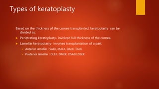
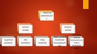
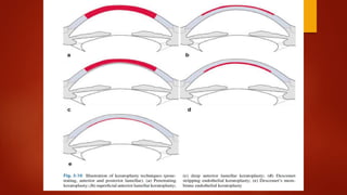
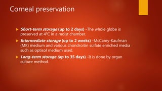
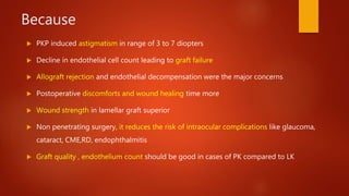
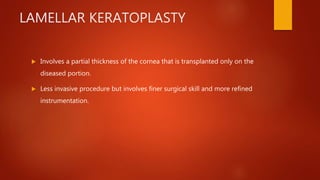
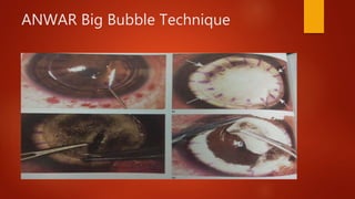
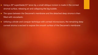
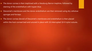
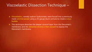
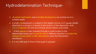
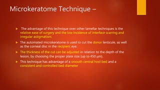
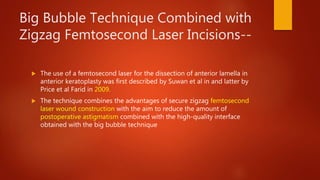
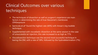
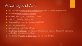
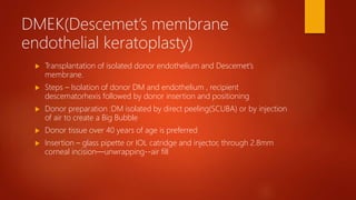
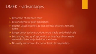
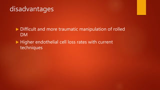
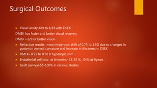
Lamellar Keratoplasty
MODERATOR- DR SUNDEEP
PRESENTER- DR AKSHAY NAYAK
DEFINITION
 The replacement of diseased cornea with autologus or heterologus cornea
is called as keratoplasty. In most of the cases the donor cornea is taken
from a deceased person.
 Either full thickness of the cornea or a part of it may be transplanted.
Introduction
 For over hundred years Penetrating keratoplasty has been the standard care for corneal
diseases
 PK involves replacing a full thickness of diseased recipient’s cornea with that of a healthy
donor cornea secured into place with 32,16 or 12 sutures
 Claesson et al studied 520 grafts, at 2 years after PK showed a visual acuity of upto
20/40 (6/12)
Layers Thick
ness(
um)
Composition
Epithelium 50 Stratified Squamous Epithelium
Bowman’s
Membrane
8-14 Compact layer of unorganised
collagen fibres Stroma
Stroma 500 Orderly arrangement of collagen
lamellae with keratocytes
Descemet’s
Membrane
10-12 Consists of basement membrane
materials
Endothelium 5 single layer of simple squamous
epithelium
Types of keratoplasty
Based on the thickness of the cornea transplanted, keratoplasty can be
divided as:
 Penetrating keratoplasty- involved full thickness of the cornea.
 Lamellar keratoplasty- involves transplantation of a part.
 Anterior lamellar : SALK, MALK, DALK, TALK
 Posterior lamellar : DLEK, DMEK, DSAEK,DSEK
. LAMELLAR
KERATOPLAST
Y
Anterior
lamellar
Superficial
ALK(SALK)
Mid
ALK(MALK)
Deep
ALK(DALK)
Posterior
lamellar
Deep Lamellar
Endothelial
Keratoplasty(D
LEK)
Descemet’s
stripping
Automated endo
thelium
keratoplasty
DSAEK
Donor tissue
 Removed as early as possible (6-12 hours of death).
 Corneas from infants (2 years and under) are rarely used -surgical,
refractive and rejection problems.
 It should be stored under sterile conditions.
 Evaluation –medical history review and donor blood screening to exclude
contraindications, and microscopic examination of the cornea including
endothelial cell count determination
Corneal preservation
 Short-term storage (up to 2 days) -The whole globe is
preserved at 40C in a moist chamber.
 Intermediate storage (up to 2 weeks) -McCarey-Kaufman
(MK) medium and various chondroitin sulfate enriched media
such as optisol medium used.
 Long-term storage (up to 35 days) -It is done by organ
culture method.
Contraindications for donation of
corneal tissue
Absolute
 Rabies
 HIV, viral hepatitis, syphilis and active malaria.
 Septicaemia
 Prior high-risk behavior for HIV and hepatitis.
 infectious diseases of the CNS.
 Creutzfeldt-Jacob disease
Relative
 Most hematological malignancies.
 Ocular disease such as inflammation and malignancies (e.g. retinoblastoma) and corneal
refractive surgery.
 Death of unknown cause.
 Congenital rubella, tuberculosis
Why there is
need for an
alternative to
PK?????
Because
 PKP induced astigmatism in range of 3 to 7 diopters
 Decline in endothelial cell count leading to graft failure
 Allograft rejection and endothelial decompensation were the major concerns
 Postoperative discomforts and wound healing time more
 Wound strength in lamellar graft superior
 Non penetrating surgery, it reduces the risk of intraocular complications like glaucoma,
cataract, CME,RD, endophthalmitis
 Graft quality , endothelium count should be good in cases of PK compared to LK
LAMELLAR KERATOPLASTY
 Involves a partial thickness of the cornea that is transplanted only on the
diseased portion.
 Less invasive procedure but involves finer surgical skill and more refined
instrumentation.
History
 In 1824 Reisinger performed the first animal graft and coined the term
‘Keratoplasty’.
 The first successful lamellar keratoplasty was performed by Arthur Von
Hippel at the end of 19th century.
 Jose Barraquer was the first to perform posterior lamellar keratoplasty in
1950
Anterior Lamellar Keratoplasty (ALK)
 Removal and replacement of deformed or diseased anterior corneal
tissue ( epithelium, Bowman’s layer, and stroma)
 Sparing the host Descemet’s membrane and endothelium
Indications
 Indicated in corneas that have a healthy endothelium
1.Optical ALK is useful in
 Anterior stromal scars after infectious keratitis or trauma
 Complications after refractive surgery
 Dystrophies like epithelial and stromal dystrophy
 Salzmann nodular degenerations, spheroidal degeneration etc
 Post inflammatory:Trachomatous kerartopathy,healed superficial keratitis
Indications (contd…)
2. Tectonic ALK is useful in
 Peripheral non inflammatory thinning
 Terrien’s marginal degeneration
 Pellucid marginal degeneration
 Peripheral ulcerative autoimmune keratitis - Mooren’s ulcer.
 Descematocoele
3. Combined indications include
 Keratoconus
 Pellucid marginal degeneration
 Iatrogenic keratoectasia after Refractive surgeries
 4.therapeutic
Infective keratitis limited to anterior corneal layers
Surgical Planning
(Surgeon to ask themselves)
 Is the Endothelium Normal?
 What level does the pathology extend?
 Will leaving minimal posterior stroma affect visual outcome?
SALK
 should be used when the anterior 30--50% of the cornea is affected with
pathology and is to be replaced with a similar amount of donor tissue.
The main indications are superficial scars resulting
 from healed infections, including trachoma, trauma (post-laser or
accidental),
 superficial corneal dystrophies and degenerations,
 or persistent epithelial defects
Surgical Techniques
1)DIRECT DISSECTION- Partial thickness(60-80%) trephination
 Corneal stromal dissection with crescent
 Partial thickness donor
 Suture
 Disadvantage – Unreliable depth of dissection
Irregular Host Bed
Interface haze
Melles Technique(closed dissection)
 To visualize the depth of lamellar dissection –
 Exchange aqueous with air- Create Air Endothelium interface – Act as convex mirror
 Specialised spatula –stromal pocket—inject visco—stroma over visco excised
 A black band is visualized in front of dissecting instrument, which represent twice the residual
stroma
 Trephination
 A full-thickness donor button stripped off its DM is sutured in place.
 Good visual results have been reported with this technique.
 There is about a 14% chance of DM perforation.
ANWAR Big Bubble Technique
Surgical technique Anwar ‘BUBBLE’
TECHNIQUE
 The technique involves trephining the anterior host corneal surface with a Hessburg-
Barron suction trephine to a depth of about 400 µm
 25-gauge disposable needle inserted into the corneal stroma,
 Air is then injected to create a big bubble (Anwar’s technique) that will detach the deep
stromal layers from the Descemet’s membrane.
 lamellar dissection and removal of the anterior stromal disk by crescent knife
 Using a 30° superblade/15° lance tip, a small oblique incision is made in the corneal
stromal surface, releasing air and collapsing the big bubble.
 The space between the Descemet’s membrane and the detached deep stroma is then
filled with viscoelastic.
 Utilizing a divide-and-conquer technique with corneal microscissors, the remaining deep
corneal stroma is excised to expose the smooth surface of the Descemet’s membrane
 The donor cornea is then trephined with a Hessburg-Barron trephine, followed by
staining of the endothelium with trypan blue.
 Descemet’s membrane and the donor endothelium are then removed using dry cellulose
sponges and forceps.
 The donor cornea devoid of Descemet’s membrane and endothelium is then placed
within the host corneal bed and sutured in place with 16 interrupted 10-0 nylon sutures.
Viscoelastic Dissection Technique –
 Viscoelastics, namely sodium hyaluronate, were forced into a previously
made stromal pocket using a 25-gauge blunt cannula to create a visco-
bubble.
 This technique detaches the deeper stromal layer from the Descemet’s
membrane, and the detached stroma is then excised to expose the
Descemet’s membrane.
Hydrodelamination Technique-
 A partial trephination and a lamellar keratectomy are carried out at a
suitable depth.
 A small cut/depression is created in the deeper stroma. A 27-gauge needle
attached to a syringe is inserted at the bottom of the depression, and
saline is injected into the stromal bed, which whitens and swells.
 A fine spatula is then inserted through a small incision in the
delaminated tissue and moved fan like in different direction to loosen the
residual stroma which is then dissected to reach the Descemet’s
membrane.
 A 5-mm DM area in front of the pupil is exposed.
Microkeratome Technique –
 The advantage of this technique over other lamellar techniques is the
relative ease of surgery and the low incidence of interface scarring and
irregular astigmatism.
 The automated microkeratome is used to cut the donor lenticule, as well
as the corneal disc in the recipient eye.
 The thickness of the cut can be adjusted in relation to the depth of the
lesion, by choosing the proper plate size (up to 450 μm).
 This technique has advantage of a smooth central host bed and a
consistent and controlled bed diameter
Big Bubble Technique Combined with
Zigzag Femtosecond Laser Incisions--
 The use of a femtosecond laser for the dissection of anterior lamella in
anterior keratoplasty was first described by Suwan et al in and latter by
Price et al Farid in 2009.
 The technique combines the advantages of secure zigzag femtosecond
laser wound construction with the aim to reduce the amount of
postoperative astigmatism combined with the high-quality interface
obtained with the big bubble technique
Clinical Outcomes over various
techniques
 The techniques of dissection as well as surgeon's experience are main
factors in determining the rate of true Descemet’s membrane
(DM)exposure
 Sarnicola et al* found the highest rate (60%) with Anwar's big-bubble
technique.
 Supplemented with viscoelastic dissection at the same session in the case
of unsuccessful air injection, this rate increased to as high as 77%
 Viscodissection technique was the second most successful technique in
baring the DM, with a rate of 58%, followed by the hydrodelamination (7%)
Complications Intraoperative
 Microperforation – Sudden softening eye and excursion of fluid or air
into the interface.
 Occurrence 39% expert hand
 Manage – Air in AC- Continue dissection peripheral to perforation.
Leaving Air bubble with supine position of patient
 Macroperforation –
Convert PKP
 Pupillary Block Glaucoma-
due to air bubble left in AC
Avoided by Pupil dilation if air left
Periodically examination eye in hour immediately after surgery
 Graft host malapposition/edge irregularity
Due to improper sizing of tissues
 Interface debris
Fibers, bleeding
COMPLICATION POSTOPERATIVE
 Double AC
Cause- Micro perforation, Entrapped Visco at interface
Manage- Accelerated intracameral Air/ SF6 and drain interface fluid
 Persistent epithelial defects
Suture related,ocular surface diseases,wound/edge problems
 Infections
Suture related, lid adnexal abnormalities,poor hygiene,steroid
use,reactivation of herpectic infections
 Graft vascularization
In cases of trachomatous keratopathy,chemical burns and SJ syndrome
 Epithelial, Subepithelial or stromal rejection
Epithelium – Line of oedema
Subepithelial- Subepithelial infiltrate
Stromal –Oedema
Advantages of ALK
 less chances of postoperative inflammation as well as secondary glaucoma.
 No risk of endothelial graft rejection.
 No need for long term steroid prophylaxis
 Rapid functional recovery of vision.
 Very good best corrected visual acuity (BCVA)
 very low astigmatism.
 No significant endothelial cell loss.
 Penetrating Keratoplasty can be done if recurrences occur or Descemet's membrane
perforation occurs intraoperatively.
 The criteria for quality of donor tissue are not very stringent
Endothelial keratoplasty
 DLEK
 DSEK
 DMEK
Endothelial Keratoplasty (EK)
 Purpose To remove the diseased recipient endothelium and replace with
healthy donor corneal endothelium.
 In 1998,Dr.Gerritt Melles et al first described this technique involved large
limbal incision and deep manual lamellar corneal dissection .
 Dr. Mark Terry modified by small incision 5mm rename the Procedure
“Deep Lamellar Endothelial Keratoplasty (DLEK)”
 Next Evolution by Melles was the substitution of the patient dissection with
Descement’s stripping and “Descement’s stripping Endo Keratoplasty (DSEK)”
coined
Why there is need
for EK ,an
alternative to
PKP?????
DLEK/DSEK/DMEK vs PKP
 EK procedure preserves the normal corneal topography to allow faster visual recovery
 Astigmatism after DLEK surgery was 1.63 ±0.97D* ,
 In contrast, after standard PKP surgery was between 4.00 and 6.00D
 After DLEK surgery and in DSEK 18-35% endothelial cell loss from preoperative donor
counts.After 5 years its was 54%.
 After PKP, the cell count has been at 6months- 34% cell loss from preoperative donor
counts and 69% at 5 years.
 Less immunological rejection rates than PK.PK>DSEK>DMEK
 Tectonically stable globe
 No suture related complication
Patients Selection
1. Endothelial Diseases
2. Fuch’s endothelial dystrophy
3. Pseudophakic bullous keratopathy
4. Aphakic bullous keratopathy
5. Iridocorneal endothelial syndrome
SURGICAL PROCEDURE LARGE
INCISION TECHNIQUE(DLEK)
 A scleral access incision is placed at superior limbal region of size 9.0
mm
 SCLERO CORNEAL LAMELLAR POCKET IS MADE USING A CRESCENT
BLADE
 STRAIGHT DEVERS DISSECTOR IS THEN USED TO EXTEND THE POCKET
TO MID PUPILLARY REGION OF THE CORNEA
 THEN A CURVED DEVERS DISSECTOR EXTENDS THE POCKET
COMPLETELY TO THE LIMBUS FOR 360 DEGREES, CREATING A TOTAL
AREA OF DEEP LAMELLAR POCKET
 THE RESECTION OF THE POSTERIOR RECIPIENT TISSUE IS DONE WITH
AN INTRASTROMAL TREPHINE (TERRY TREPHINE)
 ONCE THE BLADE IS IN POSITION IN THE POCKET, IT IS ROTATED
ALONG THE ARC OF 9.0 MM SCLERAL INCISION
 RESECTION OF THE RECIPIENTS DISK IS COMPLETED USING CINDY
SCISSORS
 ONCE THE POSTERIOR RECEIPIENT DISK HAS BEEN CUT 360 DEGREES,
THE TISSUE IS REMOVED FROM THE EYE
DONOR TISSUE PREPARATION
 THE DONOR CORNEOSCLERAL FLAP IS PLACED ON AN ARTIFICIAL ANTERIOR
CHAMBER WHICH IS COATED WITH HELON ON THE ENDOTHELIAL SIDE ARTIFICIAL
ANTERIOR CHAMBER HEALON ON THE ENDOTHELIUM
 THE DONOR TISSUE IS THEN CAPPED ONTO PLACE AND TREPHINATION IS CARRIED
OUT TO ABOUT 60% DEPTH
 LAMELLAR DISSECTION IS COMPLETED USING CRESCENT KNIFE THE DONOR
POSTERIOR DISC IS THEN PLACED ON A OUSLEY SPATULA
 THE DONOR DISC IS THEN SLOWLY INSERTED USING OUSLEY SPATULA
 THE SPATULA IS THEN GENTLY REMOVED FROM THE EYE LEAVING THE DONOR
TISSUE BEHIND SUPPORTED BY AIR BUBBLE IN ANTERIOR CHAMBER
 10-0 NYLON IS THEN USED TO CLOSE THE SCLERAL WOUND AND PREVENT THE
ESCAPE OF DONOR TISSUE
 A REVERSE SINSKEY HOOK IS THEN USED FOR ENDOTHELIAL SIDE POSITIONING
APPEARANCE AT THE END OF SURGERY
Descemet’s Membrane Stripping
Automated Endothelial Keratoplasty
(DSAEK)
 DSAEK It is a method of posterior lamellar keratoplasty in which the
recipient bed is prepared by stripping off the recipient’s Descemet's
membrane.
 Technique was popularized by Gerrit Melles in 1999
Indications of DSAEK
 Fuchs endothelial dystrophy (most common)
 Pseudophakic/ Aphakic bullous keratopathy
 Post PK endothelial graft rejection
 Iridocorneal endothelial syndromes (ICE)
 After glaucoma filtration surgeries
 Eyes with anterior chamber IOL
SURGICAL TECHNIQUE
 RECIPIENT’S CORNEA WOUND CONSTRUCTION THROUGH A 3-5 MM
SCLERO CORNEAL TUNNEL
 WITH HEALON FILLING THE ANTERIOR CHAMBER DESCEMETORHEXIS
AND REMOVAL OF DESCEMET’S MEMBRANE AS A SINGLE DISK IS
CARRIED OUT USING DEXATOME
 DESCEMETORHEXIS IS BEGUN IN THE DISTAL POINT FROM THE
ANTERIOR CHAMBER ENTRY SITE AND CONTINUED IN CLOCKWISE
FASHION.
 THE PERIPHERAL STROMA IS MADE ROUGH USING THE DSAEK
SCRUBBER TO ENHANCE DONOR DISK ATTACHMENT TO RECIPIENT
CORNEA
Graft insertion and positioning
 Forceps – charlie 2, goosey ,kelman
 Sheets glide – sheets intraocular lens
 Busin glide-reusable funnel glide
 Insertors/injectors- endosertor,endoglide,neusidl
 After graft is inserted—air BUBBLE—10-12 MINS
 Anterior chamber and wound is closed by 3 interrupted nylon sutures
 Donor disk is uniformly adherent to the patient’s cornea
DONOR PREPARATION
 MOUNTED ON AN ARTIFICIAL ANTERIOR CHAMBER
 MANUALLY OR SEMI AUTOMATED MICROKERATOME
 FEMTOSECOND LASERS
DSAEK
DSEK/DSAEK - disadvantages
 Steep learning curve
 Higher endothelial cell loss rate in initial post op period
 Graft dislocation
 Pupillary block
 Reports of graft dislocation in vitreous cavity in aphakics
 Interface haze limiting 20/20 vision
 More hyperopic shift compared to DMEK
DMEK(Descemet’s membrane
endothelial keratoplasty)
 Transplantation of isolated donor endothelium and Descemet’s
membrane.
 Steps – Isolation of donor DM and endothelium , recipient
descematorhexis followed by donor insertion and positioning
 Donor preparation :DM isolated by direct peeling(SCUBA) or by injection
of air to create a Big Bubble
 Donor tissue over 40 years of age is preferred
 Insertion – glass pipette or IOL catridge and injector, through 2.8mm
corneal incision—unwrapping--air fill
DMEK --advantages
 Reduction of interface haze
 Less incidence of graft dislocation
 Shorter visual recovery as total corneal thickness remains
same
 Larger donor surface provides more viable endothelial cells
 Less strong host graft apposition at interface allows easier
removal of failed/rejected donor lenticule
 No costly instruments for donor lenticule preparation
disadvantages
 Difficult and more traumatic manipulation of rolled
DM
 Higher endothelial cell loss rates with current
techniques
DMEK
 Not suitable for
1. Aphakics
2. Large iris defects
3. Previous pars plana vitrectomy
Surgical Outcomes
 Visual acuity-6/9 to 6/18 with DSEK
DMEK has faster and better visual recovery
DMEK – 6/9 or better vision
 Refractive results- mean hyperopic shift of 0.75 to 1.5D due to changes in
posterior corneal curvature and increase in thickness in DSEK
 DMEK– 0.25 to 0.50 D hyperopic shift
 Endothelial cell loss- at 6months- 18-35 % , 54% at 5years
 Graft survival-55-100% in various studies
Complications of EK
 Early post operative raised IOP
Pupillary block
Appositional angle closure
 Graft Detachment/Dislocation-The most common complication following DSAEK surgery
is dislocation of the graft due to difficulty in achieving air fill for required time or to
maintain a firm eye.
DMEK>DSEk
Management-scraping, venting incisions, air, supine position, rebubbling
 Epithelial down growth- donor epithelial entrapment
 Interface abnormalities- thickness irregularities due to manual dissection—
folds and wrinkles in EK, incomplete removal of visco
Haze due to proteoglycan deposition
 Infections
 Graft rejection- Lower in EK compared to PK, rejection PK>DSEK>DMEK
 Late endothelial graft Failure
 Steroid induced glaucoma
The future of keratoplasty
 Femtosecond Laser DSAEK •
 This laser is used to create flaps in LASIK and can be used to perform keratoplasty with
different shapes of stromal cut. •
 The laser uses an infrared wavelength (1053nm) to deliver closely spaced, 3 microns
spots that can be focused to a preset depth to photodisrupt the tissue within the corneal
stroma.
• Femtosecond laser is used to create a dissection plane on the donor cornea mounted on
artificial anterior chamber.
• Offers a potential advantage over microkeratome with regards to better sizing of the
posterior lenticule.
•Obtains a smooth surface and precise stromal cuts
SUTURELESS CORNEAL ADHESION
 Bioadhesive (Fibrin glue)
Kaufman et al successfully used fibrin glue in small series of lamellar
keratoplasty
 Photochemical keratodesmos is method of producing sutureless adhesion
by applying a photosensitizer to wound surfaces followed by low energy
laser irradiation.
Laser promotes cross linkage between collegen molecules to produce tight
seal without thermal damage.
Lamellar keratoplasty

Lamellar keratoplasty

  • 1.
    Lamellar Keratoplasty MODERATOR- DRSUNDEEP PRESENTER- DR AKSHAY NAYAK
  • 2.
    DEFINITION  The replacementof diseased cornea with autologus or heterologus cornea is called as keratoplasty. In most of the cases the donor cornea is taken from a deceased person.  Either full thickness of the cornea or a part of it may be transplanted.
  • 3.
    Introduction  For overhundred years Penetrating keratoplasty has been the standard care for corneal diseases  PK involves replacing a full thickness of diseased recipient’s cornea with that of a healthy donor cornea secured into place with 32,16 or 12 sutures  Claesson et al studied 520 grafts, at 2 years after PK showed a visual acuity of upto 20/40 (6/12)
  • 4.
    Layers Thick ness( um) Composition Epithelium 50Stratified Squamous Epithelium Bowman’s Membrane 8-14 Compact layer of unorganised collagen fibres Stroma Stroma 500 Orderly arrangement of collagen lamellae with keratocytes Descemet’s Membrane 10-12 Consists of basement membrane materials Endothelium 5 single layer of simple squamous epithelium
  • 5.
    Types of keratoplasty Basedon the thickness of the cornea transplanted, keratoplasty can be divided as:  Penetrating keratoplasty- involved full thickness of the cornea.  Lamellar keratoplasty- involves transplantation of a part.  Anterior lamellar : SALK, MALK, DALK, TALK  Posterior lamellar : DLEK, DMEK, DSAEK,DSEK
  • 6.
  • 8.
    Donor tissue  Removedas early as possible (6-12 hours of death).  Corneas from infants (2 years and under) are rarely used -surgical, refractive and rejection problems.  It should be stored under sterile conditions.  Evaluation –medical history review and donor blood screening to exclude contraindications, and microscopic examination of the cornea including endothelial cell count determination
  • 11.
    Corneal preservation  Short-termstorage (up to 2 days) -The whole globe is preserved at 40C in a moist chamber.  Intermediate storage (up to 2 weeks) -McCarey-Kaufman (MK) medium and various chondroitin sulfate enriched media such as optisol medium used.  Long-term storage (up to 35 days) -It is done by organ culture method.
  • 12.
    Contraindications for donationof corneal tissue Absolute  Rabies  HIV, viral hepatitis, syphilis and active malaria.  Septicaemia  Prior high-risk behavior for HIV and hepatitis.  infectious diseases of the CNS.  Creutzfeldt-Jacob disease Relative  Most hematological malignancies.  Ocular disease such as inflammation and malignancies (e.g. retinoblastoma) and corneal refractive surgery.  Death of unknown cause.  Congenital rubella, tuberculosis
  • 13.
    Why there is needfor an alternative to PK?????
  • 14.
    Because  PKP inducedastigmatism in range of 3 to 7 diopters  Decline in endothelial cell count leading to graft failure  Allograft rejection and endothelial decompensation were the major concerns  Postoperative discomforts and wound healing time more  Wound strength in lamellar graft superior  Non penetrating surgery, it reduces the risk of intraocular complications like glaucoma, cataract, CME,RD, endophthalmitis  Graft quality , endothelium count should be good in cases of PK compared to LK
  • 15.
    LAMELLAR KERATOPLASTY  Involvesa partial thickness of the cornea that is transplanted only on the diseased portion.  Less invasive procedure but involves finer surgical skill and more refined instrumentation.
  • 16.
    History  In 1824Reisinger performed the first animal graft and coined the term ‘Keratoplasty’.  The first successful lamellar keratoplasty was performed by Arthur Von Hippel at the end of 19th century.  Jose Barraquer was the first to perform posterior lamellar keratoplasty in 1950
  • 17.
    Anterior Lamellar Keratoplasty(ALK)  Removal and replacement of deformed or diseased anterior corneal tissue ( epithelium, Bowman’s layer, and stroma)  Sparing the host Descemet’s membrane and endothelium
  • 19.
    Indications  Indicated incorneas that have a healthy endothelium 1.Optical ALK is useful in  Anterior stromal scars after infectious keratitis or trauma  Complications after refractive surgery  Dystrophies like epithelial and stromal dystrophy  Salzmann nodular degenerations, spheroidal degeneration etc  Post inflammatory:Trachomatous kerartopathy,healed superficial keratitis
  • 20.
    Indications (contd…) 2. TectonicALK is useful in  Peripheral non inflammatory thinning  Terrien’s marginal degeneration  Pellucid marginal degeneration  Peripheral ulcerative autoimmune keratitis - Mooren’s ulcer.  Descematocoele 3. Combined indications include  Keratoconus  Pellucid marginal degeneration  Iatrogenic keratoectasia after Refractive surgeries  4.therapeutic Infective keratitis limited to anterior corneal layers
  • 21.
    Surgical Planning (Surgeon toask themselves)  Is the Endothelium Normal?  What level does the pathology extend?  Will leaving minimal posterior stroma affect visual outcome?
  • 22.
    SALK  should beused when the anterior 30--50% of the cornea is affected with pathology and is to be replaced with a similar amount of donor tissue. The main indications are superficial scars resulting  from healed infections, including trachoma, trauma (post-laser or accidental),  superficial corneal dystrophies and degenerations,  or persistent epithelial defects
  • 23.
    Surgical Techniques 1)DIRECT DISSECTION-Partial thickness(60-80%) trephination  Corneal stromal dissection with crescent  Partial thickness donor  Suture  Disadvantage – Unreliable depth of dissection Irregular Host Bed Interface haze
  • 24.
    Melles Technique(closed dissection) To visualize the depth of lamellar dissection –  Exchange aqueous with air- Create Air Endothelium interface – Act as convex mirror  Specialised spatula –stromal pocket—inject visco—stroma over visco excised  A black band is visualized in front of dissecting instrument, which represent twice the residual stroma  Trephination  A full-thickness donor button stripped off its DM is sutured in place.  Good visual results have been reported with this technique.  There is about a 14% chance of DM perforation.
  • 25.
  • 26.
    Surgical technique Anwar‘BUBBLE’ TECHNIQUE  The technique involves trephining the anterior host corneal surface with a Hessburg- Barron suction trephine to a depth of about 400 µm  25-gauge disposable needle inserted into the corneal stroma,  Air is then injected to create a big bubble (Anwar’s technique) that will detach the deep stromal layers from the Descemet’s membrane.  lamellar dissection and removal of the anterior stromal disk by crescent knife
  • 27.
     Using a30° superblade/15° lance tip, a small oblique incision is made in the corneal stromal surface, releasing air and collapsing the big bubble.  The space between the Descemet’s membrane and the detached deep stroma is then filled with viscoelastic.  Utilizing a divide-and-conquer technique with corneal microscissors, the remaining deep corneal stroma is excised to expose the smooth surface of the Descemet’s membrane
  • 28.
     The donorcornea is then trephined with a Hessburg-Barron trephine, followed by staining of the endothelium with trypan blue.  Descemet’s membrane and the donor endothelium are then removed using dry cellulose sponges and forceps.  The donor cornea devoid of Descemet’s membrane and endothelium is then placed within the host corneal bed and sutured in place with 16 interrupted 10-0 nylon sutures.
  • 30.
    Viscoelastic Dissection Technique–  Viscoelastics, namely sodium hyaluronate, were forced into a previously made stromal pocket using a 25-gauge blunt cannula to create a visco- bubble.  This technique detaches the deeper stromal layer from the Descemet’s membrane, and the detached stroma is then excised to expose the Descemet’s membrane.
  • 31.
    Hydrodelamination Technique-  Apartial trephination and a lamellar keratectomy are carried out at a suitable depth.  A small cut/depression is created in the deeper stroma. A 27-gauge needle attached to a syringe is inserted at the bottom of the depression, and saline is injected into the stromal bed, which whitens and swells.  A fine spatula is then inserted through a small incision in the delaminated tissue and moved fan like in different direction to loosen the residual stroma which is then dissected to reach the Descemet’s membrane.  A 5-mm DM area in front of the pupil is exposed.
  • 32.
    Microkeratome Technique – The advantage of this technique over other lamellar techniques is the relative ease of surgery and the low incidence of interface scarring and irregular astigmatism.  The automated microkeratome is used to cut the donor lenticule, as well as the corneal disc in the recipient eye.  The thickness of the cut can be adjusted in relation to the depth of the lesion, by choosing the proper plate size (up to 450 μm).  This technique has advantage of a smooth central host bed and a consistent and controlled bed diameter
  • 33.
    Big Bubble TechniqueCombined with Zigzag Femtosecond Laser Incisions--  The use of a femtosecond laser for the dissection of anterior lamella in anterior keratoplasty was first described by Suwan et al in and latter by Price et al Farid in 2009.  The technique combines the advantages of secure zigzag femtosecond laser wound construction with the aim to reduce the amount of postoperative astigmatism combined with the high-quality interface obtained with the big bubble technique
  • 34.
    Clinical Outcomes overvarious techniques  The techniques of dissection as well as surgeon's experience are main factors in determining the rate of true Descemet’s membrane (DM)exposure  Sarnicola et al* found the highest rate (60%) with Anwar's big-bubble technique.  Supplemented with viscoelastic dissection at the same session in the case of unsuccessful air injection, this rate increased to as high as 77%  Viscodissection technique was the second most successful technique in baring the DM, with a rate of 58%, followed by the hydrodelamination (7%)
  • 35.
    Complications Intraoperative  Microperforation– Sudden softening eye and excursion of fluid or air into the interface.  Occurrence 39% expert hand  Manage – Air in AC- Continue dissection peripheral to perforation. Leaving Air bubble with supine position of patient  Macroperforation – Convert PKP  Pupillary Block Glaucoma- due to air bubble left in AC Avoided by Pupil dilation if air left Periodically examination eye in hour immediately after surgery
  • 36.
     Graft hostmalapposition/edge irregularity Due to improper sizing of tissues  Interface debris Fibers, bleeding
  • 37.
    COMPLICATION POSTOPERATIVE  DoubleAC Cause- Micro perforation, Entrapped Visco at interface Manage- Accelerated intracameral Air/ SF6 and drain interface fluid  Persistent epithelial defects Suture related,ocular surface diseases,wound/edge problems  Infections Suture related, lid adnexal abnormalities,poor hygiene,steroid use,reactivation of herpectic infections
  • 38.
     Graft vascularization Incases of trachomatous keratopathy,chemical burns and SJ syndrome  Epithelial, Subepithelial or stromal rejection Epithelium – Line of oedema Subepithelial- Subepithelial infiltrate Stromal –Oedema
  • 39.
    Advantages of ALK less chances of postoperative inflammation as well as secondary glaucoma.  No risk of endothelial graft rejection.  No need for long term steroid prophylaxis  Rapid functional recovery of vision.  Very good best corrected visual acuity (BCVA)  very low astigmatism.  No significant endothelial cell loss.  Penetrating Keratoplasty can be done if recurrences occur or Descemet's membrane perforation occurs intraoperatively.  The criteria for quality of donor tissue are not very stringent
  • 40.
  • 41.
    Endothelial Keratoplasty (EK) Purpose To remove the diseased recipient endothelium and replace with healthy donor corneal endothelium.  In 1998,Dr.Gerritt Melles et al first described this technique involved large limbal incision and deep manual lamellar corneal dissection .  Dr. Mark Terry modified by small incision 5mm rename the Procedure “Deep Lamellar Endothelial Keratoplasty (DLEK)”  Next Evolution by Melles was the substitution of the patient dissection with Descement’s stripping and “Descement’s stripping Endo Keratoplasty (DSEK)” coined
  • 42.
    Why there isneed for EK ,an alternative to PKP?????
  • 43.
    DLEK/DSEK/DMEK vs PKP EK procedure preserves the normal corneal topography to allow faster visual recovery  Astigmatism after DLEK surgery was 1.63 ±0.97D* ,  In contrast, after standard PKP surgery was between 4.00 and 6.00D  After DLEK surgery and in DSEK 18-35% endothelial cell loss from preoperative donor counts.After 5 years its was 54%.  After PKP, the cell count has been at 6months- 34% cell loss from preoperative donor counts and 69% at 5 years.  Less immunological rejection rates than PK.PK>DSEK>DMEK  Tectonically stable globe  No suture related complication
  • 44.
    Patients Selection 1. EndothelialDiseases 2. Fuch’s endothelial dystrophy 3. Pseudophakic bullous keratopathy 4. Aphakic bullous keratopathy 5. Iridocorneal endothelial syndrome
  • 45.
    SURGICAL PROCEDURE LARGE INCISIONTECHNIQUE(DLEK)  A scleral access incision is placed at superior limbal region of size 9.0 mm  SCLERO CORNEAL LAMELLAR POCKET IS MADE USING A CRESCENT BLADE  STRAIGHT DEVERS DISSECTOR IS THEN USED TO EXTEND THE POCKET TO MID PUPILLARY REGION OF THE CORNEA  THEN A CURVED DEVERS DISSECTOR EXTENDS THE POCKET COMPLETELY TO THE LIMBUS FOR 360 DEGREES, CREATING A TOTAL AREA OF DEEP LAMELLAR POCKET
  • 46.
     THE RESECTIONOF THE POSTERIOR RECIPIENT TISSUE IS DONE WITH AN INTRASTROMAL TREPHINE (TERRY TREPHINE)  ONCE THE BLADE IS IN POSITION IN THE POCKET, IT IS ROTATED ALONG THE ARC OF 9.0 MM SCLERAL INCISION  RESECTION OF THE RECIPIENTS DISK IS COMPLETED USING CINDY SCISSORS  ONCE THE POSTERIOR RECEIPIENT DISK HAS BEEN CUT 360 DEGREES, THE TISSUE IS REMOVED FROM THE EYE
  • 47.
    DONOR TISSUE PREPARATION THE DONOR CORNEOSCLERAL FLAP IS PLACED ON AN ARTIFICIAL ANTERIOR CHAMBER WHICH IS COATED WITH HELON ON THE ENDOTHELIAL SIDE ARTIFICIAL ANTERIOR CHAMBER HEALON ON THE ENDOTHELIUM  THE DONOR TISSUE IS THEN CAPPED ONTO PLACE AND TREPHINATION IS CARRIED OUT TO ABOUT 60% DEPTH  LAMELLAR DISSECTION IS COMPLETED USING CRESCENT KNIFE THE DONOR POSTERIOR DISC IS THEN PLACED ON A OUSLEY SPATULA  THE DONOR DISC IS THEN SLOWLY INSERTED USING OUSLEY SPATULA  THE SPATULA IS THEN GENTLY REMOVED FROM THE EYE LEAVING THE DONOR TISSUE BEHIND SUPPORTED BY AIR BUBBLE IN ANTERIOR CHAMBER  10-0 NYLON IS THEN USED TO CLOSE THE SCLERAL WOUND AND PREVENT THE ESCAPE OF DONOR TISSUE  A REVERSE SINSKEY HOOK IS THEN USED FOR ENDOTHELIAL SIDE POSITIONING APPEARANCE AT THE END OF SURGERY
  • 49.
    Descemet’s Membrane Stripping AutomatedEndothelial Keratoplasty (DSAEK)  DSAEK It is a method of posterior lamellar keratoplasty in which the recipient bed is prepared by stripping off the recipient’s Descemet's membrane.  Technique was popularized by Gerrit Melles in 1999
  • 50.
    Indications of DSAEK Fuchs endothelial dystrophy (most common)  Pseudophakic/ Aphakic bullous keratopathy  Post PK endothelial graft rejection  Iridocorneal endothelial syndromes (ICE)  After glaucoma filtration surgeries  Eyes with anterior chamber IOL
  • 51.
    SURGICAL TECHNIQUE  RECIPIENT’SCORNEA WOUND CONSTRUCTION THROUGH A 3-5 MM SCLERO CORNEAL TUNNEL  WITH HEALON FILLING THE ANTERIOR CHAMBER DESCEMETORHEXIS AND REMOVAL OF DESCEMET’S MEMBRANE AS A SINGLE DISK IS CARRIED OUT USING DEXATOME  DESCEMETORHEXIS IS BEGUN IN THE DISTAL POINT FROM THE ANTERIOR CHAMBER ENTRY SITE AND CONTINUED IN CLOCKWISE FASHION.  THE PERIPHERAL STROMA IS MADE ROUGH USING THE DSAEK SCRUBBER TO ENHANCE DONOR DISK ATTACHMENT TO RECIPIENT CORNEA
  • 52.
    Graft insertion andpositioning  Forceps – charlie 2, goosey ,kelman  Sheets glide – sheets intraocular lens  Busin glide-reusable funnel glide  Insertors/injectors- endosertor,endoglide,neusidl  After graft is inserted—air BUBBLE—10-12 MINS  Anterior chamber and wound is closed by 3 interrupted nylon sutures  Donor disk is uniformly adherent to the patient’s cornea
  • 53.
    DONOR PREPARATION  MOUNTEDON AN ARTIFICIAL ANTERIOR CHAMBER  MANUALLY OR SEMI AUTOMATED MICROKERATOME  FEMTOSECOND LASERS
  • 54.
  • 55.
    DSEK/DSAEK - disadvantages Steep learning curve  Higher endothelial cell loss rate in initial post op period  Graft dislocation  Pupillary block  Reports of graft dislocation in vitreous cavity in aphakics  Interface haze limiting 20/20 vision  More hyperopic shift compared to DMEK
  • 56.
    DMEK(Descemet’s membrane endothelial keratoplasty) Transplantation of isolated donor endothelium and Descemet’s membrane.  Steps – Isolation of donor DM and endothelium , recipient descematorhexis followed by donor insertion and positioning  Donor preparation :DM isolated by direct peeling(SCUBA) or by injection of air to create a Big Bubble  Donor tissue over 40 years of age is preferred  Insertion – glass pipette or IOL catridge and injector, through 2.8mm corneal incision—unwrapping--air fill
  • 57.
    DMEK --advantages  Reductionof interface haze  Less incidence of graft dislocation  Shorter visual recovery as total corneal thickness remains same  Larger donor surface provides more viable endothelial cells  Less strong host graft apposition at interface allows easier removal of failed/rejected donor lenticule  No costly instruments for donor lenticule preparation
  • 58.
    disadvantages  Difficult andmore traumatic manipulation of rolled DM  Higher endothelial cell loss rates with current techniques
  • 59.
    DMEK  Not suitablefor 1. Aphakics 2. Large iris defects 3. Previous pars plana vitrectomy
  • 60.
    Surgical Outcomes  Visualacuity-6/9 to 6/18 with DSEK DMEK has faster and better visual recovery DMEK – 6/9 or better vision  Refractive results- mean hyperopic shift of 0.75 to 1.5D due to changes in posterior corneal curvature and increase in thickness in DSEK  DMEK– 0.25 to 0.50 D hyperopic shift  Endothelial cell loss- at 6months- 18-35 % , 54% at 5years  Graft survival-55-100% in various studies
  • 61.
    Complications of EK Early post operative raised IOP Pupillary block Appositional angle closure  Graft Detachment/Dislocation-The most common complication following DSAEK surgery is dislocation of the graft due to difficulty in achieving air fill for required time or to maintain a firm eye. DMEK>DSEk Management-scraping, venting incisions, air, supine position, rebubbling  Epithelial down growth- donor epithelial entrapment
  • 62.
     Interface abnormalities-thickness irregularities due to manual dissection— folds and wrinkles in EK, incomplete removal of visco Haze due to proteoglycan deposition  Infections  Graft rejection- Lower in EK compared to PK, rejection PK>DSEK>DMEK  Late endothelial graft Failure  Steroid induced glaucoma
  • 63.
    The future ofkeratoplasty  Femtosecond Laser DSAEK •  This laser is used to create flaps in LASIK and can be used to perform keratoplasty with different shapes of stromal cut. •  The laser uses an infrared wavelength (1053nm) to deliver closely spaced, 3 microns spots that can be focused to a preset depth to photodisrupt the tissue within the corneal stroma. • Femtosecond laser is used to create a dissection plane on the donor cornea mounted on artificial anterior chamber. • Offers a potential advantage over microkeratome with regards to better sizing of the posterior lenticule. •Obtains a smooth surface and precise stromal cuts
  • 64.
    SUTURELESS CORNEAL ADHESION Bioadhesive (Fibrin glue) Kaufman et al successfully used fibrin glue in small series of lamellar keratoplasty  Photochemical keratodesmos is method of producing sutureless adhesion by applying a photosensitizer to wound surfaces followed by low energy laser irradiation. Laser promotes cross linkage between collegen molecules to produce tight seal without thermal damage.