Downloaded 1,142 times
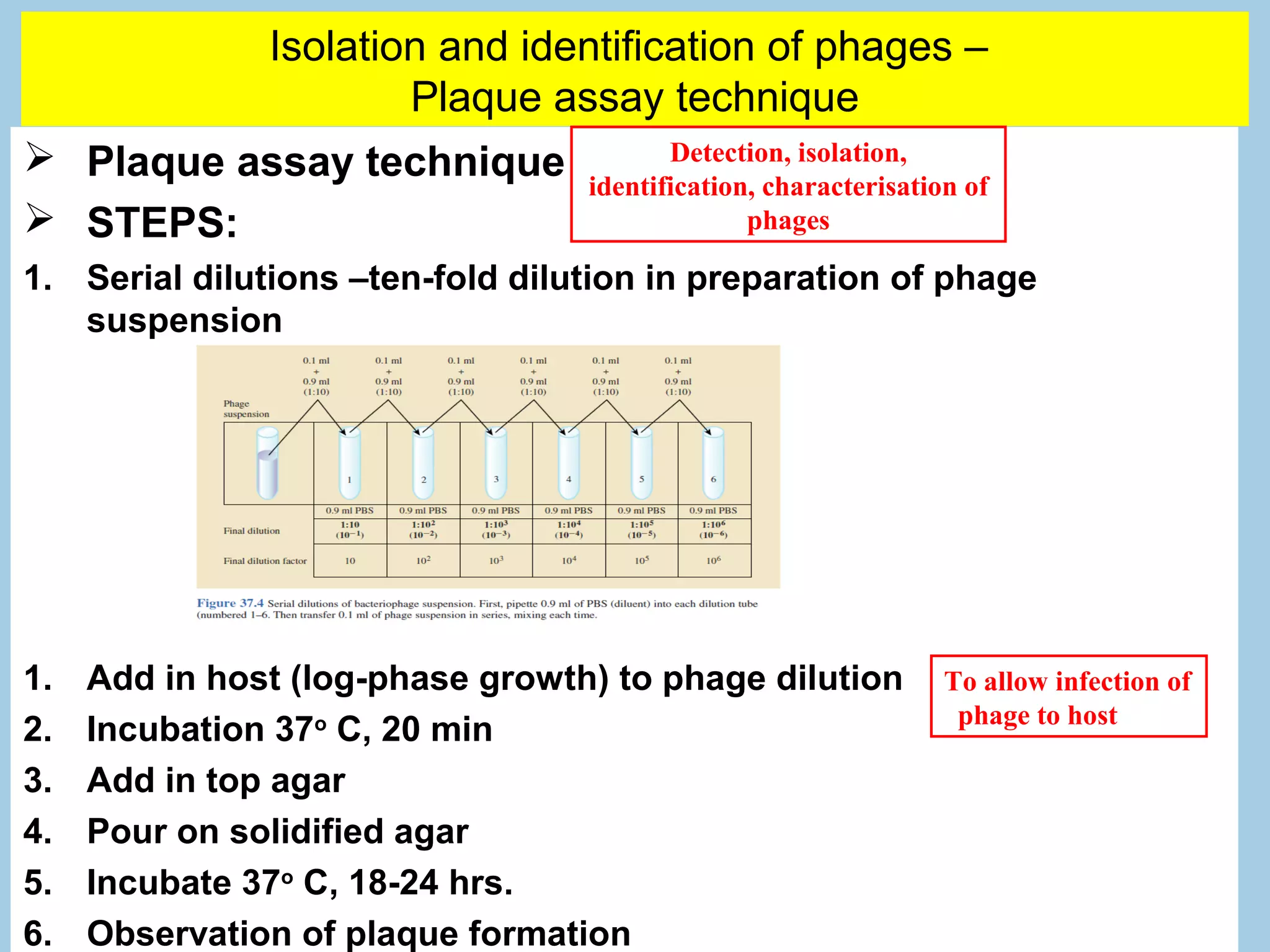

![Identification of phages –
Plaque assay technique
Basis of plaque formation:
Plaque assay – also to
calculate number of phages
present.
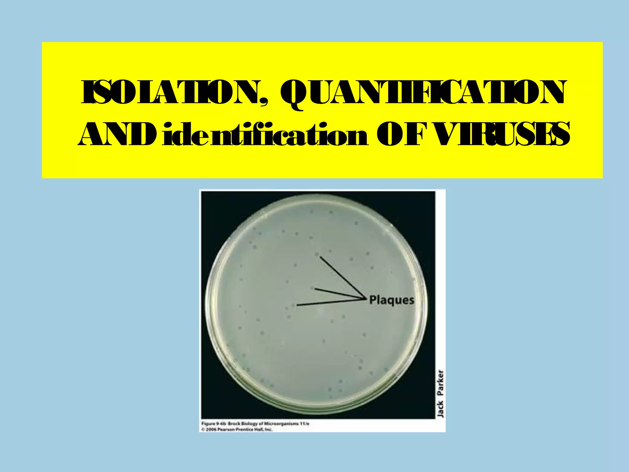
The titer of a phage
suspension, is determined
by counting the number of
plaques that form from a given
volume of suspension. Phage
titer is expressed as plaque
forming units (PFU) per
milliliter (ml).
[pfu/ml] * measurement of the
number of viable, infectious](https://image.slidesharecdn.com/chapter4isolation-identification-and-cultivation-130408155641-phpapp02/75/Chapter-4-isolation-identification-and-cultivation-12-2048.jpg)

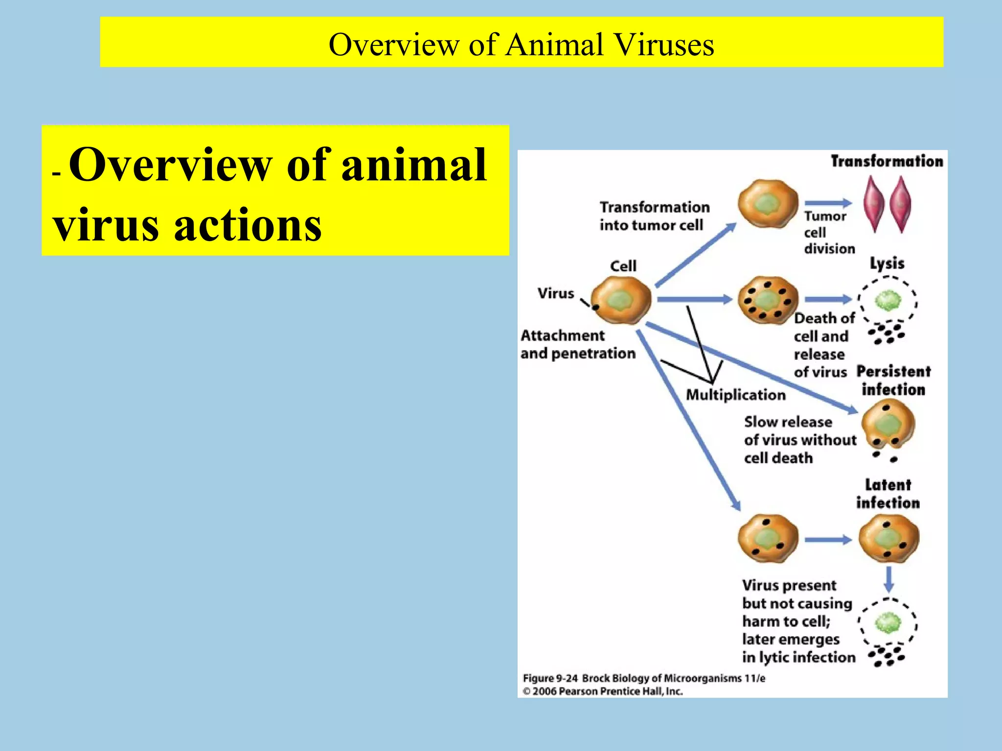
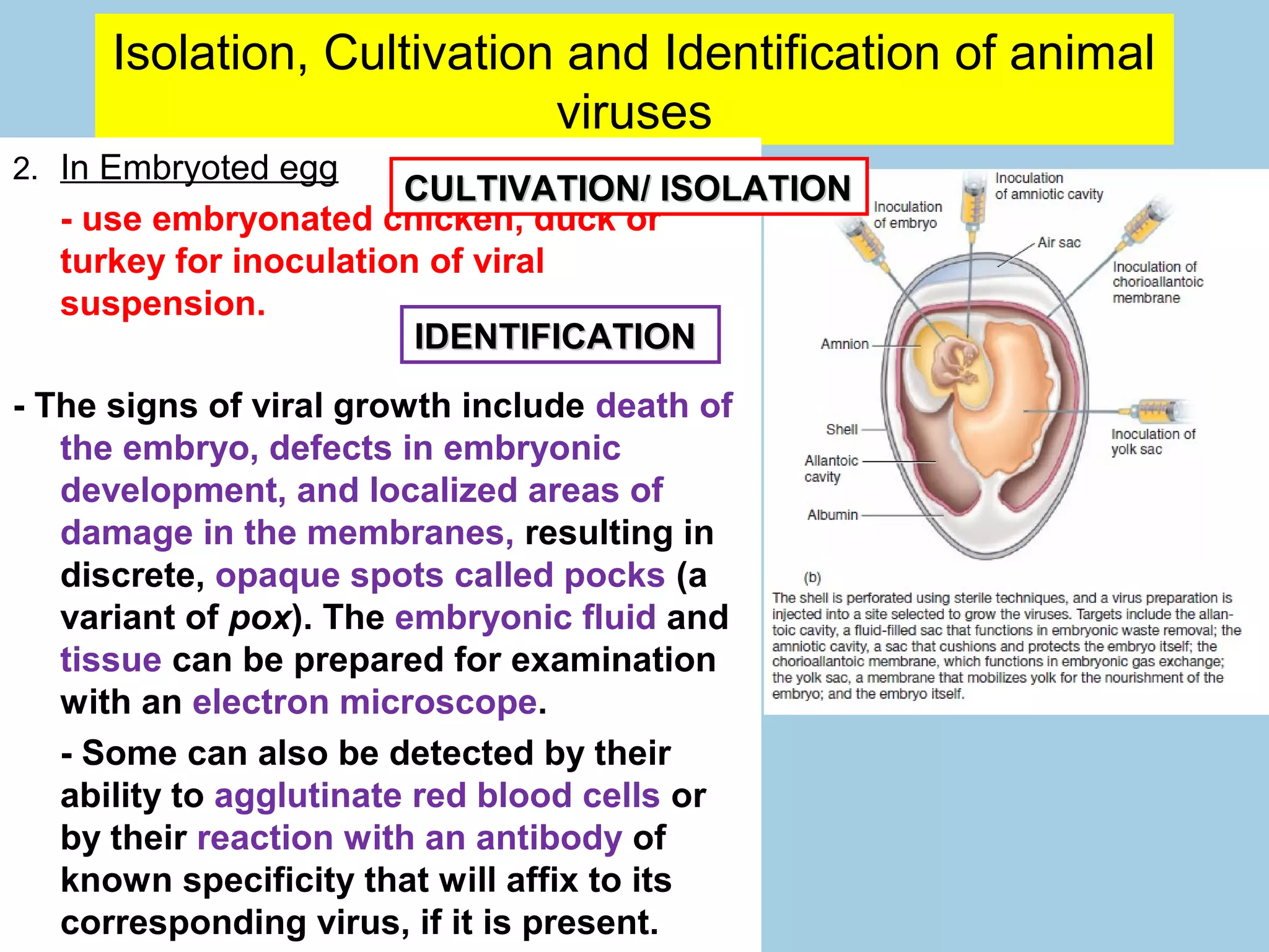
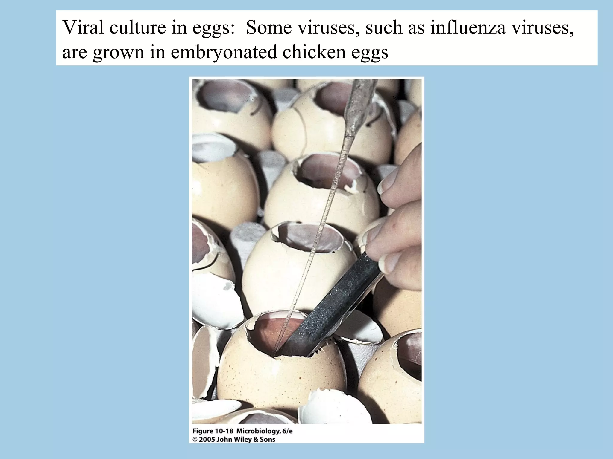
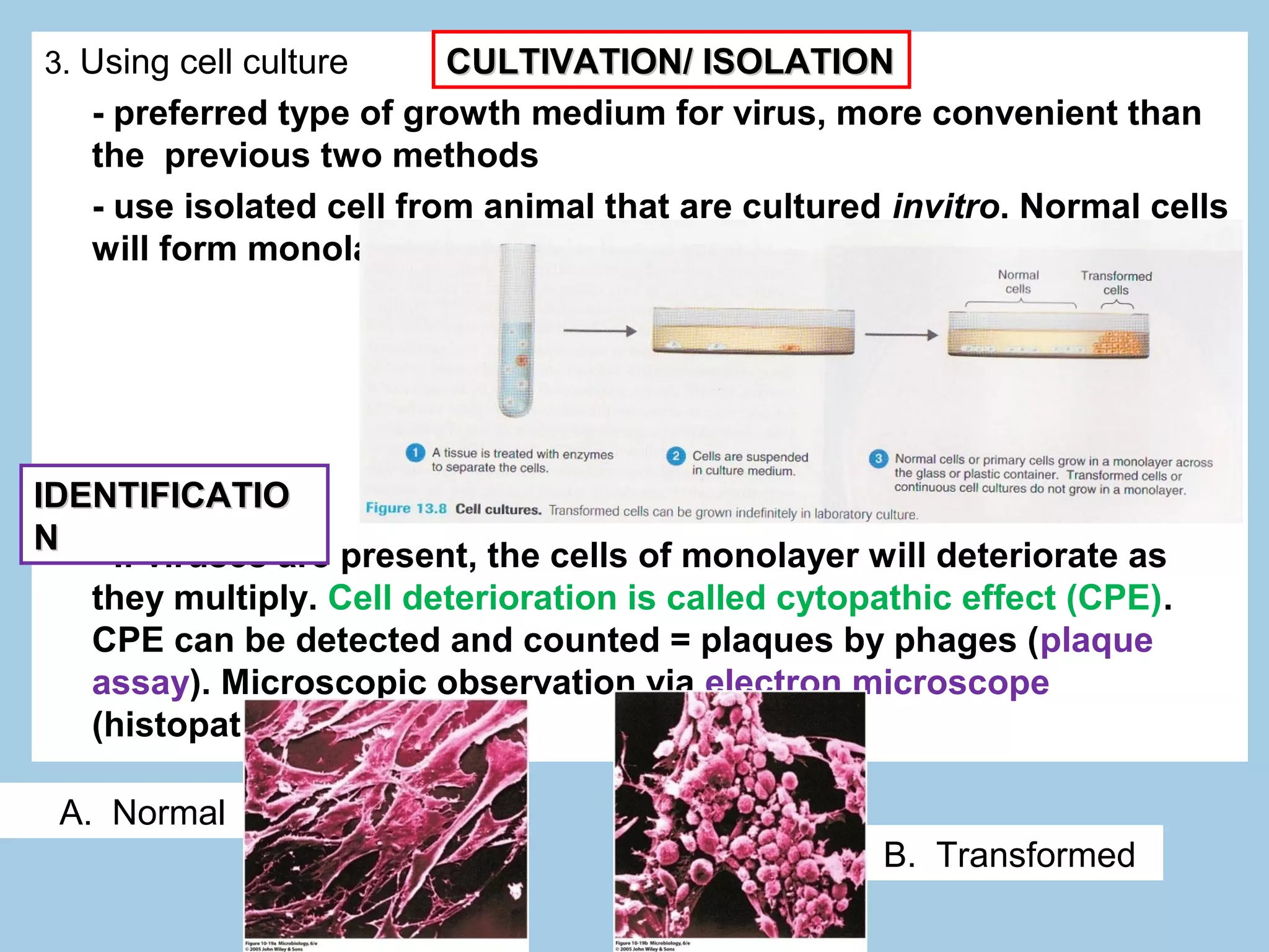
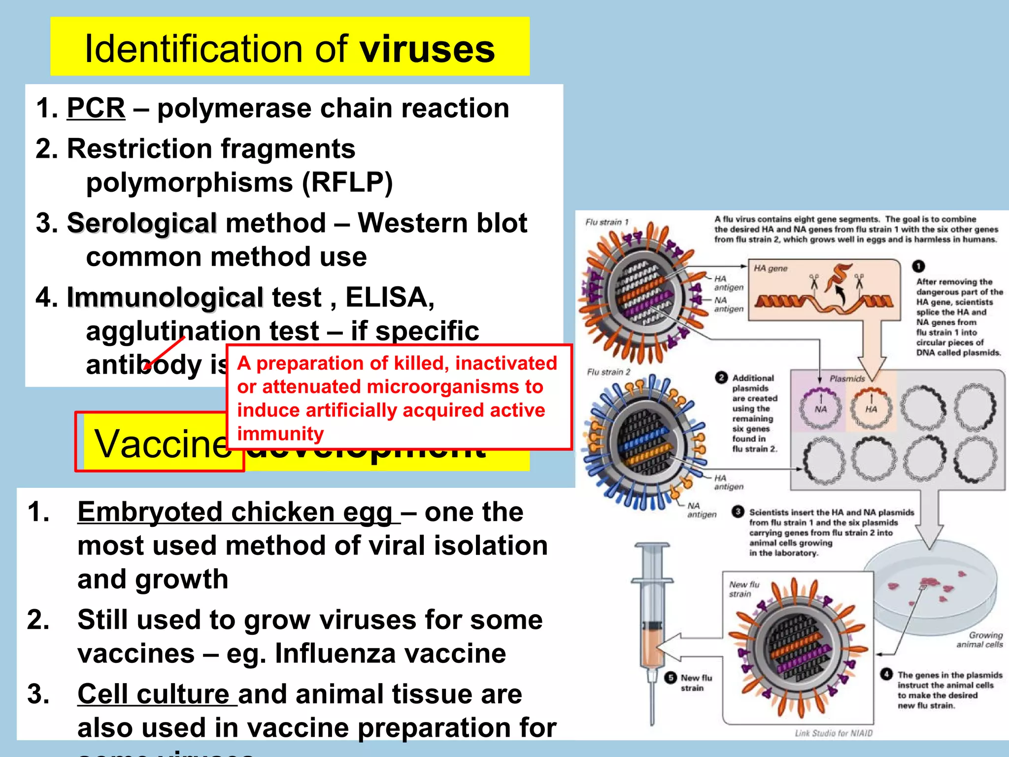

1. Viruses are cultivated in living animals, embryonated eggs, or cell cultures. 2. Signs of viral growth include death, defects, or cytopathic effects in the host. 3. Infected tissues or fluids are examined microscopically or using techniques like PCR to identify the virus.
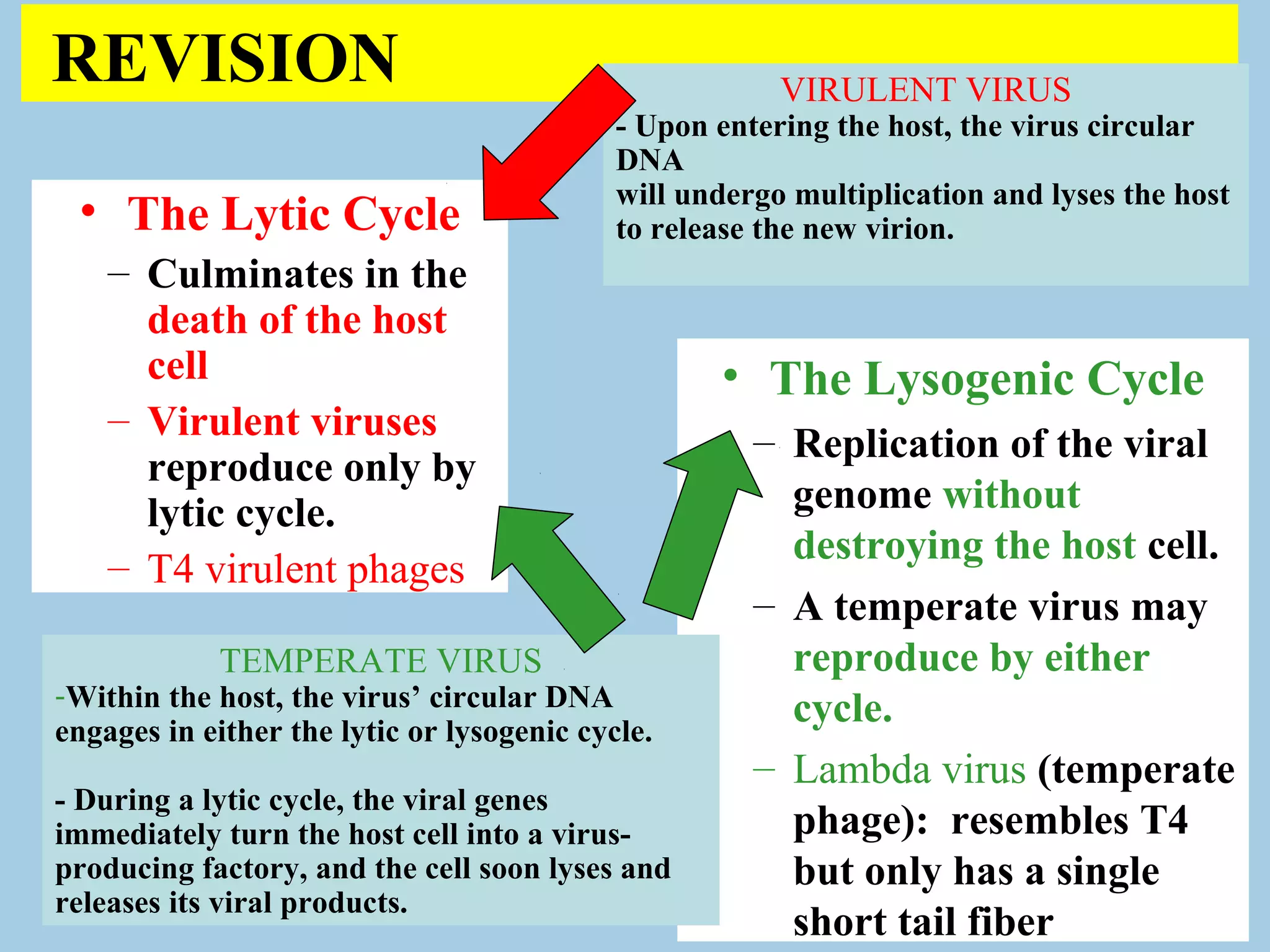
Introduction to virus replication cycles: lytic and lysogenic. Virulent and temperate viruses, and their multiplication impact on host cells.
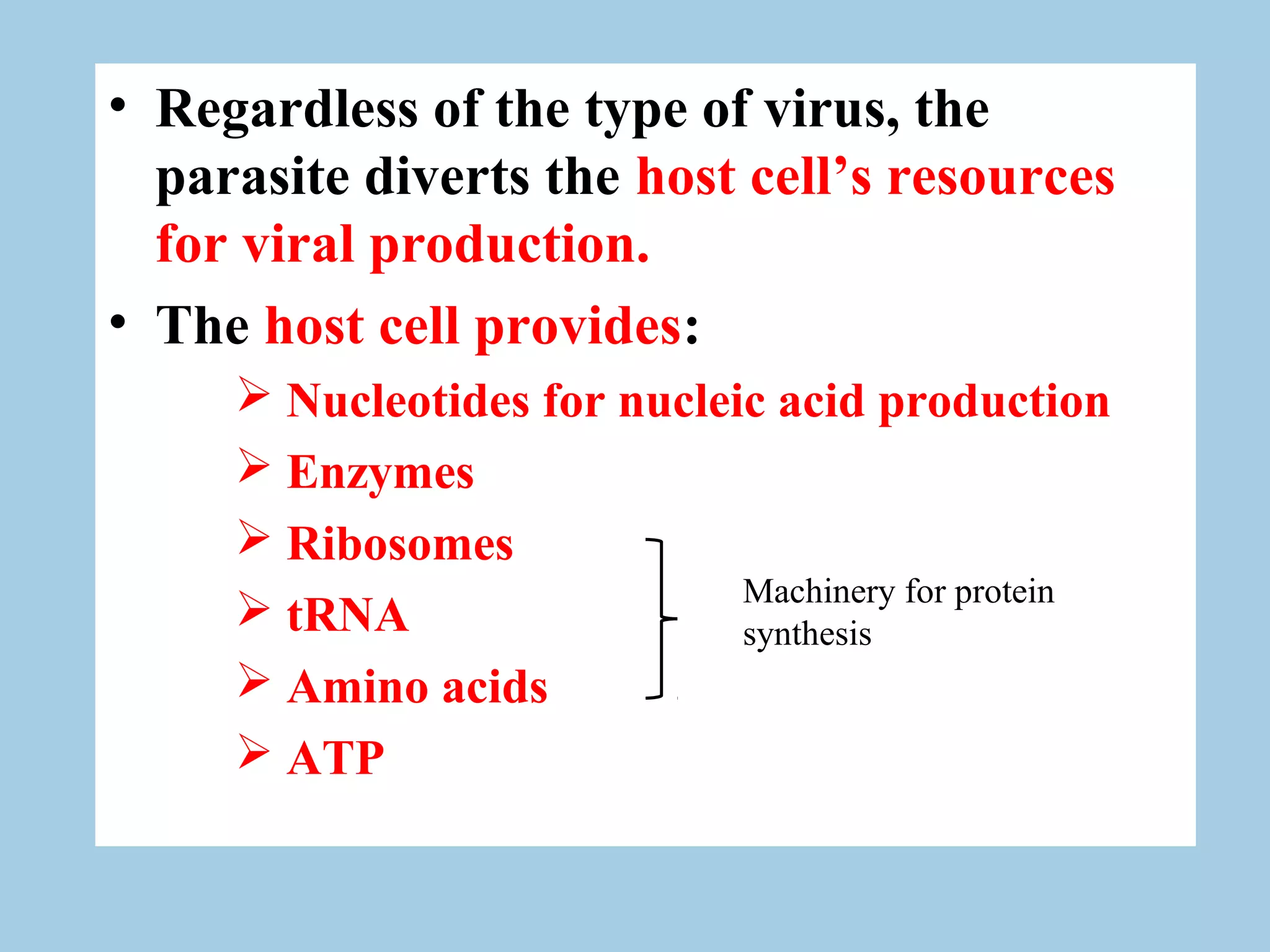
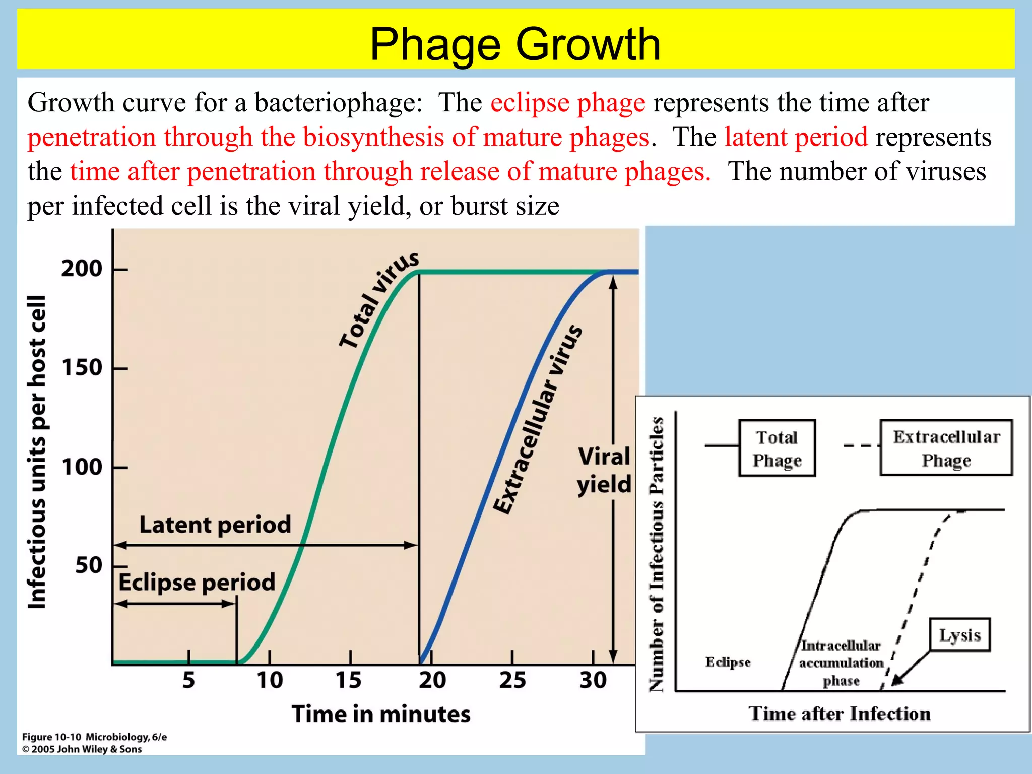
Discussion on host cellular resources used for viral production and the growth curve of bacteriophages.
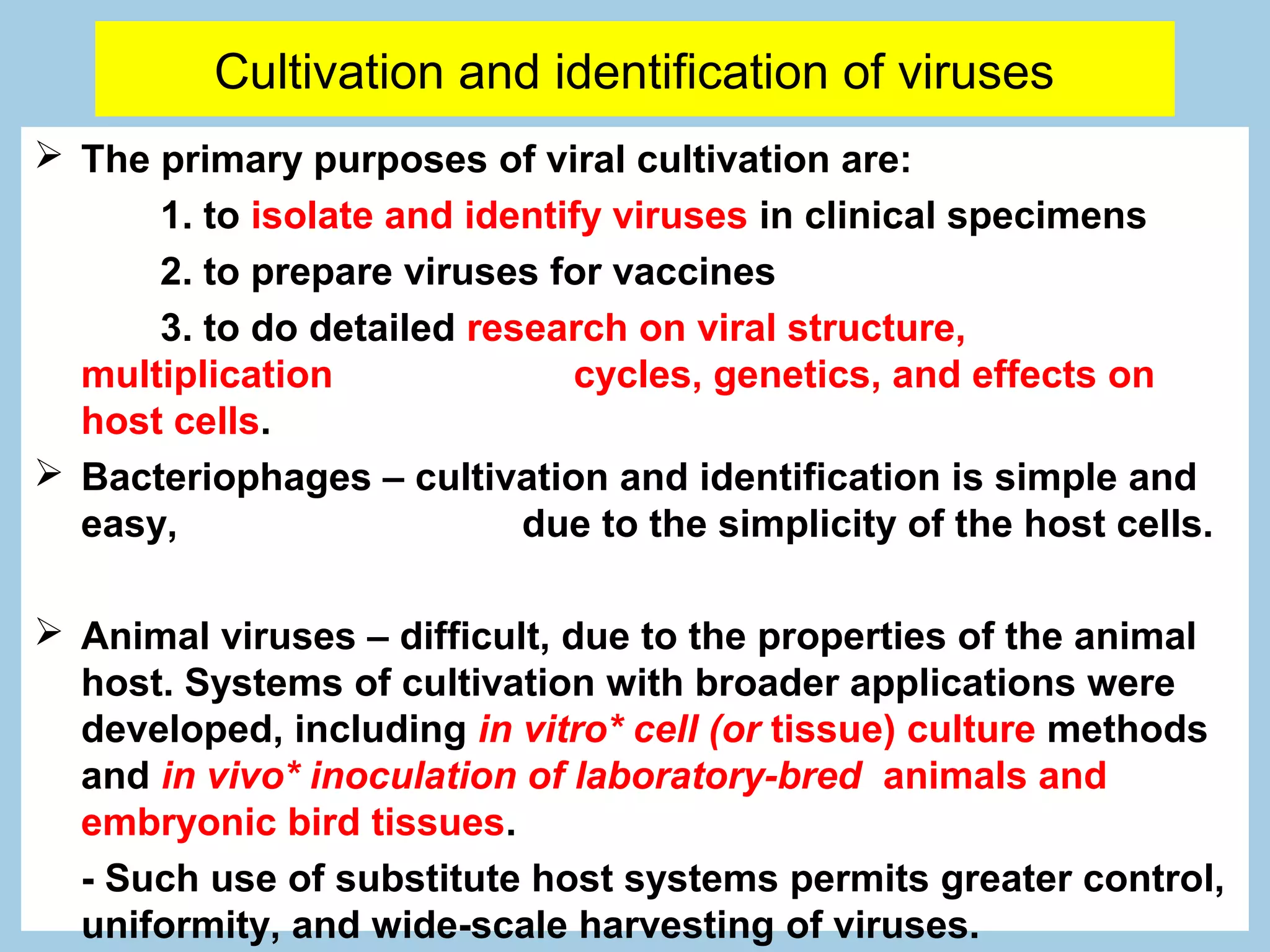
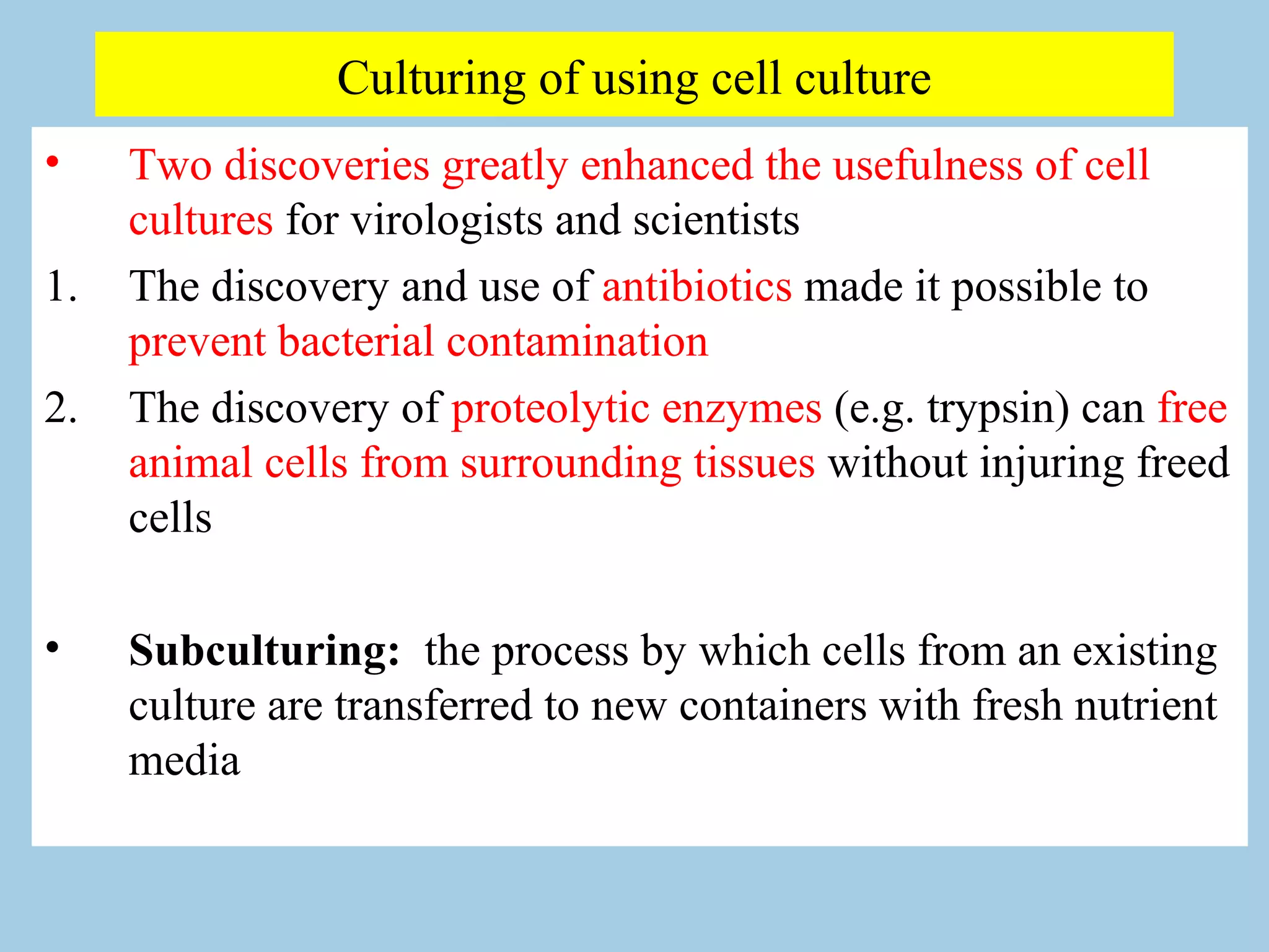
Techniques for cultivating viruses focused on isolation, vaccine preparation, and research; differences between bacteriophages and animal viruses.
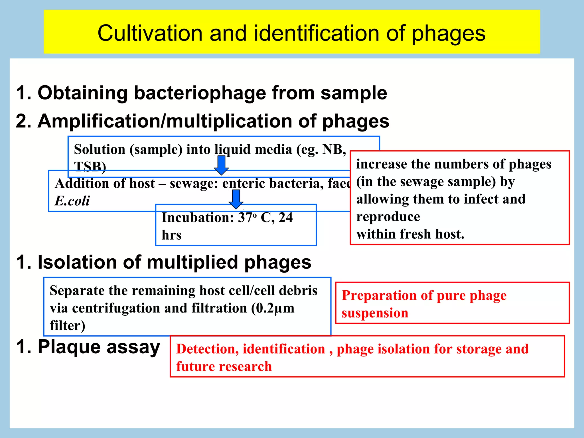
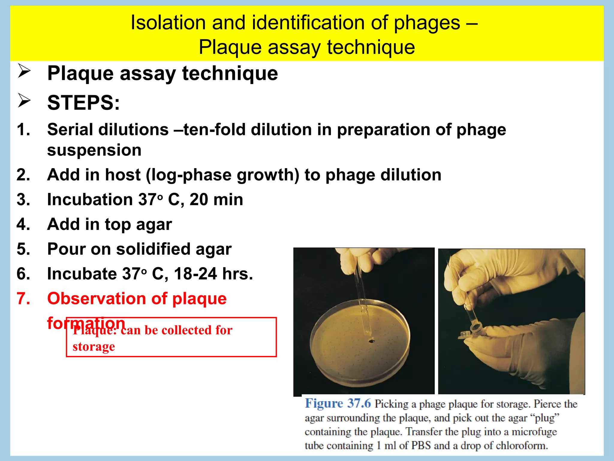
Steps for obtaining and culturing bacteriophages, including amplification using bacterial hosts and plaque assay methods for isolation.
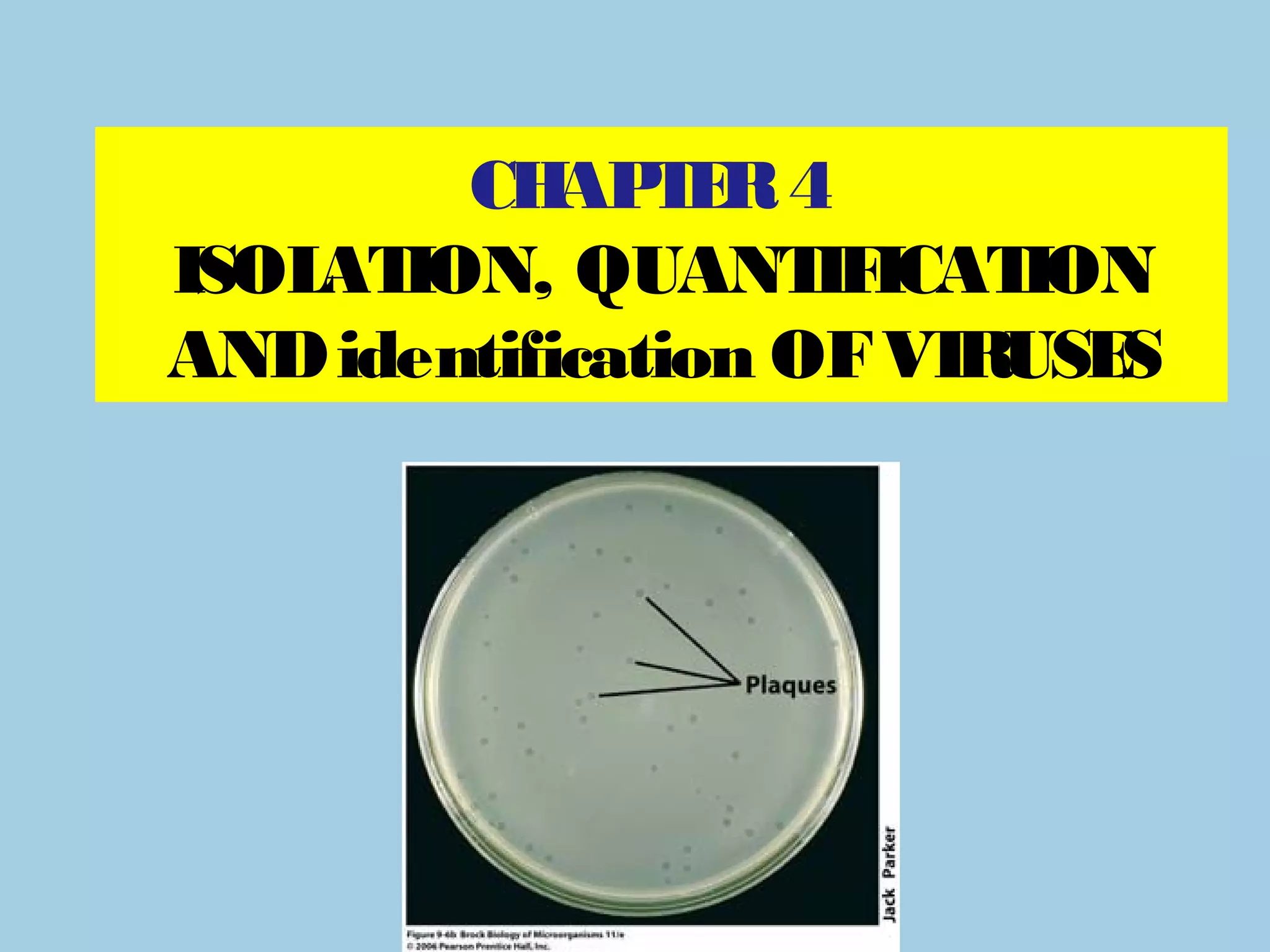
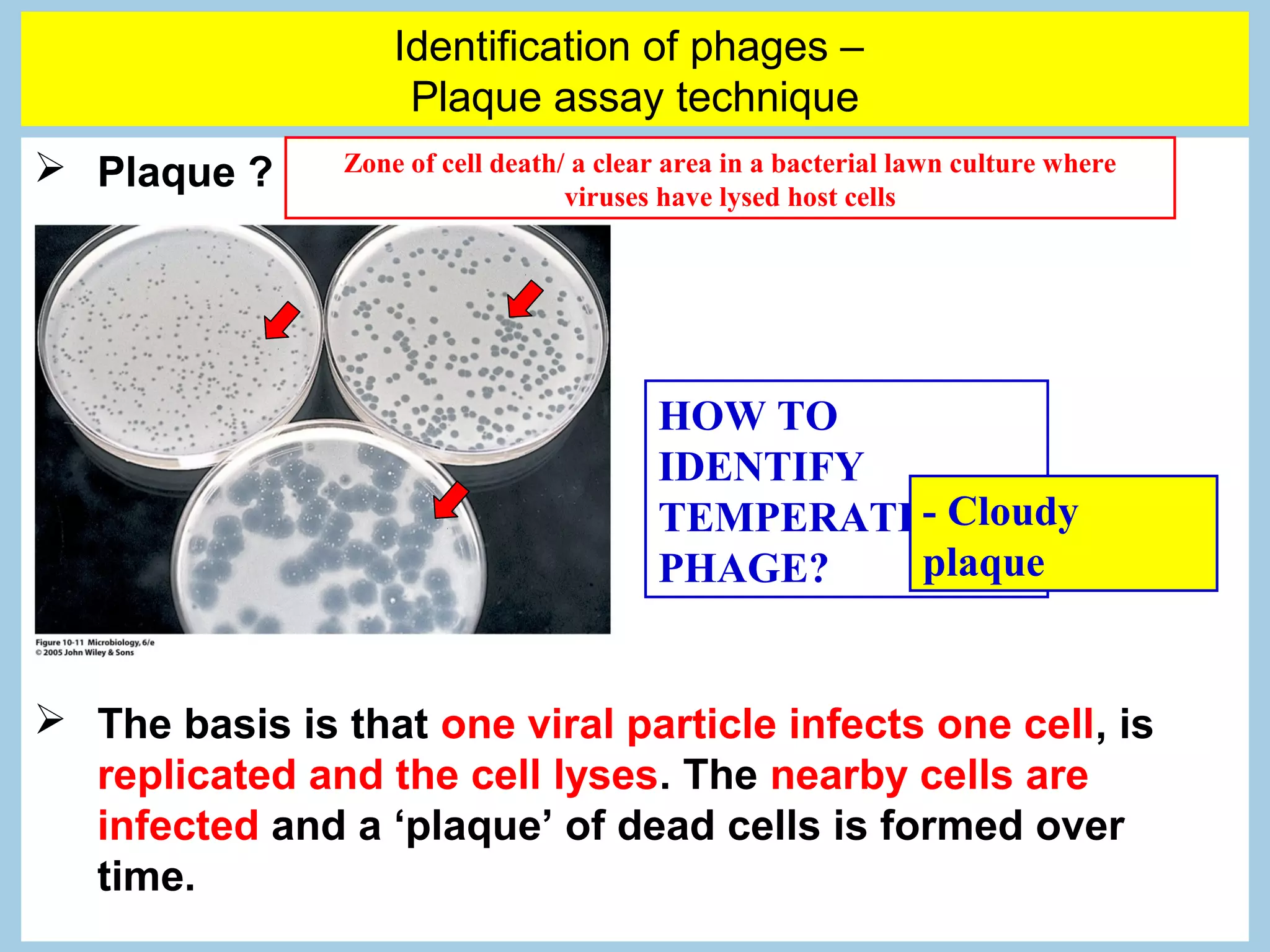
In-depth steps and concepts on plaque assays, including identification and quantification of phages through plaque formation.
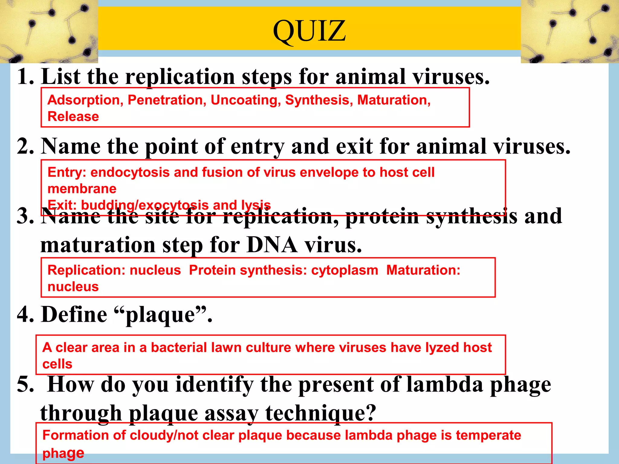
Review questions covering replication steps of animal viruses, identification of plaques, and methods to observe viral presence.
Insights on the isolation and identification of animal viruses using live animals, embryonated eggs, and cell cultures, emphasizing techniques like PCR and serological methods.


