Downloaded 5,158 times
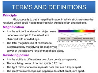
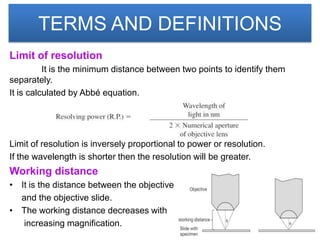
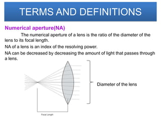
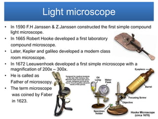
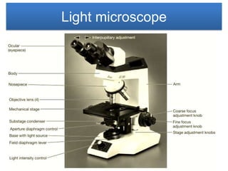
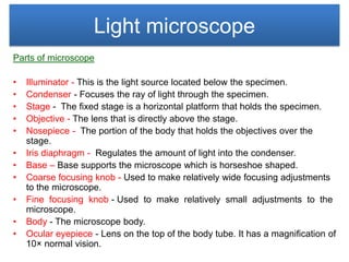
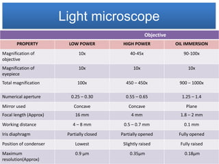
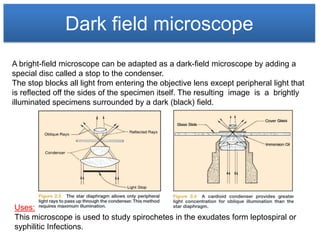
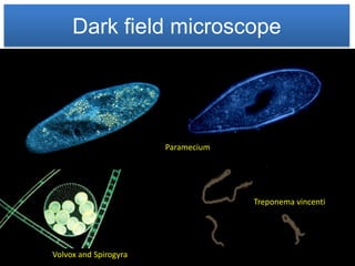
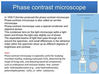
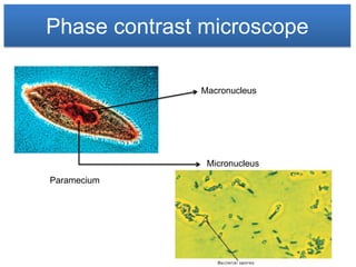
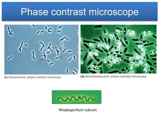
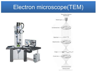
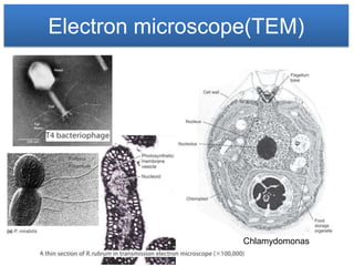
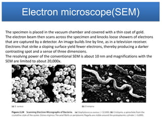
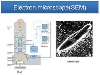
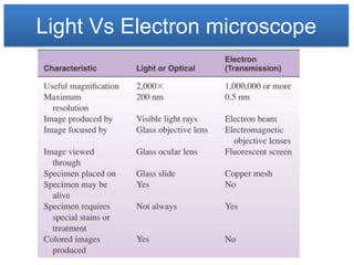
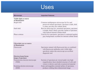


The document provides an overview of microscopy, detailing its principles such as magnification, resolving power, and various types of microscopes including light, dark field, phase contrast, fluorescence, and electron microscopes. Key definitions, components, and functions of these microscopes are explained, with emphasis on their historical development and applications in microbiology. Additionally, the document outlines the trade-offs between different microscopy techniques, such as resolution capabilities and specimen preparation requirements.