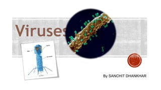
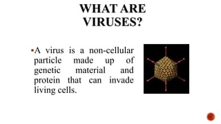
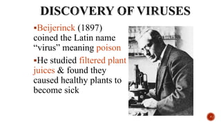
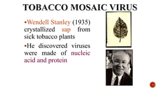
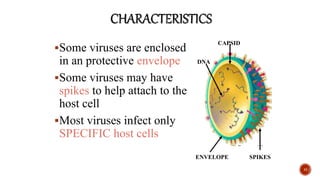
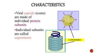
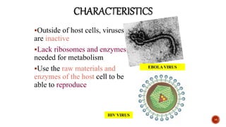
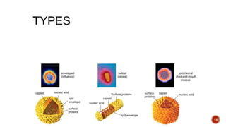
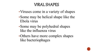
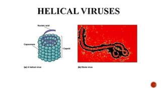
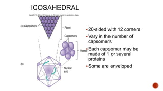
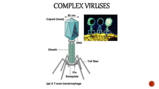
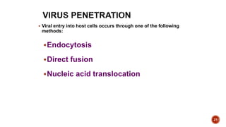
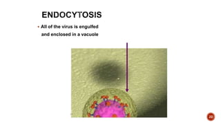
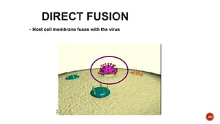
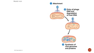
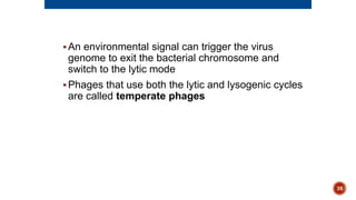
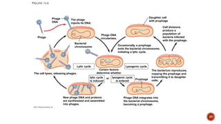
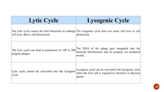
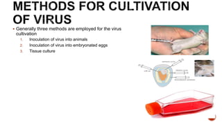
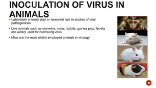
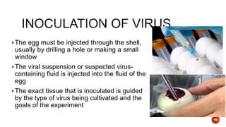
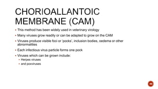
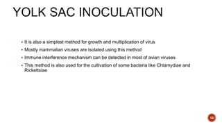
By SANCHIT DHANKHAR
T4 phage
 M13 (General properties and structure,classification,reproduction)
 Retrovirus
 Herpes simplex virus (Classification, reproduction )
 TMV (Morphology, taxonomy, and reproduction)

2
WHAT ARE
VIRUSES?
A virus is a non-cellular
particle made up of
genetic material and
protein that can invade
living cells.
3
VIRAL HISTORY
Beijerinck (1897)
coined the Latin name
“virus” meaning poison
He studied filtered plant
juices & found they
caused healthy plants to
become sick
5
Wendell Stanley (1935)
crystallized sap from
sick tobacco plants
He discovered viruses
were made of nucleic
acid and protein
6
Edward Jenner (1796)
developed a smallpox
vaccine using milder
cowpox viruses
Deadly viruses are said to
be virulent
Smallpox has been
eradicated in the world
today
7
VIEWING VIRUSES
Viruses are smaller than
the smallest cell
Measured in nanometers
Viruses couldn’t be seen
until the electron
microscope was invented
in the 20th century
8
9
VIRAL
STRUCTURE
Non living structures
Noncellular
Contain a protein coat called the capsid
Have a nucleic acid core containing DNA or
RNA
Capable of reproducing only when inside a
HOST cell
11
Some viruses are enclosed
in an protective envelope
Some viruses may have
spikes to help attach to the
host cell
Most viruses infect only
SPECIFIC host cells
12
CAPSID
ENVELOPE
DNA
SPIKES
Viral capsids (coats)
are made of
individual protein
subunits
Individual subunits
are called
capsomeres
13
CAPSOMERES
Outside of host cells, viruses
are inactive
Lack ribosomes and enzymes
needed for metabolism
Use the raw materials and
enzymes of the host cell to be
able to reproduce
14
EBOLA VIRUS
HIV VIRUS
capsid nucleic acid
lipid
envelope
surface
proteins
capsid
nucleic acid
lipid envelope
Surface proteins capsid
surface
proteins
nucleic acid
helical
(rabies)
polyhedral
(foot-and-mouth
disease)
enveloped
(influenza)
15
CHARACTERISTICS
•Some viruses cause disease
•Smallpox, measles,
mononucleosis, influenza,
colds, warts, AIDS, Ebola
•Some viruses may cause some
cancers like leukemia
•Virus-free cells are rare
16
MEASLES
•Viruses come in a variety of shapes
•Some may be helical shape like the
Ebola virus
•Some may be polyhedral shapes
like the influenza virus
•Others have more complex shapes
like bacteriophages
17
18
19
20-sided with 12 corners
Vary in the number of
capsomers
Each capsomer may be
made of 1 or several
proteins
Some are enveloped
20
21
 Viral entry into host cells occurs through one of the following
methods:
Endocytosis
Direct fusion
Nucleic acid translocation
22
 All of the virus is engulfed
and enclosed in a vacuole
23
 Host cell membrane fuses with the virus
24
Non enveloped virus injecting its
nucleic acid to the host cell
Viruses are obligate intracellular parasites, which
means they can replicate only within a host cell
Each virus has a host range, a limited number of
host cells that it can infect
25
Once a viral genome has entered a cell, the cell
begins to manufacture viral proteins
The virus makes use of host enzymes, ribosomes,
tRNAs, amino acids, ATP, and other molecules
Viral nucleic acid molecules and capsomeres
spontaneously self-assemble into new viruses
26
Phages are the best understood of all viruses
Phages have two reproductive mechanisms: the
lytic cycle and the lysogenic cycle
27
The lytic cycle is a phage replicative cycle that
culminates in the death of the host cell
The lytic cycle produces new phages and lyses
(breaks open) the host’s cell wall, releasing the
progeny viruses
A phage that reproduces only by the lytic cycle is
called a virulent phage
Bacteria have defenses against phages, including
restriction enzymes that recognize and cut up
certain phage DNA
28
Attachment
1
29
Attachment
2
1
Entry of phage
DNA and
degradation
of host DNA
30
Attachment
2
1
3
Entry of phage
DNA and
degradation
of host DNA
Synthesis of
viral genomes
and proteins
31
Attachment
2
1
4
3
Entry of phage
DNA and
degradation
of host DNA
Synthesis of
viral genomes
and proteins
Assembly
Phage assembly
Head Tail Tail
fibers
32
Attachment
2
1
5
4
3
Entry of phage
DNA and
degradation
of host DNA
Release
Synthesis of
viral genomes
and proteins
Assembly
Phage assembly
Head Tail Tail
fibers
33
The lysogenic cycle replicates the phage genome
without destroying the host
The viral DNA molecule is incorporated into the
host cell’s chromosome
This integrated viral DNA is known as a prophage
Every time the host divides, it copies the phage
DNA and passes the copies to daughter cells
34
An environmental signal can trigger the virus
genome to exit the bacterial chromosome and
switch to the lytic mode
Phages that use both the lytic and lysogenic cycles
are called temperate phages
35
lytic cycle
is induced
or
Phage DNA
circularizes.
Certain factors
determine whether
lysogenic cycle
is entered
Lysogenic cycle
Prophage
Daughter cell
with prophage
Occasionally, a prophage
exits the bacterial chromosome,
initiating a lytic cycle.
Cell divisions
produce a
population of
bacteria infected
with the prophage.
The bacterium reproduces,
copying the prophage and
transmitting it to daughter
cells.
Phage DNA integrates into
the bacterial chromosome,
becoming a prophage. 36
New phage DNA and proteins
are synthesized and assembled
into phages.
The cell lyses, releasing phages.
Phage
Phage
DNA
The phage
injects its DNA.
Bacterial
chromosome
Lytic cycle
lytic cycle
is induced
or
Phage DNA
circularizes.
Certain factors
determine whether
lysogenic cycle
is entered
Lysogenic cycle
Prophage
Daughter cell
with prophage
Occasionally, a prophage
exits the bacterial chromosome,
initiating a lytic cycle.
Cell divisions
produce a
population of
bacteria infected
with the prophage.
The bacterium reproduces,
copying the prophage and
transmitting it to daughter
cells.
Phage DNA integrates into
the bacterial chromosome,
becoming a prophage.
37
The lytic cycle of the bacteriophage: a lunar analogy
Lytic Cycle Lysogenic Cycle
The lytic cycle causes the host bacterium to undergo
cell lysis, that is, cell destruction.
The lysogenic cycle does not cause cell lysis or cell
destruction.
The lytic cycle can lead to production of 100 to 200
progeny phages.
The DNA of the phage gets integrated into the
bacterial chromosome and no progeny are produced
mostly.
Lytic cycle cannot be converted into the lysogenic
cycle.
Lysogenic cycle can be converted into lysogenic cycle
when the host cell is exposed to chemical or physical
agents.
38
 Generally three methods are employed for the virus
cultivation
1. Inoculation of virus into animals
2. Inoculation of virus into embryonated eggs
3. Tissue culture
39
 Laboratory animals play an essential role in studies of viral
pathogenesis
 Live animals such as monkeys, mice, rabbits, guinea pigs, ferrets
are widely used for cultivating virus
 Mice are the most widely employed animals in virology
40
 The different routes of inoculation in mice are:
 intracerebral
 subcutaneous
 intraperitoneal
 or intranasal
 After the animal is inoculated with the virus suspension, the animal
is:
 observed for signs of disease
 visible lesions
 or is killed so that infected tissues can be examined for virus
41
 Prior to the advent of cell culture, animal viruses could be propagated only on whole
animals or embryonated chicken eggs
 Cell cultures have replaced embryonated eggs as the preferred type of growth
medium for many viruses
 Cell culture consists of cells grown in culture media in the laboratory
 These cultures can be propagated and handled like bacterial cultures; they are more
convenient to work with than whole animals or embryonated eggs
 Primary cell lines
 Secondary cell lines
42
 Goodpasture and Burnet in 1931 first used the embryonated hen’s egg
for the cultivation of virus
 The process of cultivation of viruses in embryonated eggs depends on
the type of egg being used
 Eggs provide a suitable means for:
 the primary isolation and identification of viruses
 the maintenance of stock cultures
 and the production of vaccines
43
 An embryo is an early developmental stage of animals marked by rapid differentiation
of cells
 Birds undergo their embryonic period within the closed protective case of an egg,
which makes an incubating bird egg a nearly perfect system for viral propagation
 It is an intact and self-supporting unit, complete with its own sterile environment and
nourishment
 It furnishes several embryonic tissues that readily support viral multiplication
 Defense mechanisms are not involved in embryonated eggs
 Cost- much less, Maintenance-easier, Less labor and Readily available
44
 Chicken, duck, and turkey eggs are the most common choices for inoculation
 The egg used for cultivation must be sterile and the shell should be intact and healthy
 Rigorous sterile techniques must be used to prevent contamination by bacteria and
fungi from the air and the outer surface of the shell
45
The egg must be injected through the shell,
usually by drilling a hole or making a small
window
The viral suspension or suspected virus-
containing fluid is injected into the fluid of the
egg
The exact tissue that is inoculated is guided
by the type of virus being cultivated and the
goals of the experiment
46
 An embryonated egg offers various sites for the
cultivation of viruses
 The different sites of viral inoculation in
embryonated eggs are:
1. Chorioallantoic membrane(CAM)
2. Amniotic Cavity
3. Allantoic Cavity
4. Yolk sac
47
 The chosen route of inoculation and age of the embryo are determined by the given
virus selectivity for a certain membrane or developmental stage of the embryo
 For example Infectious bronchitis virus is propagated in the yolk sac of a 5-6 day old embryo
 whereas Rous-sarcoma virus is inoculated on the chorioallantoic membrane of a 9-11 day
old embryo and will produce pocks 5-10 days post-infection
48
 This method has been widely used in veterinary virology
 Many viruses grow readily or can be adapted to grow on the CAM
 Viruses produce visible foci or ‘pocks’, inclusion bodies, oedema or other
abnormalities
 Each infectious virus particle forms one pock
 Viruses which can be grown include:
 Herpes viruses
 and poxviruses
49
 The virus is introduced directly into the amniotic fluid that bathes the developing embryo
 The volume of fluid in the infected amniotic sac is small (1-2 ml)
 The amniotic route is recommended for the primary isolation of human viruses:
 mumps virus
 and influenza A, B and C viruses
 has little application in veterinary virology
 Newly isolated influenza viruses may require several passages before they adapt to growth by
other routes, such as allantoic
50
 Many viruses such as Newcastle disease virus can grow readily
 Other viruses such as influenza, may require repeated amniotic passages before
becoming adapted to the egg and grown in the allantoic cavity
 Allantoic inoculation is a quick and easy method that yields large amounts (8–15 ml)
of virus-infected egg fluids
51
 It is also a simplest method for growth and multiplication of virus
 Mostly mammalian viruses are isolated using this method
 Immune interference mechanism can be detected in most of avian viruses
 This method is also used for the cultivation of some bacteria like Chlamydiae and
Rickettsiae
52
THANKYOU
53

Viruses- Pharmaceutical Microbiology

  • 1.
  • 2.
    T4 phage  M13(General properties and structure,classification,reproduction)  Retrovirus  Herpes simplex virus (Classification, reproduction )  TMV (Morphology, taxonomy, and reproduction)  2
  • 3.
    WHAT ARE VIRUSES? A virusis a non-cellular particle made up of genetic material and protein that can invade living cells. 3
  • 4.
  • 5.
    Beijerinck (1897) coined theLatin name “virus” meaning poison He studied filtered plant juices & found they caused healthy plants to become sick 5
  • 6.
    Wendell Stanley (1935) crystallizedsap from sick tobacco plants He discovered viruses were made of nucleic acid and protein 6
  • 7.
    Edward Jenner (1796) developeda smallpox vaccine using milder cowpox viruses Deadly viruses are said to be virulent Smallpox has been eradicated in the world today 7
  • 8.
    VIEWING VIRUSES Viruses aresmaller than the smallest cell Measured in nanometers Viruses couldn’t be seen until the electron microscope was invented in the 20th century 8
  • 9.
  • 10.
  • 11.
    Non living structures Noncellular Containa protein coat called the capsid Have a nucleic acid core containing DNA or RNA Capable of reproducing only when inside a HOST cell 11
  • 12.
    Some viruses areenclosed in an protective envelope Some viruses may have spikes to help attach to the host cell Most viruses infect only SPECIFIC host cells 12 CAPSID ENVELOPE DNA SPIKES
  • 13.
    Viral capsids (coats) aremade of individual protein subunits Individual subunits are called capsomeres 13 CAPSOMERES
  • 14.
    Outside of hostcells, viruses are inactive Lack ribosomes and enzymes needed for metabolism Use the raw materials and enzymes of the host cell to be able to reproduce 14 EBOLA VIRUS HIV VIRUS
  • 15.
    capsid nucleic acid lipid envelope surface proteins capsid nucleicacid lipid envelope Surface proteins capsid surface proteins nucleic acid helical (rabies) polyhedral (foot-and-mouth disease) enveloped (influenza) 15
  • 16.
    CHARACTERISTICS •Some viruses causedisease •Smallpox, measles, mononucleosis, influenza, colds, warts, AIDS, Ebola •Some viruses may cause some cancers like leukemia •Virus-free cells are rare 16 MEASLES
  • 17.
    •Viruses come ina variety of shapes •Some may be helical shape like the Ebola virus •Some may be polyhedral shapes like the influenza virus •Others have more complex shapes like bacteriophages 17
  • 18.
  • 19.
    19 20-sided with 12corners Vary in the number of capsomers Each capsomer may be made of 1 or several proteins Some are enveloped
  • 20.
  • 21.
    21  Viral entryinto host cells occurs through one of the following methods: Endocytosis Direct fusion Nucleic acid translocation
  • 22.
    22  All ofthe virus is engulfed and enclosed in a vacuole
  • 23.
    23  Host cellmembrane fuses with the virus
  • 24.
    24 Non enveloped virusinjecting its nucleic acid to the host cell
  • 25.
    Viruses are obligateintracellular parasites, which means they can replicate only within a host cell Each virus has a host range, a limited number of host cells that it can infect 25
  • 26.
    Once a viralgenome has entered a cell, the cell begins to manufacture viral proteins The virus makes use of host enzymes, ribosomes, tRNAs, amino acids, ATP, and other molecules Viral nucleic acid molecules and capsomeres spontaneously self-assemble into new viruses 26
  • 27.
    Phages are thebest understood of all viruses Phages have two reproductive mechanisms: the lytic cycle and the lysogenic cycle 27
  • 28.
    The lytic cycleis a phage replicative cycle that culminates in the death of the host cell The lytic cycle produces new phages and lyses (breaks open) the host’s cell wall, releasing the progeny viruses A phage that reproduces only by the lytic cycle is called a virulent phage Bacteria have defenses against phages, including restriction enzymes that recognize and cut up certain phage DNA 28
  • 29.
  • 30.
    Attachment 2 1 Entry of phage DNAand degradation of host DNA 30
  • 31.
    Attachment 2 1 3 Entry of phage DNAand degradation of host DNA Synthesis of viral genomes and proteins 31
  • 32.
    Attachment 2 1 4 3 Entry of phage DNAand degradation of host DNA Synthesis of viral genomes and proteins Assembly Phage assembly Head Tail Tail fibers 32
  • 33.
    Attachment 2 1 5 4 3 Entry of phage DNAand degradation of host DNA Release Synthesis of viral genomes and proteins Assembly Phage assembly Head Tail Tail fibers 33
  • 34.
    The lysogenic cyclereplicates the phage genome without destroying the host The viral DNA molecule is incorporated into the host cell’s chromosome This integrated viral DNA is known as a prophage Every time the host divides, it copies the phage DNA and passes the copies to daughter cells 34
  • 35.
    An environmental signalcan trigger the virus genome to exit the bacterial chromosome and switch to the lytic mode Phages that use both the lytic and lysogenic cycles are called temperate phages 35
  • 36.
    lytic cycle is induced or PhageDNA circularizes. Certain factors determine whether lysogenic cycle is entered Lysogenic cycle Prophage Daughter cell with prophage Occasionally, a prophage exits the bacterial chromosome, initiating a lytic cycle. Cell divisions produce a population of bacteria infected with the prophage. The bacterium reproduces, copying the prophage and transmitting it to daughter cells. Phage DNA integrates into the bacterial chromosome, becoming a prophage. 36
  • 37.
    New phage DNAand proteins are synthesized and assembled into phages. The cell lyses, releasing phages. Phage Phage DNA The phage injects its DNA. Bacterial chromosome Lytic cycle lytic cycle is induced or Phage DNA circularizes. Certain factors determine whether lysogenic cycle is entered Lysogenic cycle Prophage Daughter cell with prophage Occasionally, a prophage exits the bacterial chromosome, initiating a lytic cycle. Cell divisions produce a population of bacteria infected with the prophage. The bacterium reproduces, copying the prophage and transmitting it to daughter cells. Phage DNA integrates into the bacterial chromosome, becoming a prophage. 37
  • 38.
    The lytic cycleof the bacteriophage: a lunar analogy Lytic Cycle Lysogenic Cycle The lytic cycle causes the host bacterium to undergo cell lysis, that is, cell destruction. The lysogenic cycle does not cause cell lysis or cell destruction. The lytic cycle can lead to production of 100 to 200 progeny phages. The DNA of the phage gets integrated into the bacterial chromosome and no progeny are produced mostly. Lytic cycle cannot be converted into the lysogenic cycle. Lysogenic cycle can be converted into lysogenic cycle when the host cell is exposed to chemical or physical agents. 38
  • 39.
     Generally threemethods are employed for the virus cultivation 1. Inoculation of virus into animals 2. Inoculation of virus into embryonated eggs 3. Tissue culture 39
  • 40.
     Laboratory animalsplay an essential role in studies of viral pathogenesis  Live animals such as monkeys, mice, rabbits, guinea pigs, ferrets are widely used for cultivating virus  Mice are the most widely employed animals in virology 40
  • 41.
     The differentroutes of inoculation in mice are:  intracerebral  subcutaneous  intraperitoneal  or intranasal  After the animal is inoculated with the virus suspension, the animal is:  observed for signs of disease  visible lesions  or is killed so that infected tissues can be examined for virus 41
  • 42.
     Prior tothe advent of cell culture, animal viruses could be propagated only on whole animals or embryonated chicken eggs  Cell cultures have replaced embryonated eggs as the preferred type of growth medium for many viruses  Cell culture consists of cells grown in culture media in the laboratory  These cultures can be propagated and handled like bacterial cultures; they are more convenient to work with than whole animals or embryonated eggs  Primary cell lines  Secondary cell lines 42
  • 43.
     Goodpasture andBurnet in 1931 first used the embryonated hen’s egg for the cultivation of virus  The process of cultivation of viruses in embryonated eggs depends on the type of egg being used  Eggs provide a suitable means for:  the primary isolation and identification of viruses  the maintenance of stock cultures  and the production of vaccines 43
  • 44.
     An embryois an early developmental stage of animals marked by rapid differentiation of cells  Birds undergo their embryonic period within the closed protective case of an egg, which makes an incubating bird egg a nearly perfect system for viral propagation  It is an intact and self-supporting unit, complete with its own sterile environment and nourishment  It furnishes several embryonic tissues that readily support viral multiplication  Defense mechanisms are not involved in embryonated eggs  Cost- much less, Maintenance-easier, Less labor and Readily available 44
  • 45.
     Chicken, duck,and turkey eggs are the most common choices for inoculation  The egg used for cultivation must be sterile and the shell should be intact and healthy  Rigorous sterile techniques must be used to prevent contamination by bacteria and fungi from the air and the outer surface of the shell 45
  • 46.
    The egg mustbe injected through the shell, usually by drilling a hole or making a small window The viral suspension or suspected virus- containing fluid is injected into the fluid of the egg The exact tissue that is inoculated is guided by the type of virus being cultivated and the goals of the experiment 46
  • 47.
     An embryonatedegg offers various sites for the cultivation of viruses  The different sites of viral inoculation in embryonated eggs are: 1. Chorioallantoic membrane(CAM) 2. Amniotic Cavity 3. Allantoic Cavity 4. Yolk sac 47
  • 48.
     The chosenroute of inoculation and age of the embryo are determined by the given virus selectivity for a certain membrane or developmental stage of the embryo  For example Infectious bronchitis virus is propagated in the yolk sac of a 5-6 day old embryo  whereas Rous-sarcoma virus is inoculated on the chorioallantoic membrane of a 9-11 day old embryo and will produce pocks 5-10 days post-infection 48
  • 49.
     This methodhas been widely used in veterinary virology  Many viruses grow readily or can be adapted to grow on the CAM  Viruses produce visible foci or ‘pocks’, inclusion bodies, oedema or other abnormalities  Each infectious virus particle forms one pock  Viruses which can be grown include:  Herpes viruses  and poxviruses 49
  • 50.
     The virusis introduced directly into the amniotic fluid that bathes the developing embryo  The volume of fluid in the infected amniotic sac is small (1-2 ml)  The amniotic route is recommended for the primary isolation of human viruses:  mumps virus  and influenza A, B and C viruses  has little application in veterinary virology  Newly isolated influenza viruses may require several passages before they adapt to growth by other routes, such as allantoic 50
  • 51.
     Many virusessuch as Newcastle disease virus can grow readily  Other viruses such as influenza, may require repeated amniotic passages before becoming adapted to the egg and grown in the allantoic cavity  Allantoic inoculation is a quick and easy method that yields large amounts (8–15 ml) of virus-infected egg fluids 51
  • 52.
     It isalso a simplest method for growth and multiplication of virus  Mostly mammalian viruses are isolated using this method  Immune interference mechanism can be detected in most of avian viruses  This method is also used for the cultivation of some bacteria like Chlamydiae and Rickettsiae 52
  • 53.