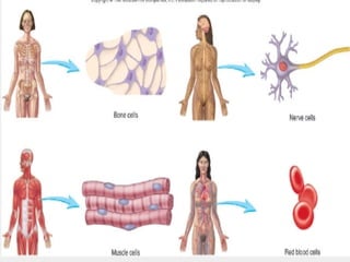
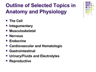
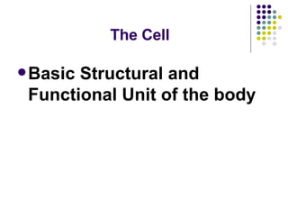
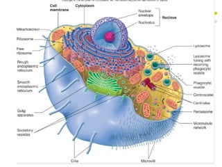
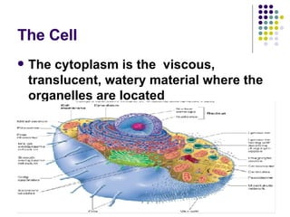
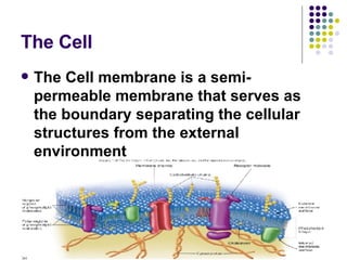
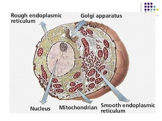
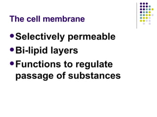
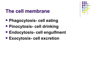
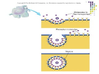
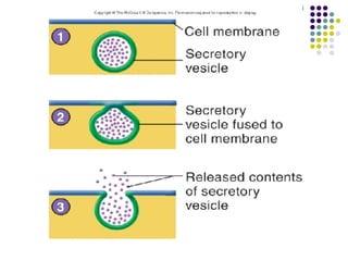
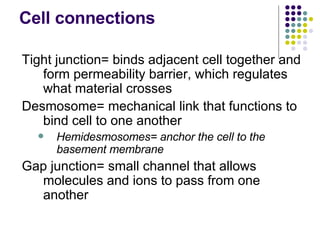
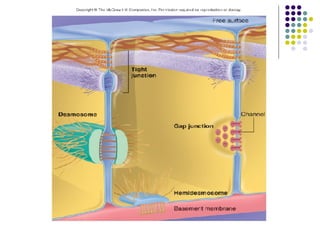
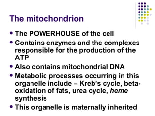
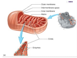
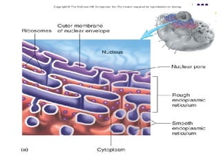
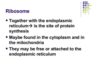
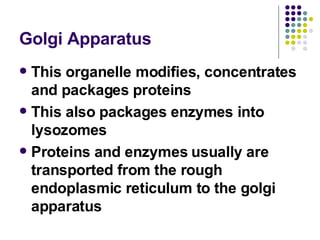
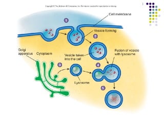
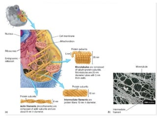
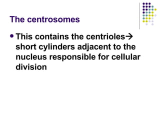
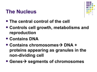
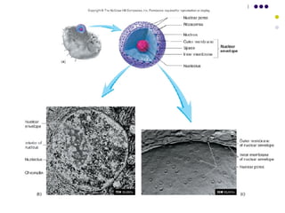
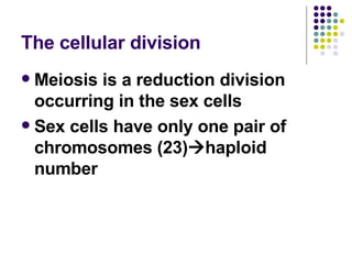
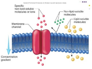
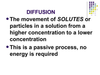
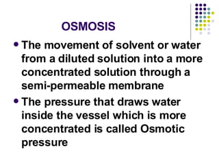
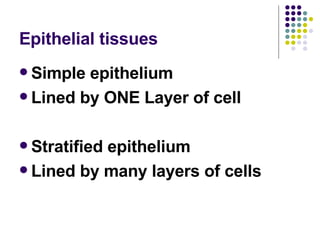
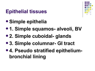
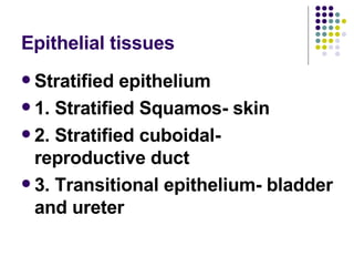
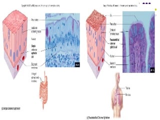
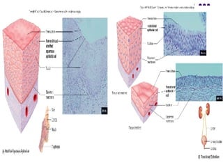
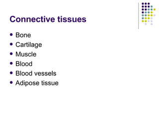
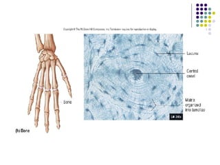
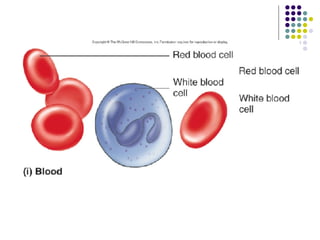
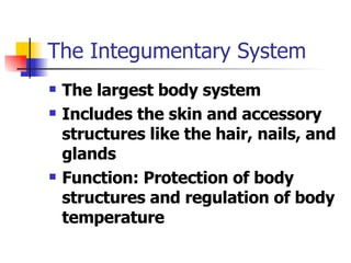
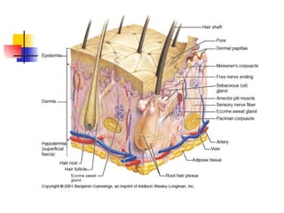
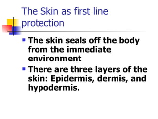
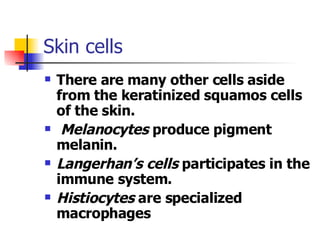
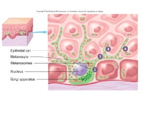
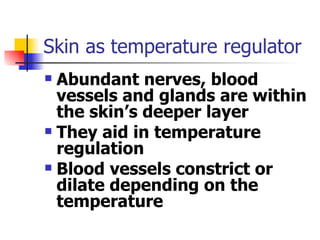
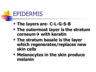
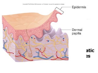
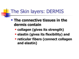
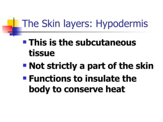
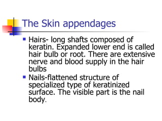
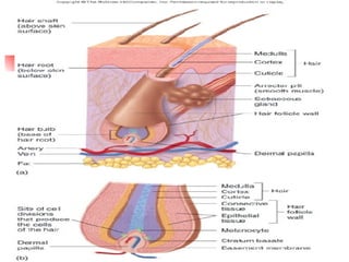
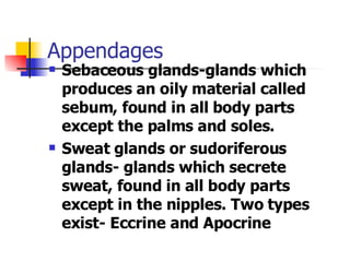
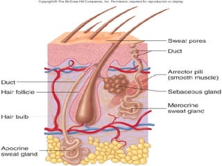
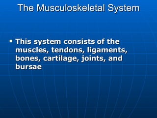
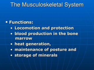
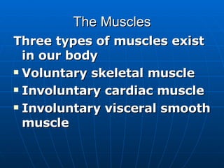
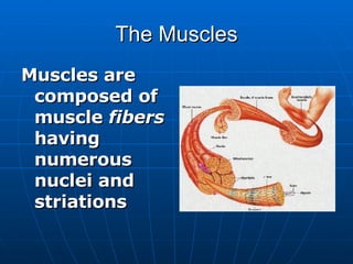
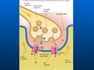
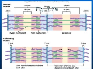
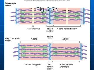
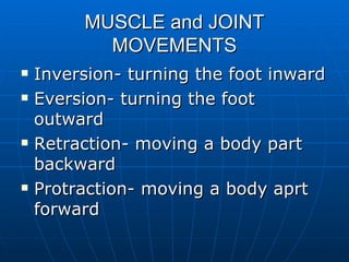
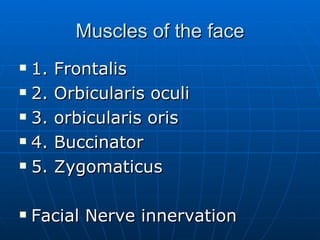
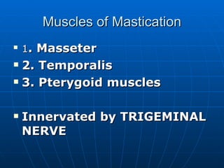
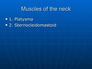
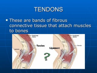
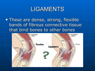
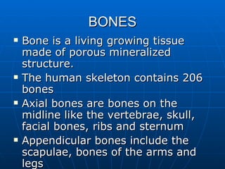
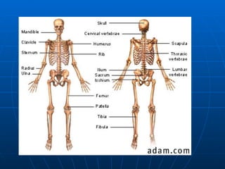
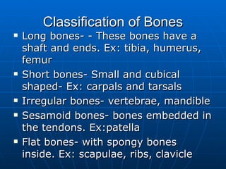
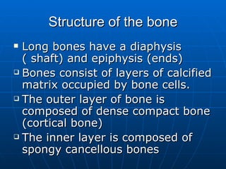
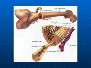
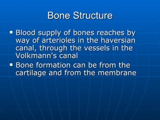
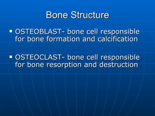
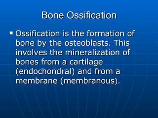
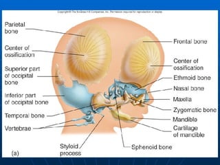
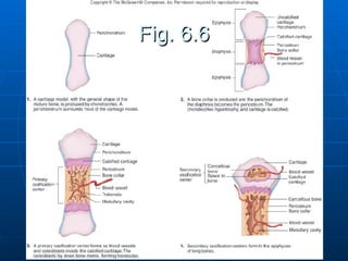
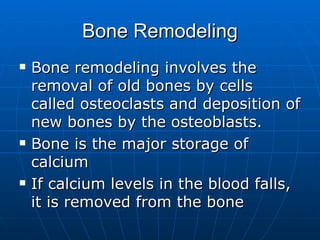
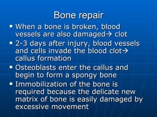
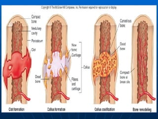
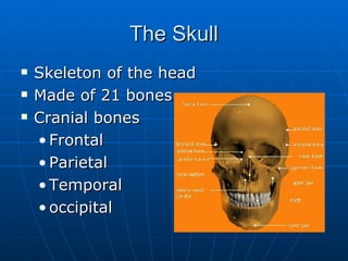
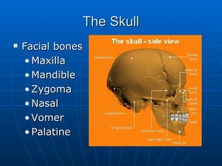
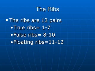
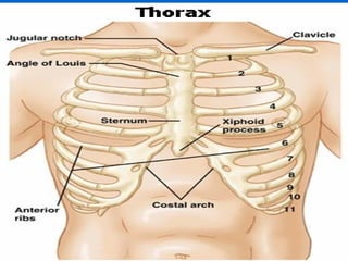
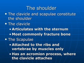
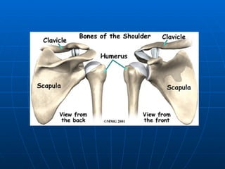
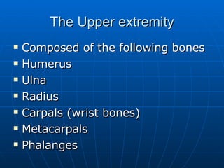
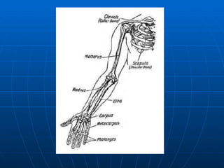
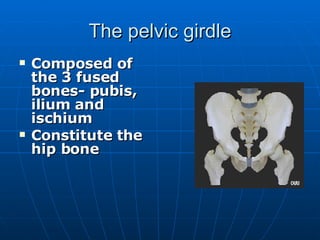
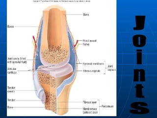
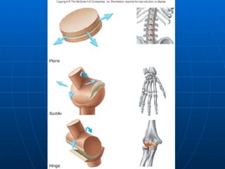
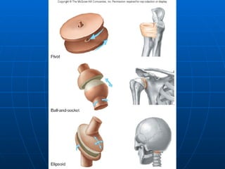
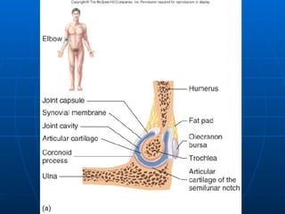
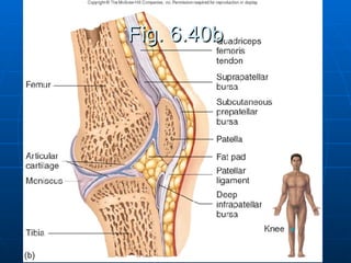
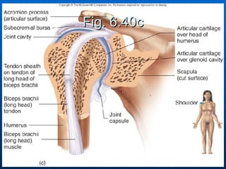
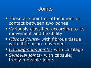
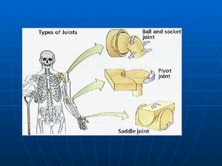
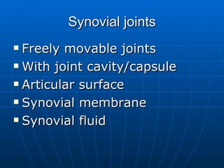
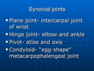
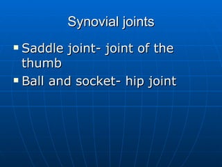
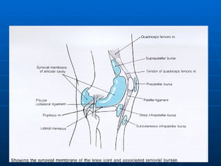
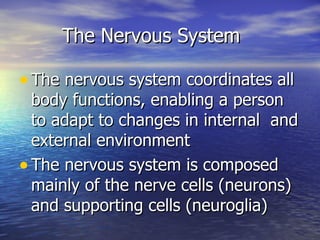
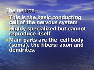
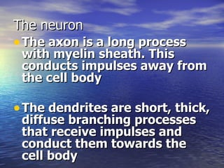
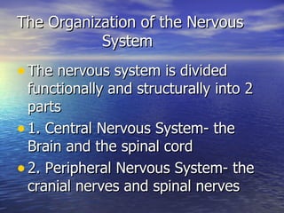
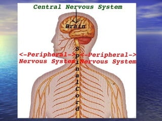
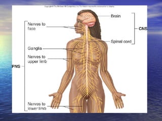
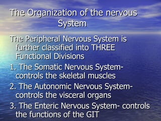
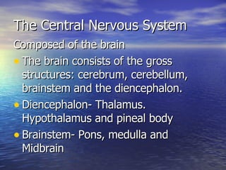
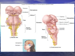
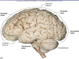
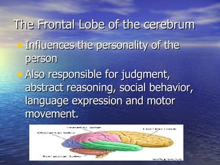
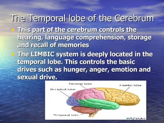
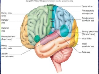
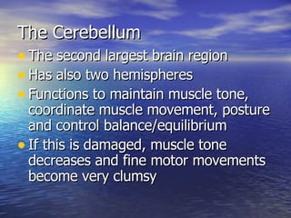
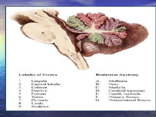
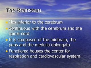
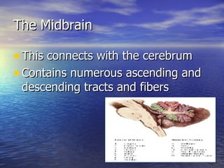
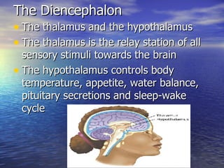
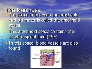
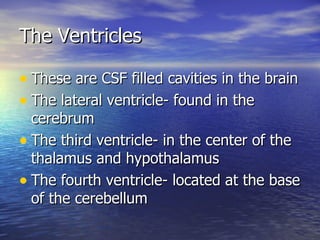
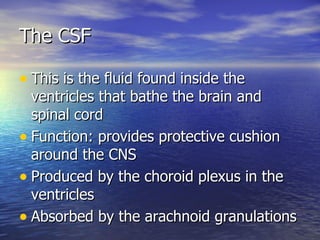
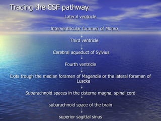
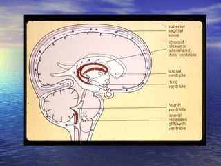
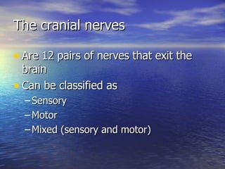
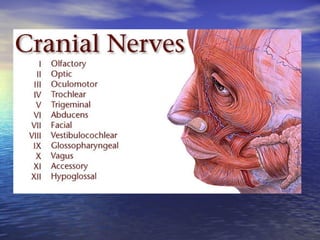
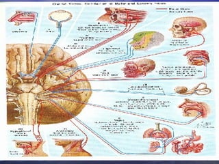
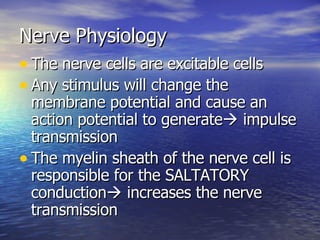
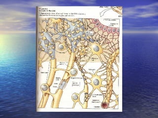
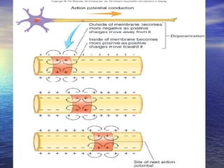
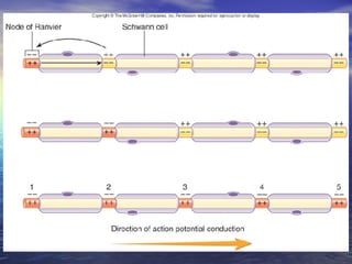
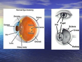
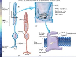
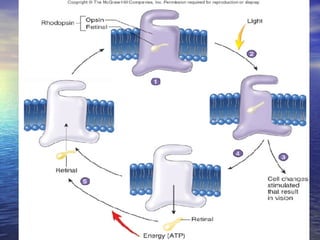
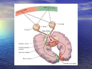
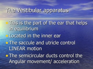
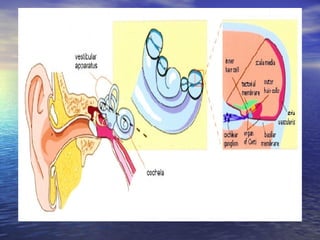
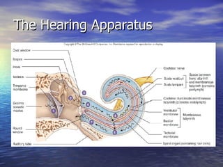
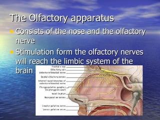
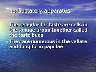
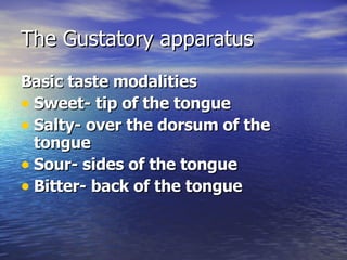
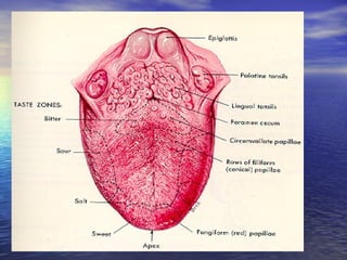
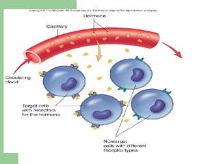
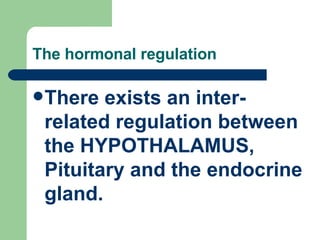
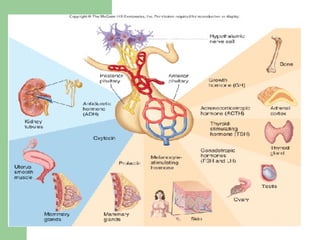
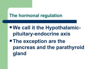
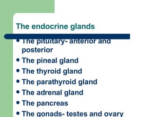
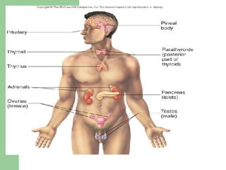
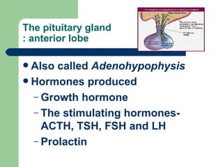
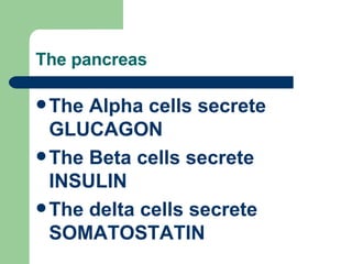
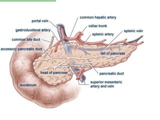
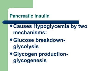
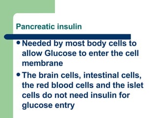
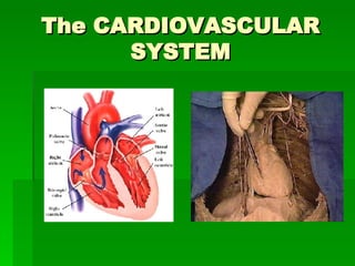
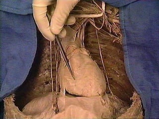
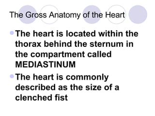
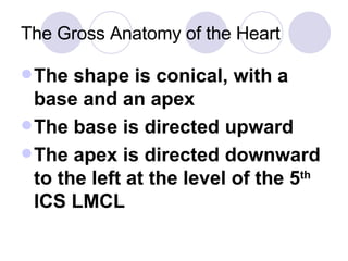
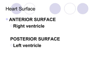
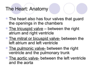
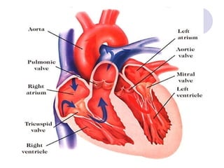
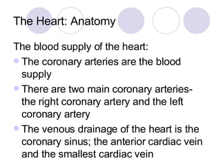
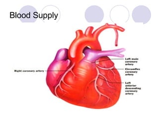
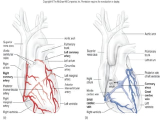
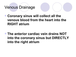
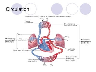
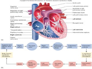
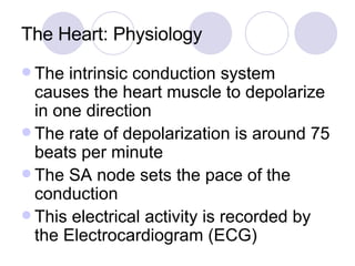
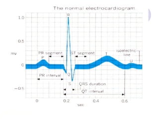
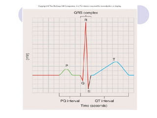
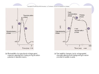
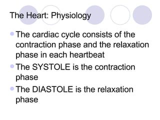
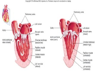
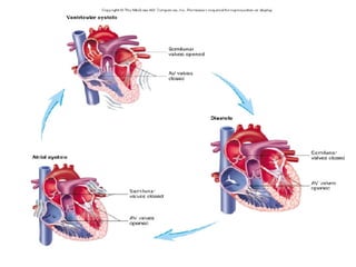
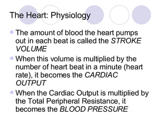
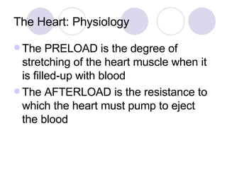
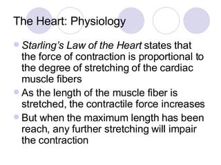
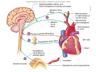
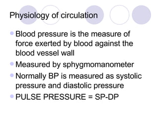
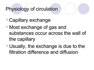
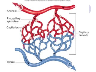
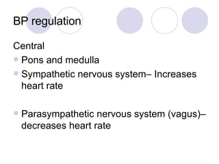
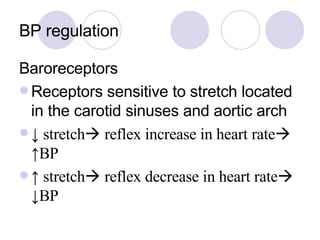
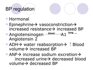
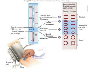
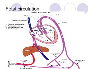
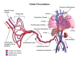
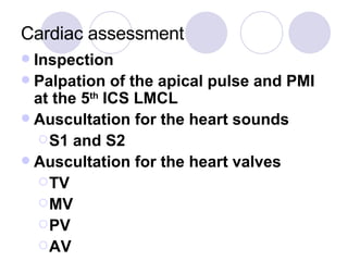
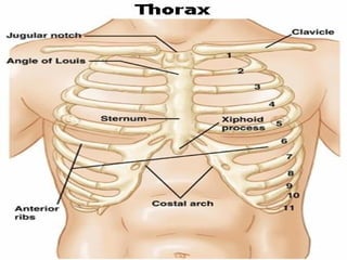
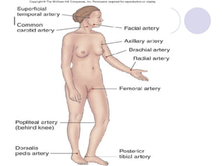
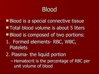
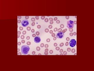
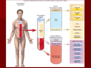
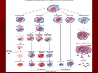
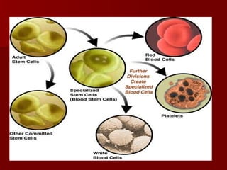
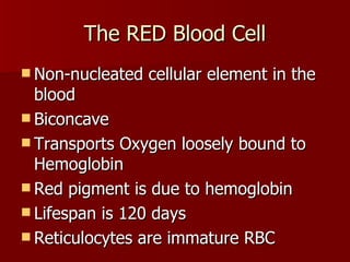
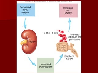
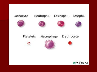
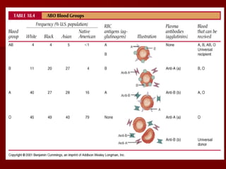
This document provides a summary of key topics in anatomy and physiology that are relevant for the Philippine Nursing Licensure Examination, including the cell, integumentary system, musculoskeletal system, nervous system, endocrine system, cardiovascular and hematologic systems, gastrointestinal system, urinary system, and reproductive system. The cell, its structures and functions, and the process of cellular division are described. An overview is given of the skin and its layers, functions of temperature regulation and protection.


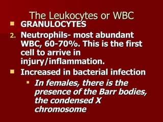

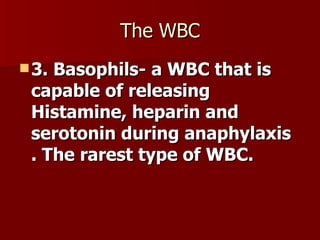
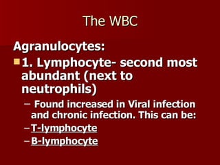
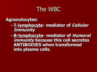


![anatomy introductory [Autosaved].pptx](https://cdn.slidesharecdn.com/ss_thumbnails/anatomyintroductoryautosaved-220930180143-87a00045-thumbnail.jpg?width=640&height=640&fit=bounds)


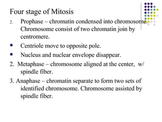
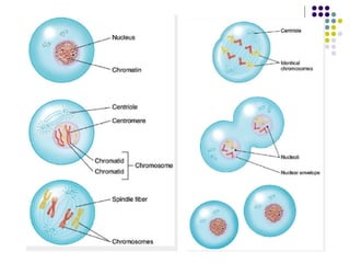
![Getting Started with Apache Spark: Big Data Made Simple [Free Meetup]](https://cdn.slidesharecdn.com/ss_thumbnails/apachesparkgettingstarted-260203175547-8361bcc3-thumbnail.jpg?width=640&height=640&fit=bounds)


