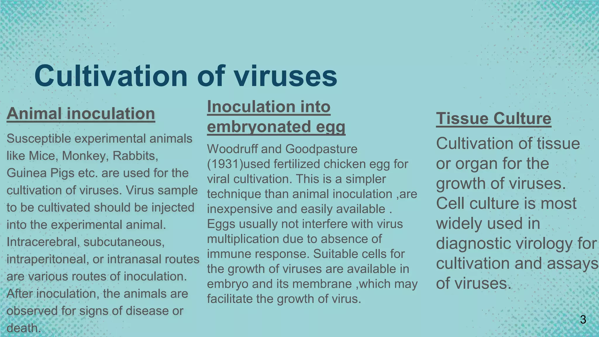
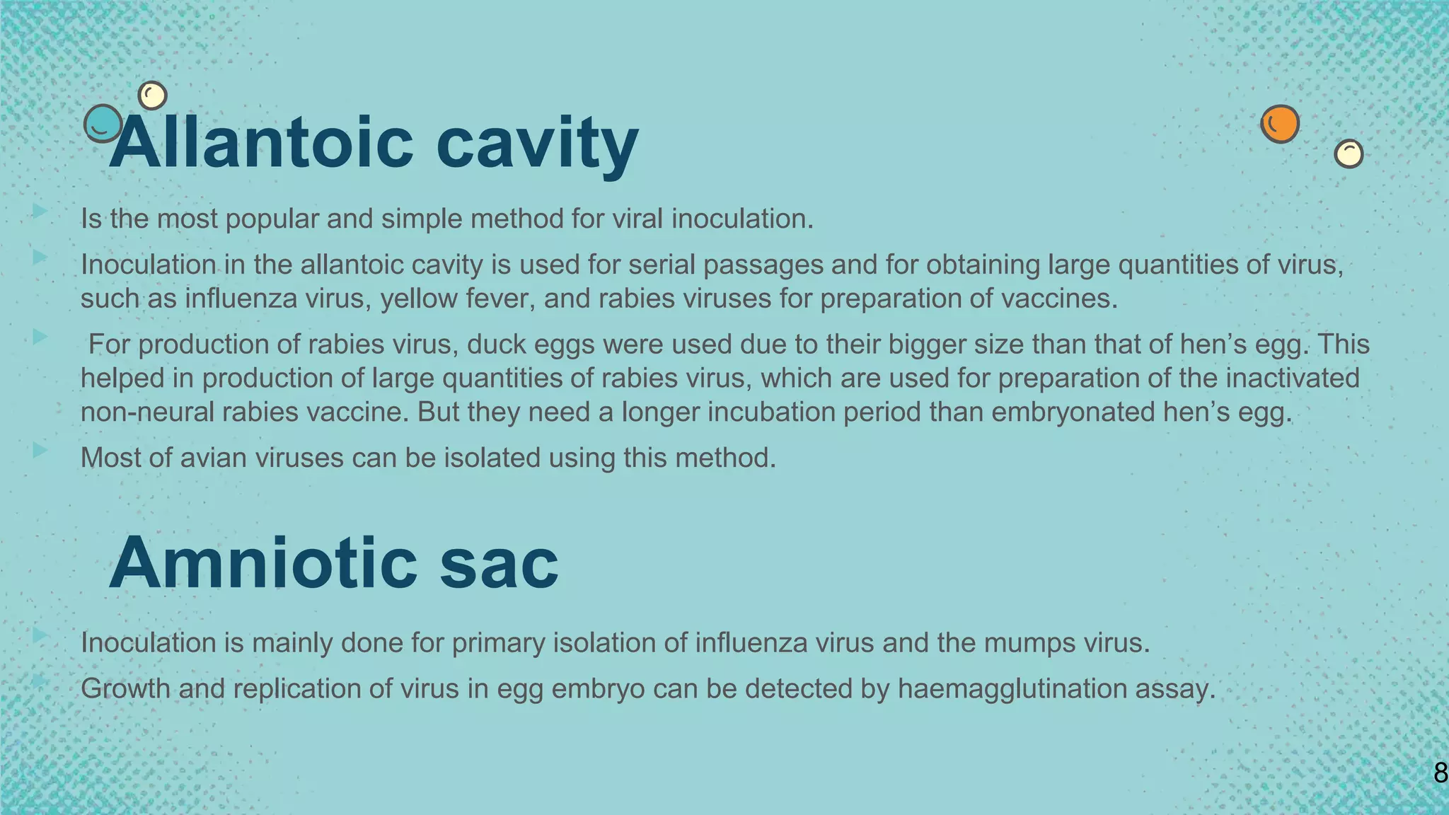
The document discusses the cultivation of viruses using embryonated eggs. It notes that embryonated eggs are a simpler technique than animal inoculation for growing viruses, as eggs do not have an immune response and contain suitable cells for virus growth. Viruses can be inoculated into different areas of embryonated eggs, including the chorioallantoic membrane, allantoic cavity, amniotic sac, and yolk sac. The allantoic cavity is most commonly used due to its simplicity and ability to produce large quantities of viruses like influenza. Embryonated eggs remain an important method for growing stocks of viruses for research and vaccine production.
Serological test for virus identificationPlock Ghosh
This presentation consist of detailed study of serological method of virus identification. Basically ELISA is vastly used for virus detection. Western blot method is used for HIV identification.
Serological test for virus identificationPlock Ghosh
This presentation consist of detailed study of serological method of virus identification. Basically ELISA is vastly used for virus detection. Western blot method is used for HIV identification.
Largest viruses that infect vertebrates
Can be seen under light microscope
Poxvirus diseases are characterized by skin lesions – localized or generalized
Important diseases caused by poxviruses are-
Smallpox
Monkeypox
Cowpox
Tanapox
Molluscum contagiosum
Poxviruses are brick or oval-shaped viruses with large double-stranded DNA genomes. Poxviruses exist throughout the world and cause disease in humans and many other types of animals. Poxvirus infections typically result in the formation of lesions, skin nodules, or disseminated rash.
A bacteriophage (informally, phage) is a virus that infects and replicates within a bacterium. The term is derived from "bacteria" and the Greek (phagein), "to devour". Bacteriophages are composed of proteins that encapsulate a DNA or RNA genome, and may have relatively simple or elaborate structures. Their genomes may encode as few as four genes, and as many as hundreds of genes. Phages replicate within the bacterium following the injection of their genome into its cytoplasm. Bacteriophages are among the most common and diverse entities in the biosphere.
Phages are widely distributed in locations populated by bacterial hosts, such as soil or the intestines of animals. One of the densest natural sources for phages and other viruses is sea water, where up to 9×10⁸ virions per milliliter have been found in microbial mats at the surface,] and up to 70% of marine bacteria may be infected by phages. They have been used for over 90 years as an alternative to antibiotics in the former Soviet Union and Central Europe, as well as in France. They are seen as a possible therapy against multi-drug-resistant strains of many bacteria (see phage therapy). Nevertheless, phages of Inoviridae have been shown to complicate biofilms involved in pneumonia and cystic fibrosis, shelter the bacteria from drugs meant to eradicate disease and promote persistent infection
Viruses are obligate intracellular parasites so they depend on host for their survival. They cannot be grown in non-living culture media or on agar plates alone, they must require living cells to support their replication.Cultivation of viruses can be discussed under following headings:
Animal Inoculation
Inoculation into embryonated egg
Cell Culture
VIRAL VACCINES
Since viruses are intracellular parasites they will grow only within other living cells.
Methods of viral vaccine production:
Cultivation of virus using free living animals
Fertile eggs
Tissue cultures
Viruses are Different From Other Microbes
Viruses are obligate intracellular parasites. They depend totally on their host cells for their existence. Their total host dependence makes it, extremely difficult to get good insight of them natural conditions, because the internal characteristics of the host cells are likely to interfere with the observations. Due to these reasons, it has been found desirable that viruses are cultivated or grown in the laboratory itself.
Laboratory animals
Fertilized Hen’s Egg
Chorioallantoic membrane
Allantoic cavity
Amniotic cavity
Yolk sac
Organ/Tissue/Cell Culture
Growth identified by serological method like neutralization.
Embryonated Egg Chorioallantioc membrane (CAM)
Allantoic cavity
Amniotic cavity
Yolk Sac
Cell Lines/ Tissue cultures Primary
Diploid/ Secondary
Continuous
Animal inoculation Suckling
Embryonated Hen’s Egg
Cultivation of Viruses and Bacteria
Chorioallantoic membrane (CAM) – visible lesions called pocks. Each infectious virus particle forms one pock. e.g. Variola, Vaccinia virus
Allantoic cavity – Influenza virus (vaccine production) & paramyxoviruses
Amniotic cavity – primary isolation of Influenza virus
Yolk sac – Chlamydia, Rickettsia & some viruses
Embryonated eggs:
The Embryonated hen’s egg was first used for cultivation of viruses by Good Pasteur and Burnet (1931). Cultivation of viruses in organized tissues like chick embryo necessitates a different type of approach.. For all practical purposes they all themselves behave as tissue cultures. The process of cultivation of viruses in embryonated eggs depends on the type of egg which is used. The egg used for cultivation must be sterile and the shell should be intact and healthy.
F.M. Burnet in the laboratory
in the early 1950's,
was experimenting
on influenza virus genetics,
using the developing hen's egg
Inoculated eggs are candled
daily to see the chicken
embryos inside.
Animals and chick embryo
were the first method that was used
to cultivate virus. This method is rarely
used as it is not convenient.
However, when preparing for bulk virus,
(e.g. antigen or vaccine production)
the usage of chick embryo is useful.
Fertile chicken eggs provide
a convenient, space-saving incubator
for many kinds of animal viruses.
Different viruses can be injected
into an egg at different sites and
the egg can be easily observed
for viral replication throughout
the development of the chicken embryo.
Isolation and cultivation of many avian
Viruses are obligate intracellular parasites which means they can only grow or reproduce inside a host cell.
The primary purpose of virus cultivation:
To isolate and identify viruses in clinical samples.
To do research on the viral structure, replication, genetics, and effects on the host cell.
To prepare viruses for vaccine production.
Isolation of the virus is always considered a gold standard for establishing the viral origin of the disease
topics covered
CULTIVATION OF VIRUSES
Animal inoculation
Embryonated eggs
CAM
Allantoic cavity
Amniotic cavity
Yolk sac
Tissue culture
Organ culture
Explant culture
Cell culture
Primary cell culture
diploid cell culture
Continues cell lines
Introduction:
RNA interference (RNAi) or Post-Transcriptional Gene Silencing (PTGS) is an important biological process for modulating eukaryotic gene expression.
It is highly conserved process of posttranscriptional gene silencing by which double stranded RNA (dsRNA) causes sequence-specific degradation of mRNA sequences.
dsRNA-induced gene silencing (RNAi) is reported in a wide range of eukaryotes ranging from worms, insects, mammals and plants.
This process mediates resistance to both endogenous parasitic and exogenous pathogenic nucleic acids, and regulates the expression of protein-coding genes.
What are small ncRNAs?
micro RNA (miRNA)
short interfering RNA (siRNA)
Properties of small non-coding RNA:
Involved in silencing mRNA transcripts.
Called “small” because they are usually only about 21-24 nucleotides long.
Synthesized by first cutting up longer precursor sequences (like the 61nt one that Lee discovered).
Silence an mRNA by base pairing with some sequence on the mRNA.
Discovery of siRNA?
The first small RNA:
In 1993 Rosalind Lee (Victor Ambros lab) was studying a non- coding gene in C. elegans, lin-4, that was involved in silencing of another gene, lin-14, at the appropriate time in the
development of the worm C. elegans.
Two small transcripts of lin-4 (22nt and 61nt) were found to be complementary to a sequence in the 3' UTR of lin-14.
Because lin-4 encoded no protein, she deduced that it must be these transcripts that are causing the silencing by RNA-RNA interactions.
Types of RNAi ( non coding RNA)
MiRNA
Length (23-25 nt)
Trans acting
Binds with target MRNA in mismatch
Translation inhibition
Si RNA
Length 21 nt.
Cis acting
Bind with target Mrna in perfect complementary sequence
Piwi-RNA
Length ; 25 to 36 nt.
Expressed in Germ Cells
Regulates trnasposomes activity
MECHANISM OF RNAI:
First the double-stranded RNA teams up with a protein complex named Dicer, which cuts the long RNA into short pieces.
Then another protein complex called RISC (RNA-induced silencing complex) discards one of the two RNA strands.
The RISC-docked, single-stranded RNA then pairs with the homologous mRNA and destroys it.
THE RISC COMPLEX:
RISC is large(>500kD) RNA multi- protein Binding complex which triggers MRNA degradation in response to MRNA
Unwinding of double stranded Si RNA by ATP independent Helicase
Active component of RISC is Ago proteins( ENDONUCLEASE) which cleave target MRNA.
DICER: endonuclease (RNase Family III)
Argonaute: Central Component of the RNA-Induced Silencing Complex (RISC)
One strand of the dsRNA produced by Dicer is retained in the RISC complex in association with Argonaute
ARGONAUTE PROTEIN :
1.PAZ(PIWI/Argonaute/ Zwille)- Recognition of target MRNA
2.PIWI (p-element induced wimpy Testis)- breaks Phosphodiester bond of mRNA.)RNAse H activity.
MiRNA:
The Double-stranded RNAs are naturally produced in eukaryotic cells during development, and they have a key role in regulating gene expression .
This presentation explores a brief idea about the structural and functional attributes of nucleotides, the structure and function of genetic materials along with the impact of UV rays and pH upon them.
(May 29th, 2024) Advancements in Intravital Microscopy- Insights for Preclini...Scintica Instrumentation
Intravital microscopy (IVM) is a powerful tool utilized to study cellular behavior over time and space in vivo. Much of our understanding of cell biology has been accomplished using various in vitro and ex vivo methods; however, these studies do not necessarily reflect the natural dynamics of biological processes. Unlike traditional cell culture or fixed tissue imaging, IVM allows for the ultra-fast high-resolution imaging of cellular processes over time and space and were studied in its natural environment. Real-time visualization of biological processes in the context of an intact organism helps maintain physiological relevance and provide insights into the progression of disease, response to treatments or developmental processes.
In this webinar we give an overview of advanced applications of the IVM system in preclinical research. IVIM technology is a provider of all-in-one intravital microscopy systems and solutions optimized for in vivo imaging of live animal models at sub-micron resolution. The system’s unique features and user-friendly software enables researchers to probe fast dynamic biological processes such as immune cell tracking, cell-cell interaction as well as vascularization and tumor metastasis with exceptional detail. This webinar will also give an overview of IVM being utilized in drug development, offering a view into the intricate interaction between drugs/nanoparticles and tissues in vivo and allows for the evaluation of therapeutic intervention in a variety of tissues and organs. This interdisciplinary collaboration continues to drive the advancements of novel therapeutic strategies.
Nutraceutical market, scope and growth: Herbal drug technologyLokesh Patil
As consumer awareness of health and wellness rises, the nutraceutical market—which includes goods like functional meals, drinks, and dietary supplements that provide health advantages beyond basic nutrition—is growing significantly. As healthcare expenses rise, the population ages, and people want natural and preventative health solutions more and more, this industry is increasing quickly. Further driving market expansion are product formulation innovations and the use of cutting-edge technology for customized nutrition. With its worldwide reach, the nutraceutical industry is expected to keep growing and provide significant chances for research and investment in a number of categories, including vitamins, minerals, probiotics, and herbal supplements.
Professional air quality monitoring systems provide immediate, on-site data for analysis, compliance, and decision-making.
Monitor common gases, weather parameters, particulates.
Deep Behavioral Phenotyping in Systems Neuroscience for Functional Atlasing a...Ana Luísa Pinho
Functional Magnetic Resonance Imaging (fMRI) provides means to characterize brain activations in response to behavior. However, cognitive neuroscience has been limited to group-level effects referring to the performance of specific tasks. To obtain the functional profile of elementary cognitive mechanisms, the combination of brain responses to many tasks is required. Yet, to date, both structural atlases and parcellation-based activations do not fully account for cognitive function and still present several limitations. Further, they do not adapt overall to individual characteristics. In this talk, I will give an account of deep-behavioral phenotyping strategies, namely data-driven methods in large task-fMRI datasets, to optimize functional brain-data collection and improve inference of effects-of-interest related to mental processes. Key to this approach is the employment of fast multi-functional paradigms rich on features that can be well parametrized and, consequently, facilitate the creation of psycho-physiological constructs to be modelled with imaging data. Particular emphasis will be given to music stimuli when studying high-order cognitive mechanisms, due to their ecological nature and quality to enable complex behavior compounded by discrete entities. I will also discuss how deep-behavioral phenotyping and individualized models applied to neuroimaging data can better account for the subject-specific organization of domain-general cognitive systems in the human brain. Finally, the accumulation of functional brain signatures brings the possibility to clarify relationships among tasks and create a univocal link between brain systems and mental functions through: (1) the development of ontologies proposing an organization of cognitive processes; and (2) brain-network taxonomies describing functional specialization. To this end, tools to improve commensurability in cognitive science are necessary, such as public repositories, ontology-based platforms and automated meta-analysis tools. I will thus discuss some brain-atlasing resources currently under development, and their applicability in cognitive as well as clinical neuroscience.
This pdf is about the Schizophrenia.
For more details visit on YouTube; @SELF-EXPLANATORY;
https://www.youtube.com/channel/UCAiarMZDNhe1A3Rnpr_WkzA/videos
Thanks...!
2. Introduction
▸ Viruses are obligate intracellular parasites so they depend on host for their
survival.
▸ They cannot be grown in non-living culture media or on agar plates alone, they
must require living cells to support their replication.
▸ In vivo-laboratory bred animal and embryonic bird tissue
▸ In vitro-cell or tissue culture method
▸ The primary purpose of virus cultivation is:
▸ To isolate and identify viruses in clinical samples.
▸ To do research on viral structure, replication, genetics and effects on host cell.
▸ To prepare viruses for vaccine production.
2
3. Cultivation of viruses
Inoculation into
embryonated egg
Woodruff and Goodpasture
(1931)used fertilized chicken egg for
viral cultivation. This is a simpler
technique than animal inoculation ,are
inexpensive and easily available .
Eggs usually not interfere with virus
multiplication due to absence of
immune response. Suitable cells for
the growth of viruses are available in
embryo and its membrane ,which may
facilitate the growth of virus.
Tissue Culture
Cultivation of tissue
or organ for the
growth of viruses.
Cell culture is most
widely used in
diagnostic virology for
cultivation and assays
of viruses.
3
Animal inoculation
Susceptible experimental animals
like Mice, Monkey, Rabbits,
Guinea Pigs etc. are used for the
cultivation of viruses. Virus sample
to be cultivated should be injected
into the experimental animal.
Intracerebral, subcutaneous,
intraperitoneal, or intranasal routes
are various routes of inoculation.
After inoculation, the animals are
observed for signs of disease or
death.
4. Embryonated eggs
▸ Woodruff and Goodpasture in 1931 first used the embryonated hen’s egg for the
cultivation of virus.
▸ The method further developed by Burnet was used for cultivation of viruses in
different sites of the egg.
▸ Many of these viruses cause well-defined and characteristic foci, providing a
method for identification, quantification, or assessing virus pathogenicity.
▸ The embryonated egg is also used for growing higher stocks of some viruses in
research laboratories and for vaccine production.
▸ Viruses can be cultivated in various parts of egg like Chorioallantoic membrane,
Allantoic cavity, Amniotic sac and Yolk sac.
4
5. Continue….
▸ Viruses are inoculated into chick embryo of 7-12 days old.
▸ For inoculation, eggs are first prepared for cultivation, the shell surface is first
disinfected with iodine, checked in ovoscope if it is alive and penetrated with a
small sterile drill.
▸ After inoculation, the opening is sealed with gelatin or paraffin and incubated at
36°c for 2-3 days.
▸ After incubation, the egg is broken and virus is isolated from tissue of egg.
▸ Viral growth and multiplication in the egg embryo is indicated by the death of the
embryo, by embryo cell damage, or by the formation of typical pocks or lesions on
the egg membranes
5
7. Chorioallantoic Membrane (CAM)
▸ Inoculation is mainly for growing poxvirus.
▸ After incubation, visible lesions called pocks are observed, which is grey white area in
transparent CAM.
▸ Each infectious virus particle produces one pock.
▸ The morphology of the pocks may vary depending on the nature of the virus.
▸ The pox viruses, such as variola or vaccinia are identified by demonstration of typical pocks
on the CAM inoculated with the pox virus.
▸ Herpes simplex virus is also grown.
▸ This method is suitable for plaque studies.
▸ Nowadays, in a virology laboratory, chick embryo inoculation has been replaced by cell
cultures for routine isolation of viruses.
7
8. Allantoic cavity
▸ Is the most popular and simple method for viral inoculation.
▸ Inoculation in the allantoic cavity is used for serial passages and for obtaining large quantities of virus,
such as influenza virus, yellow fever, and rabies viruses for preparation of vaccines.
▸ For production of rabies virus, duck eggs were used due to their bigger size than that of hen’s egg. This
helped in production of large quantities of rabies virus, which are used for preparation of the inactivated
non-neural rabies vaccine. But they need a longer incubation period than embryonated hen’s egg.
▸ Most of avian viruses can be isolated using this method.
8
Amniotic sac
▸ Inoculation is mainly done for primary isolation of influenza virus and the mumps virus.
▸ Growth and replication of virus in egg embryo can be detected by haemagglutination assay.
9. Yolk sac inoculation:
▸ It is also a simplest method for growth and multiplication of virus.
▸ Mostly mammalian viruses are isolated using this method.
▸ Yolk sac inoculation is used for cultivation of Japanese encephalitis, Saint Louis encephalitis, and West
Nile virus.
▸ It is inoculated for cultivation of some viruses and some bacteria (Chlamydia, Rickettsiae)
▸ Immune interference mechanism can be detected in most of avian viruses.
9
10. Procedure :
▸ 1. Candling: For propagation of influenza virus,
pathogen-free eggs are used 11-12 days after
fertilization. The egg is placed in front of a light
source to locate a non-veined area of the
allantoic cavity just below the air sac. This is
marked with a pencil. After all the eggs have
been ‘candled’ in this way, a small nick is made
in the shell at this position using a jeweler’s
scribe.
10
11. Procedure :
▸ 2. Drill the hole: Next, a hole is drilled at the
top of the egg with a Dremel motorized tool.
If this is not done, when virus is injected, the
pressure in the air sac will simply force out
the inoculum.
11
Drilling pin- point hole.
12. Procedure :
3. Inject the specimen: After all the eggs
have been nicked and drilled, they are
inoculated with virus using a tuberculin
syringe. The needle passes through the hole
in the shell, through the chorioallantoic
membrane, and the virus is placed in the
allantoic cavity, which is filled with allantoic
fluid.
4. Hole sealed with paraffin wax: The two
holes in the shell are sealed with melted
paraffin,
5. Incubation: The eggs are placed at 37
degrees C for 48 hours. 12
13. Advantages
▸ Cost effective and maintenance is much easier.
▸ Less labor is needed.
▸ The embryonated eggs are readily available.
▸ Sterile and wide range of tissues and fluids
▸ They are free from contaminating bacteria and many
latent viruses.
▸ Defense factors are not involved in embryonated eggs.
▸ Widely used method to grow virus for some vaccine
production
▸ Chick embryos are packed in their shells and have
natural resistant against bacterial contamination.
▸ Chick embryo method is cheaper and easy to handle.
13
Disadvantages
▸ The site of inoculation for varies
with different virus. That is, each
virus have different sites for their
growth and replication.
▸ Some viruses do not show growth
on primary inoculation into the
eggs.
▸ Slight amount of bacterial
contamination in the inoculum may
kill the embryo.