Microscopes are instruments designed to produce magnified images of small objects. They must accomplish three tasks: produce a magnified image, separate details in the image, and render details visible. There are different types of microscopes including simple, compound, stereoscopic, electron, scanning electron, and transmission electron microscopes. Electron microscopes use a beam of electrons instead of light to magnify images and can achieve higher magnifications than light microscopes. Confocal laser scanning microscopes use a laser beam to generate 3D images of thick specimens.
Microscopes magnify small objects. They produce, separate, and render visible details.
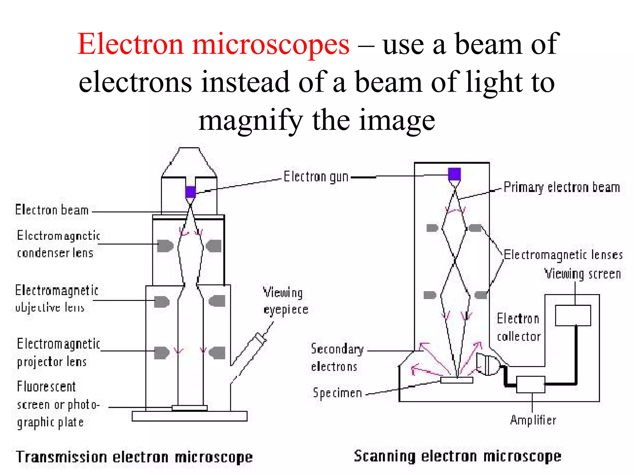
Overview of different microscope types: Simple, Compound, Stereoscopic, Electron (SEM & TEM), and Confocal Laser Scanning. Each type varies in imaging capability and detail.
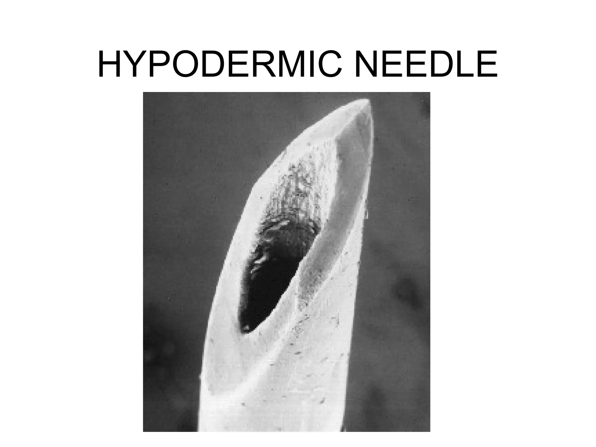
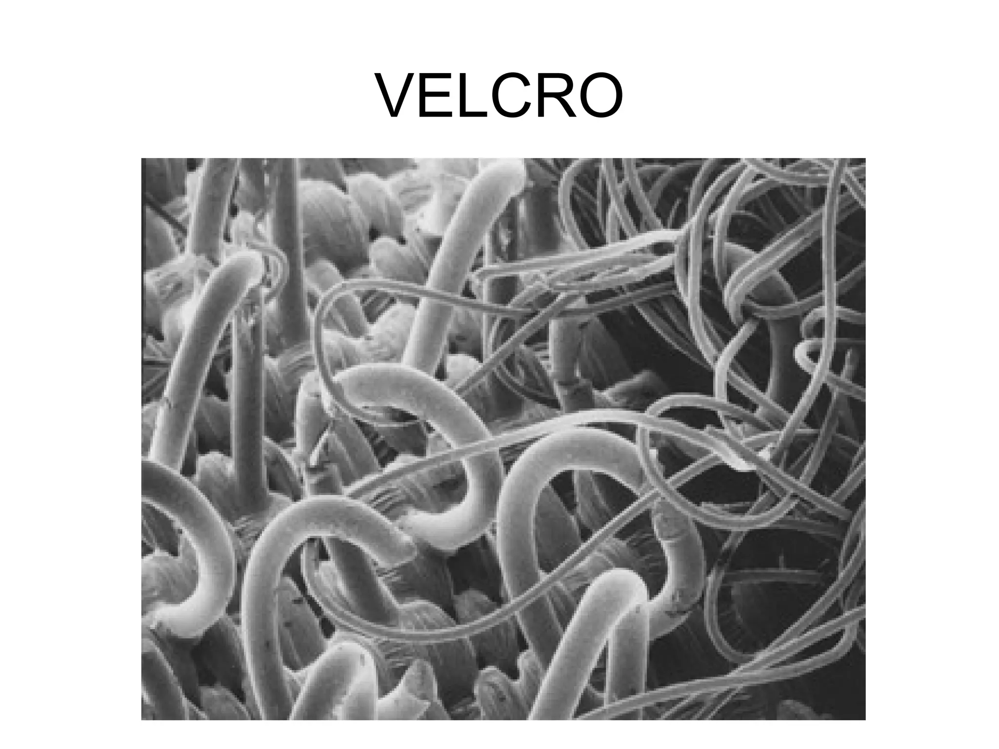
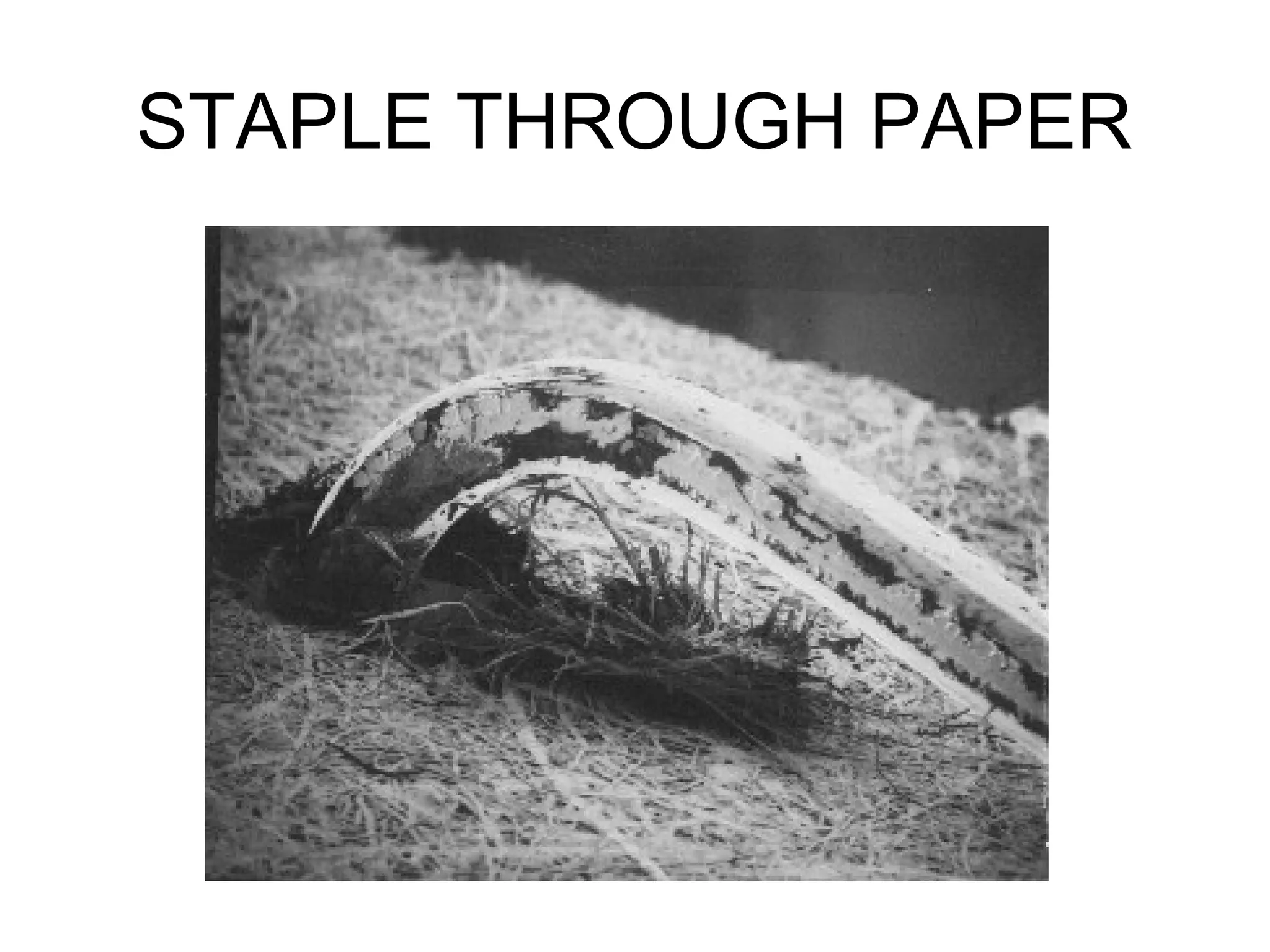
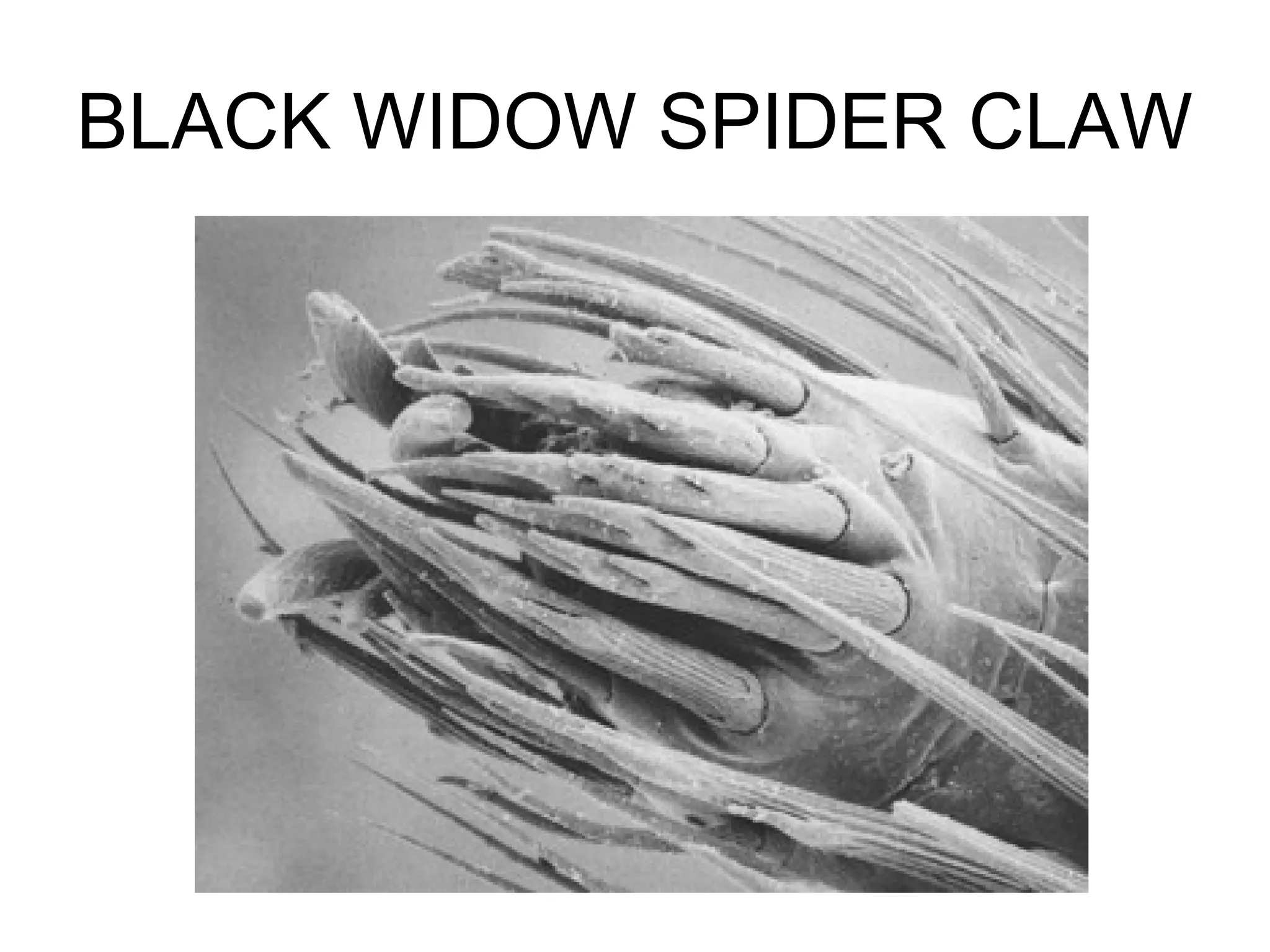
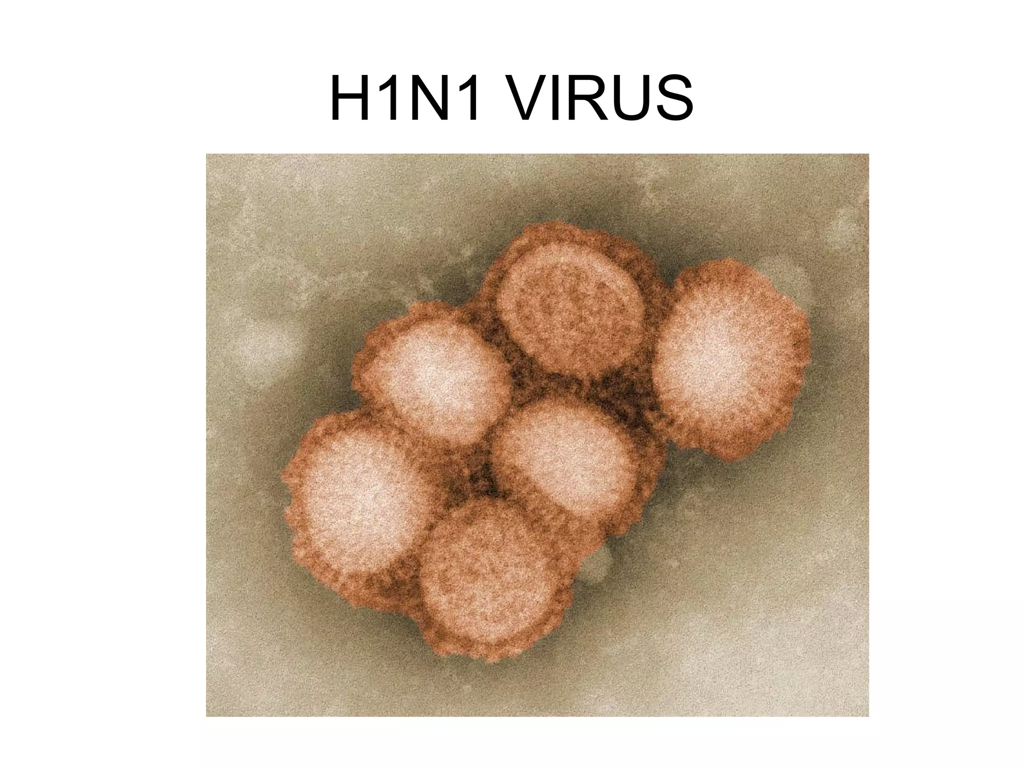
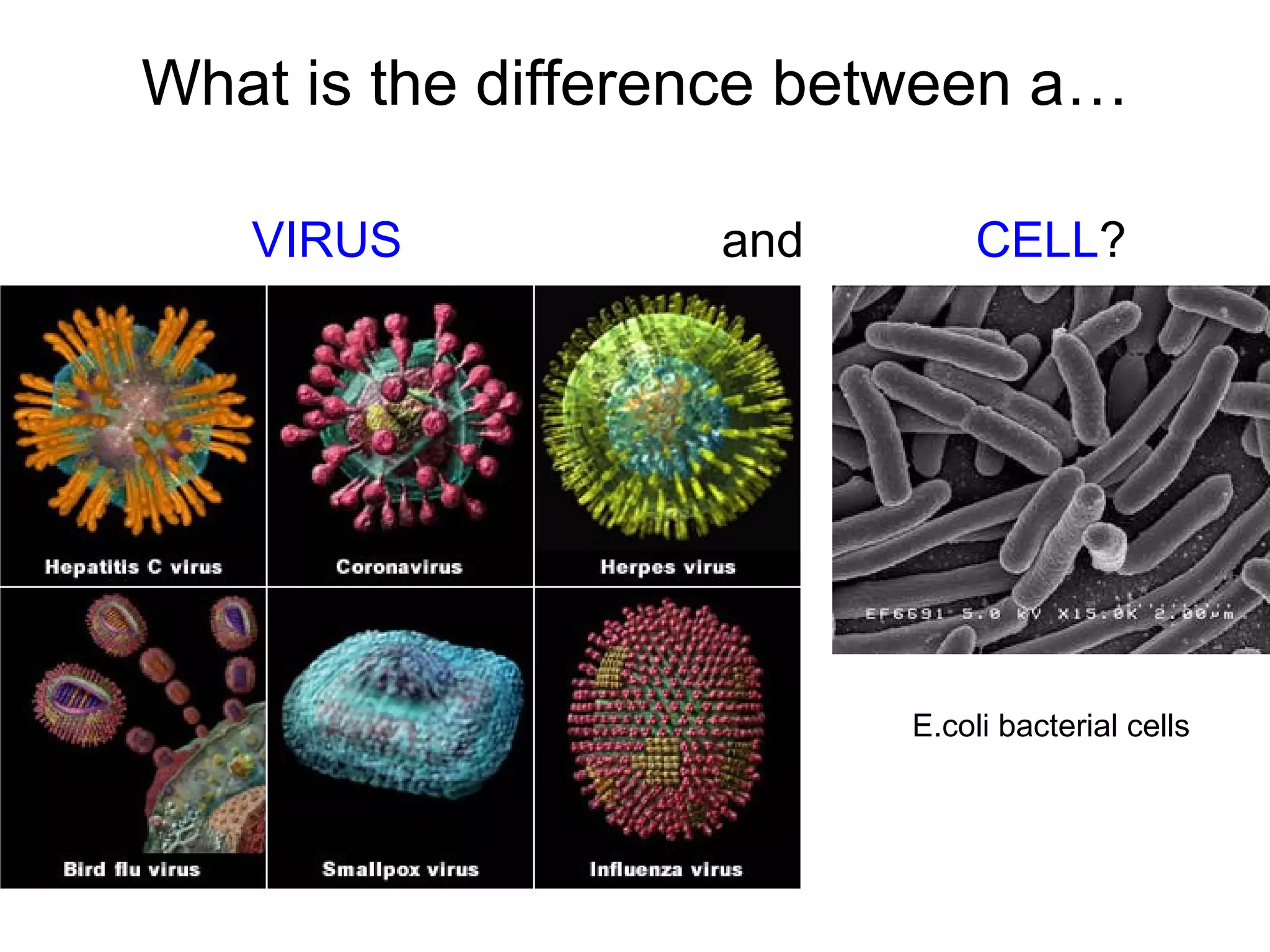
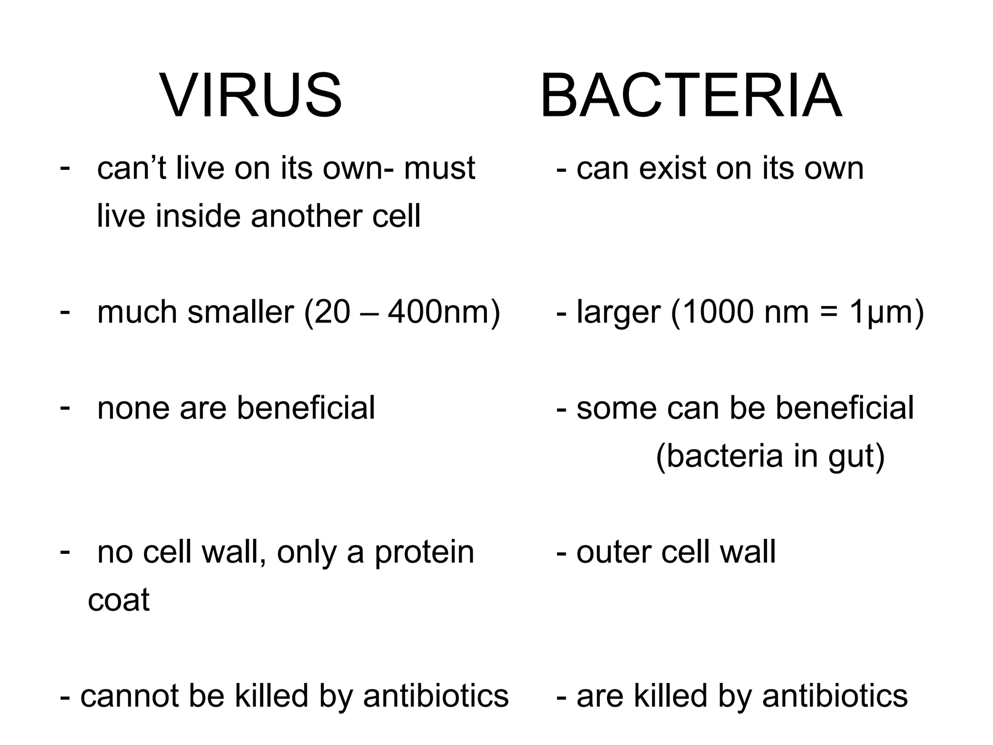
Engagement with micrographs displaying everyday objects and organisms. Comparison between viruses and bacteria highlighting key differences.
Microscopy - anintroduction
• Microscopes are instruments
designed to produce magnified
visual or photographic images of
small objects.
The microscope must accomplish three tasks
1. produce a magnified image of the specimen
2. separate the details in the image,
3. render the details visible to the human eye or camera.
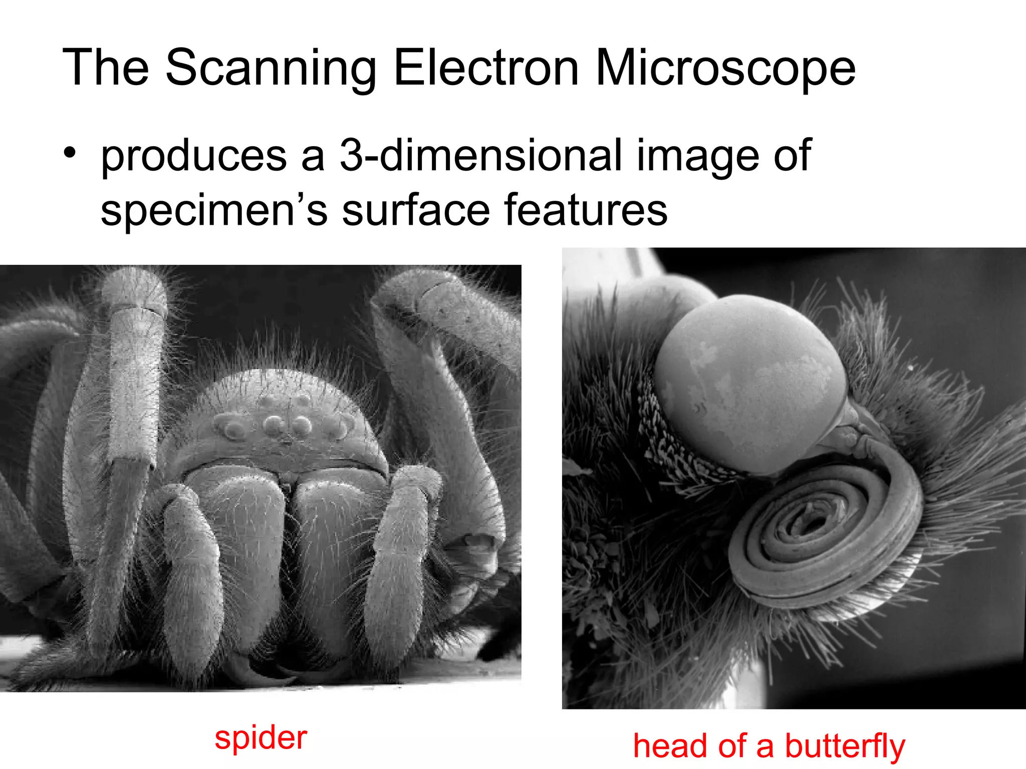
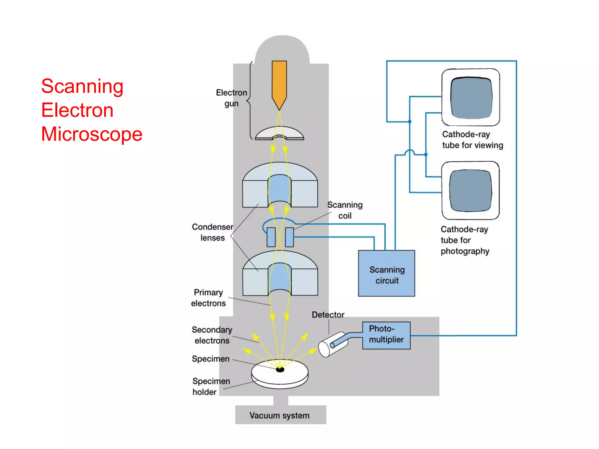
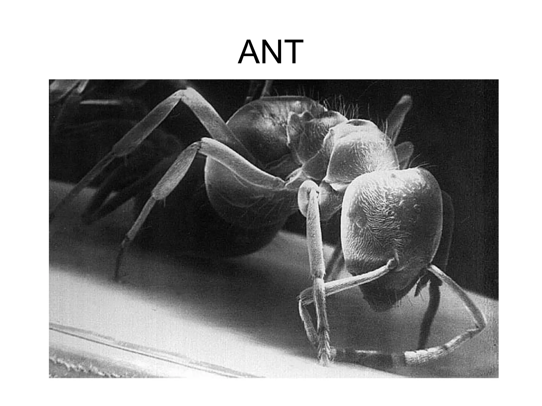
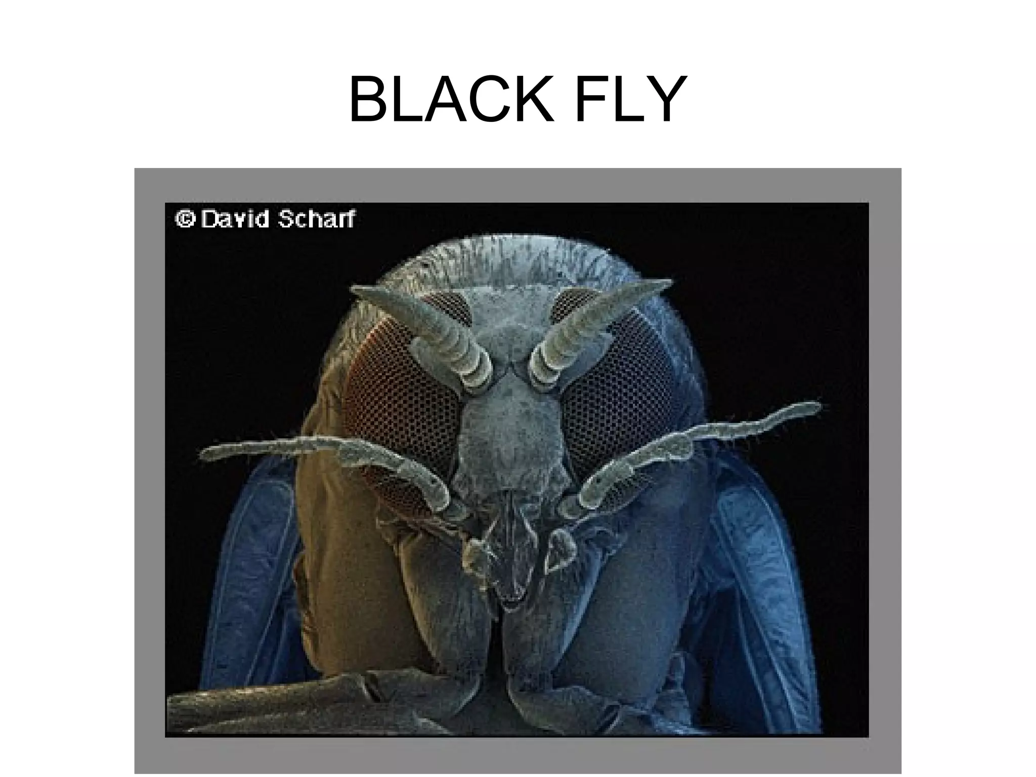
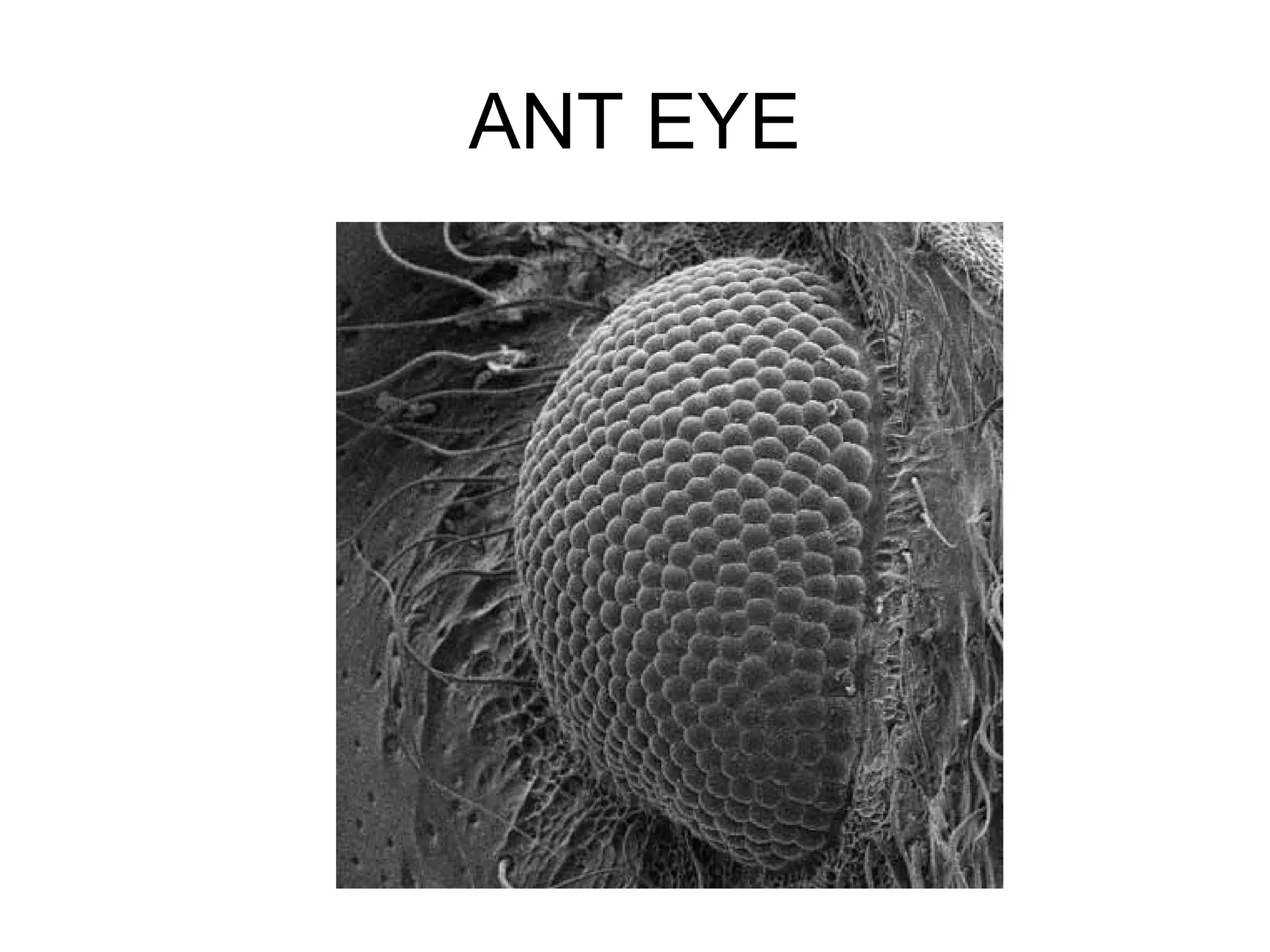
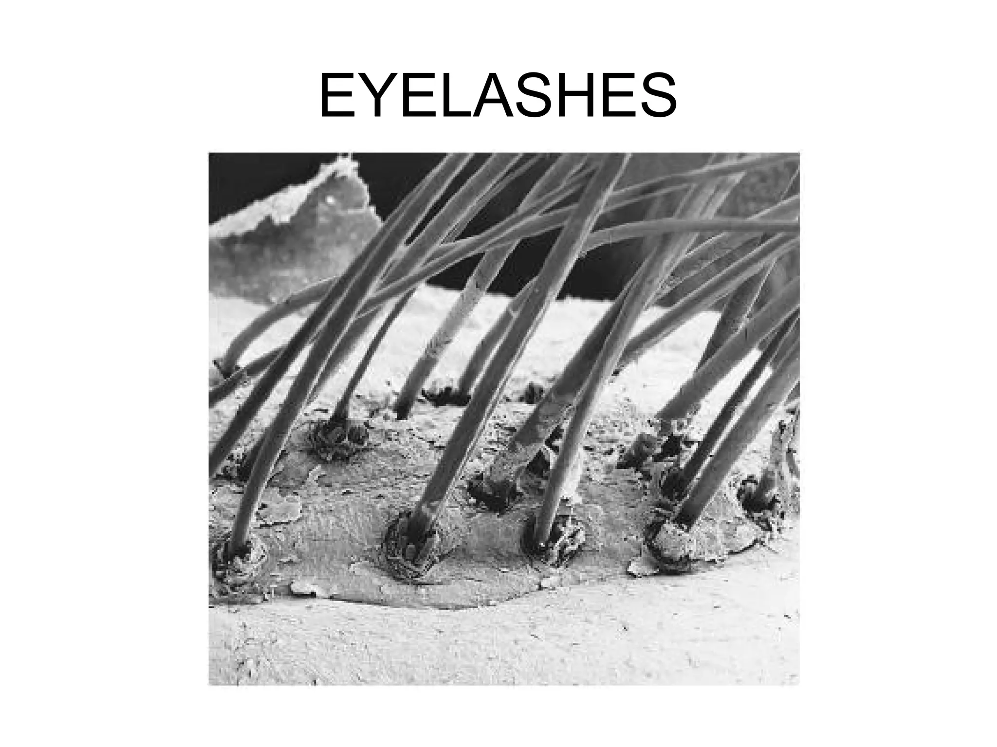
The Scanning ElectronMicroscope
• produces a 3-dimensional image of
specimen’s surface features
spider head of a butterfly
10.
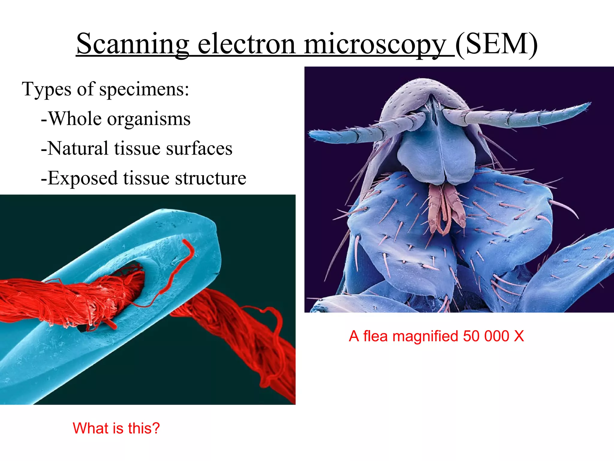
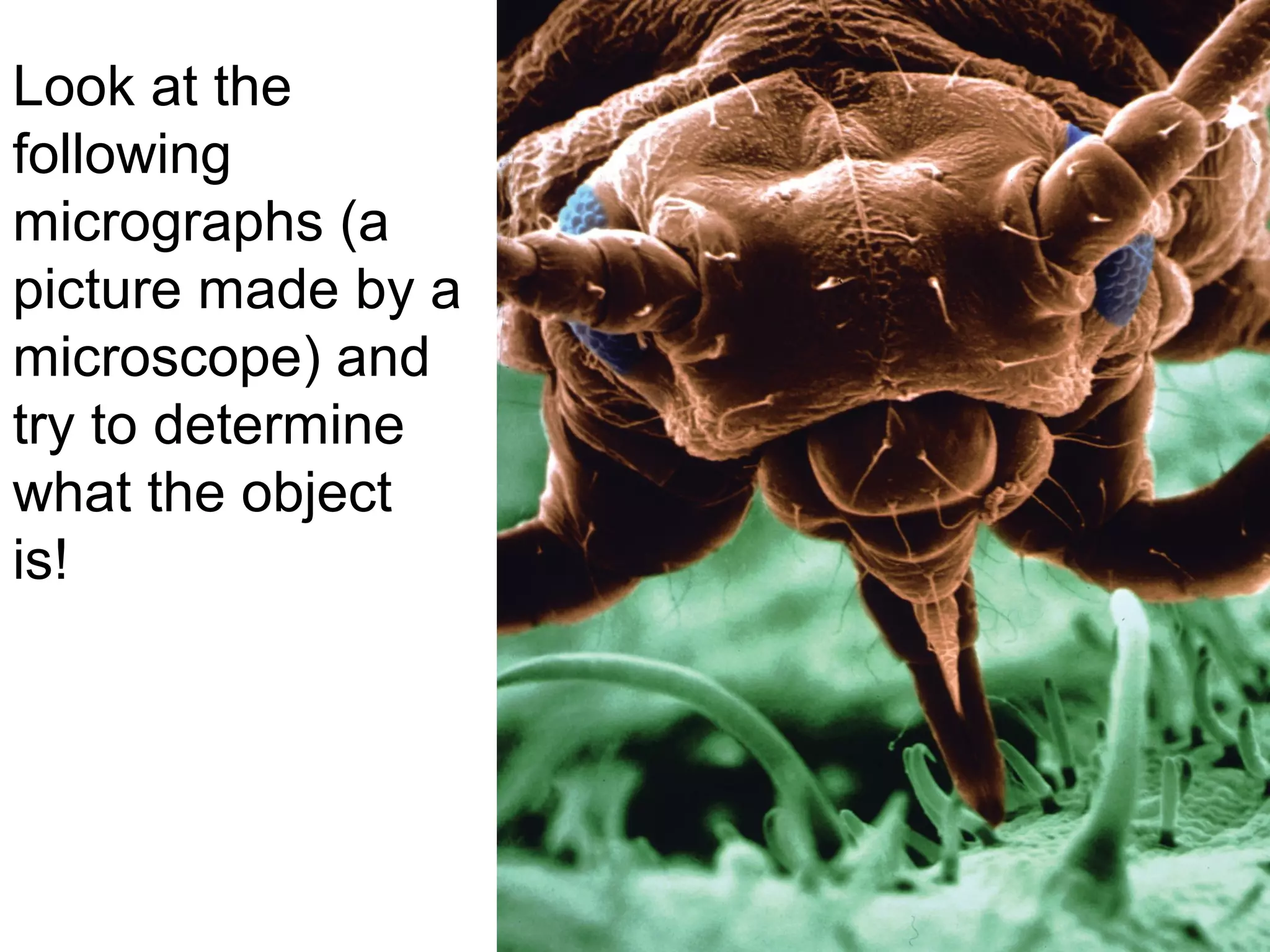
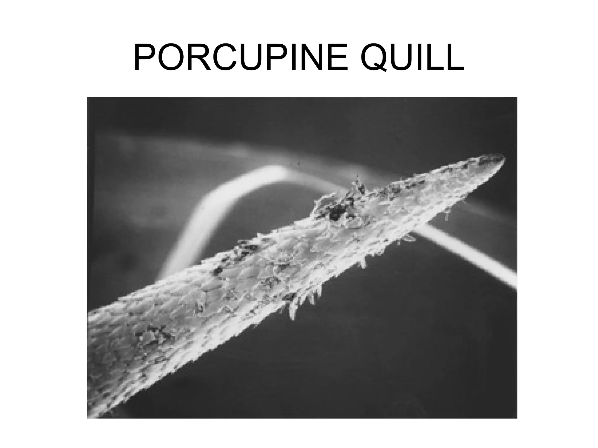
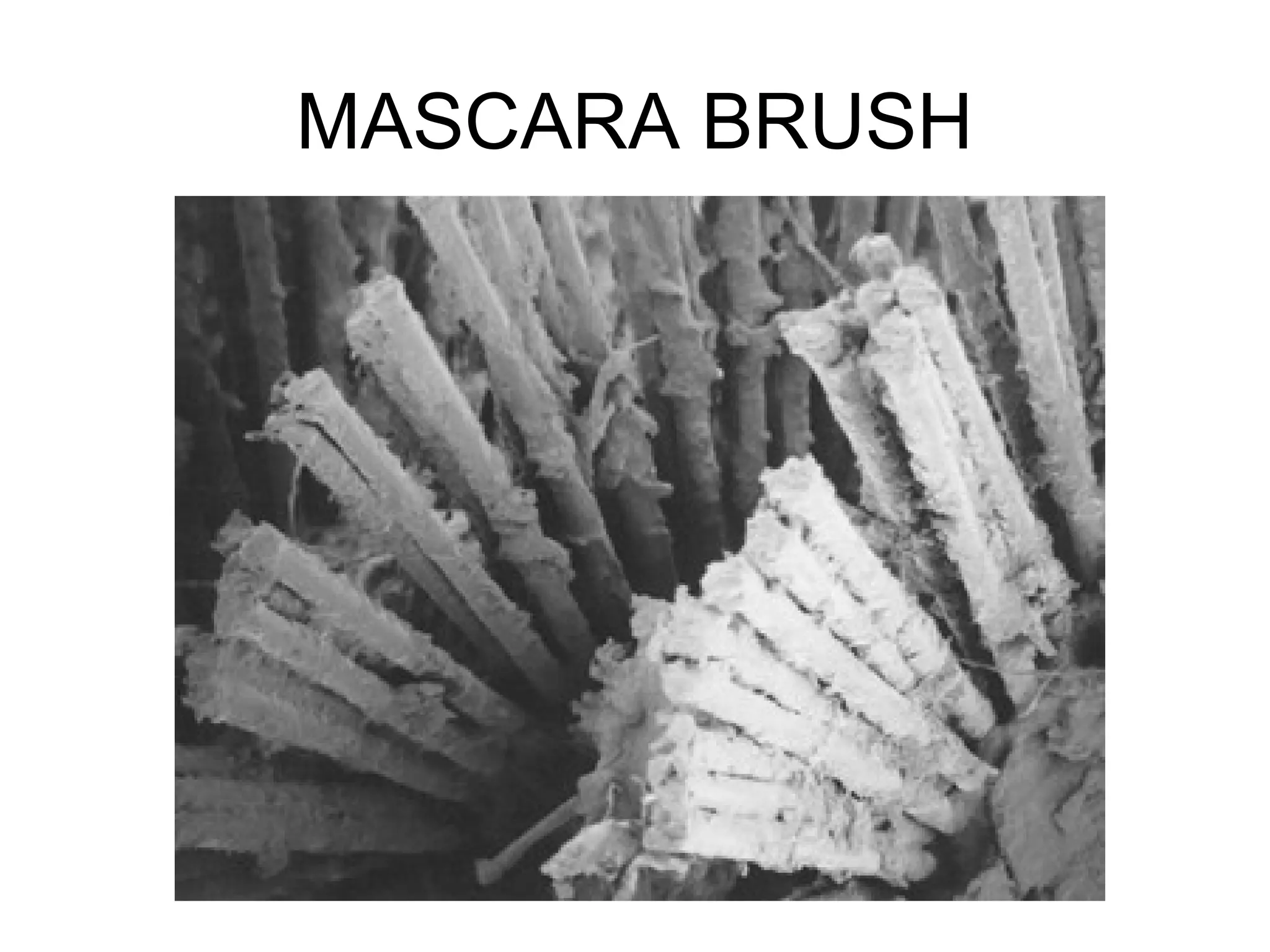
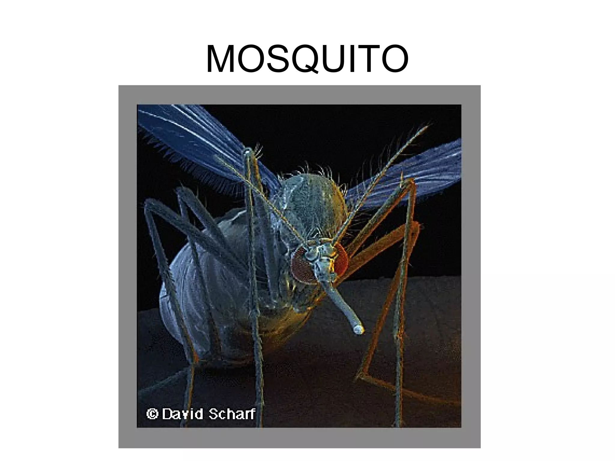
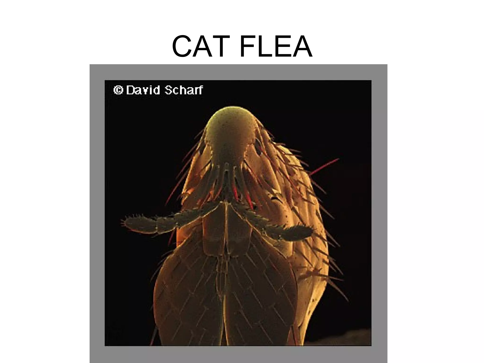
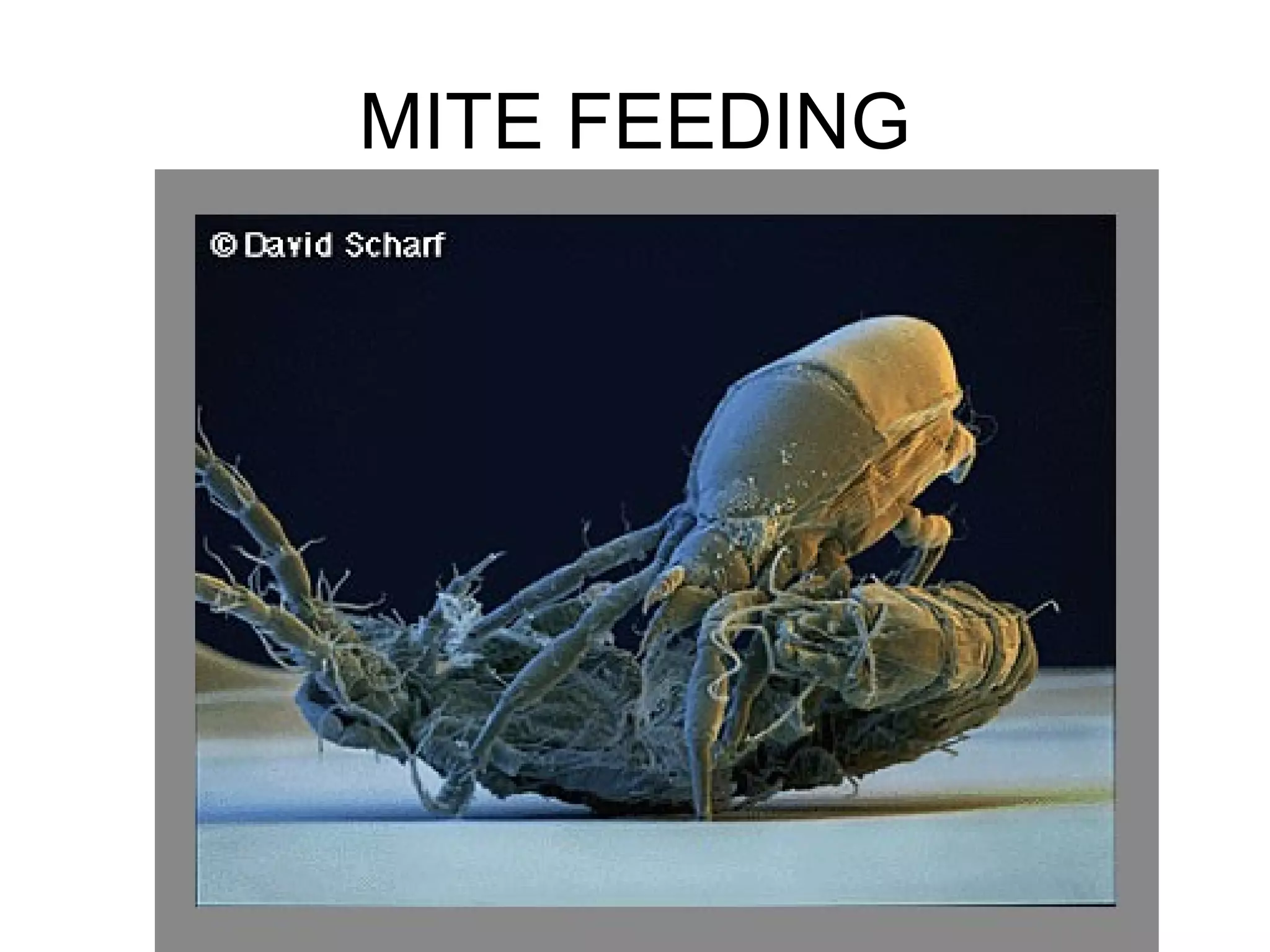
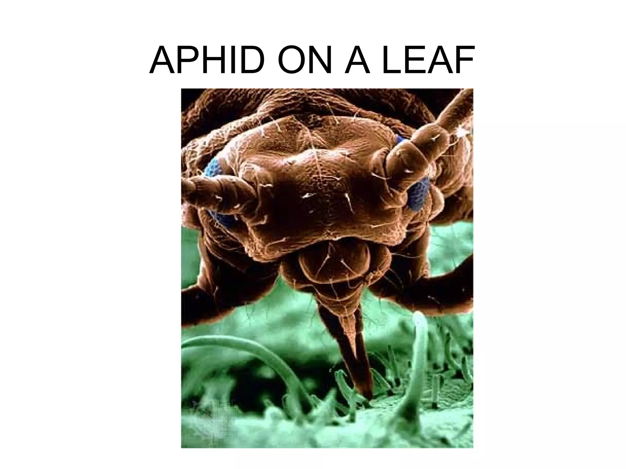
Scanning electron microscopy(SEM)
Types of specimens:
-Whole organisms
-Natural tissue surfaces
-Exposed tissue structure
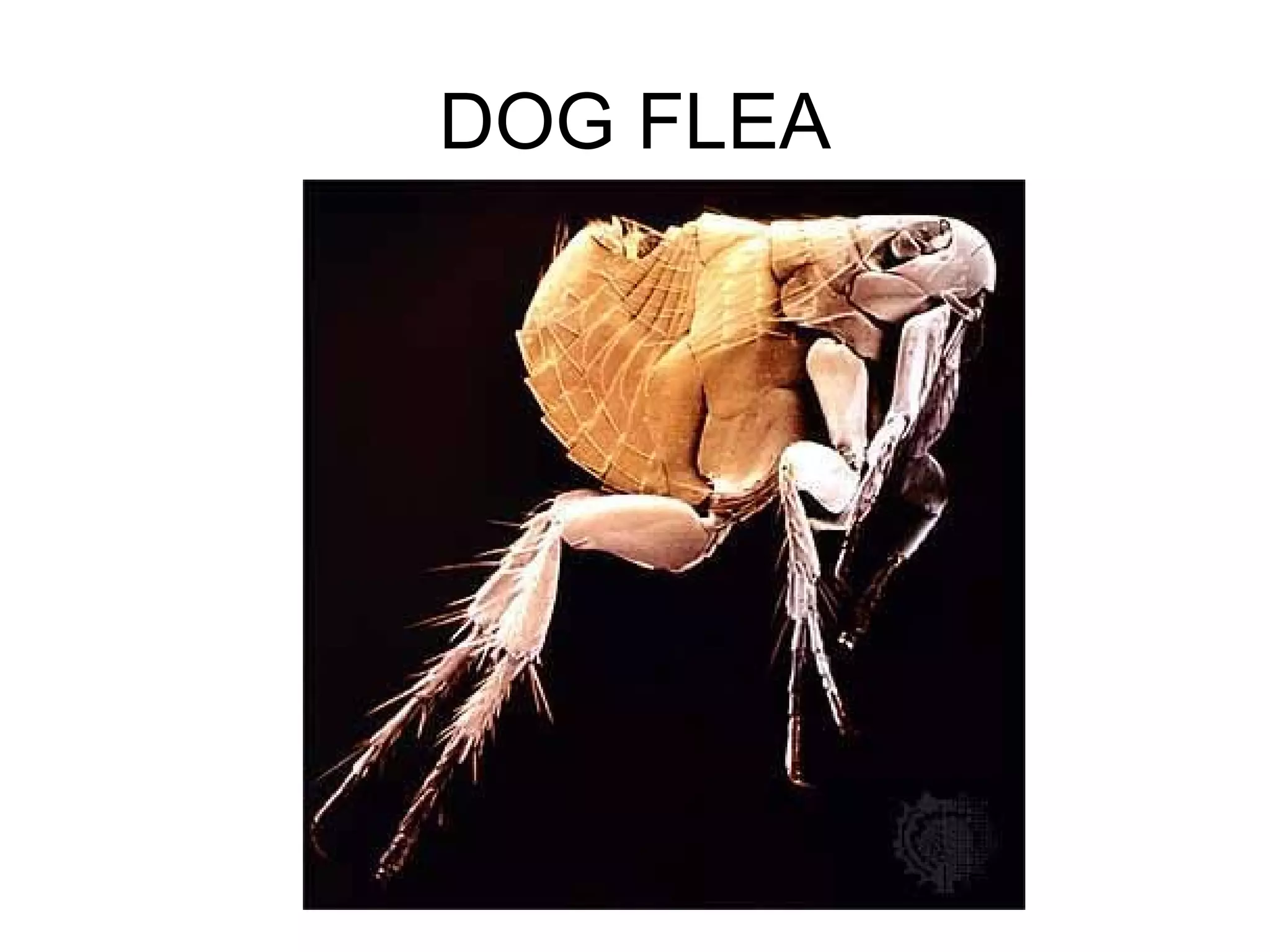
A flea magnified 50 000 X
What is this?
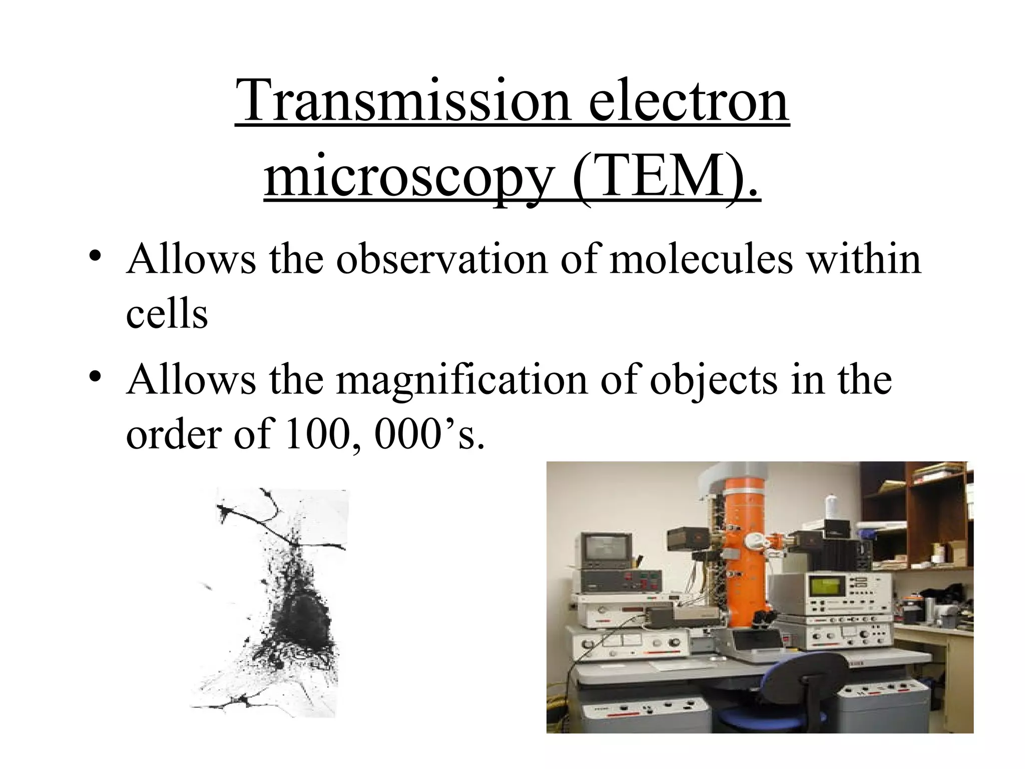
Transmission electron
microscopy (TEM).
• Allows the observation of molecules within
cells
• Allows the magnification of objects in the
order of 100, 000’s.
13.
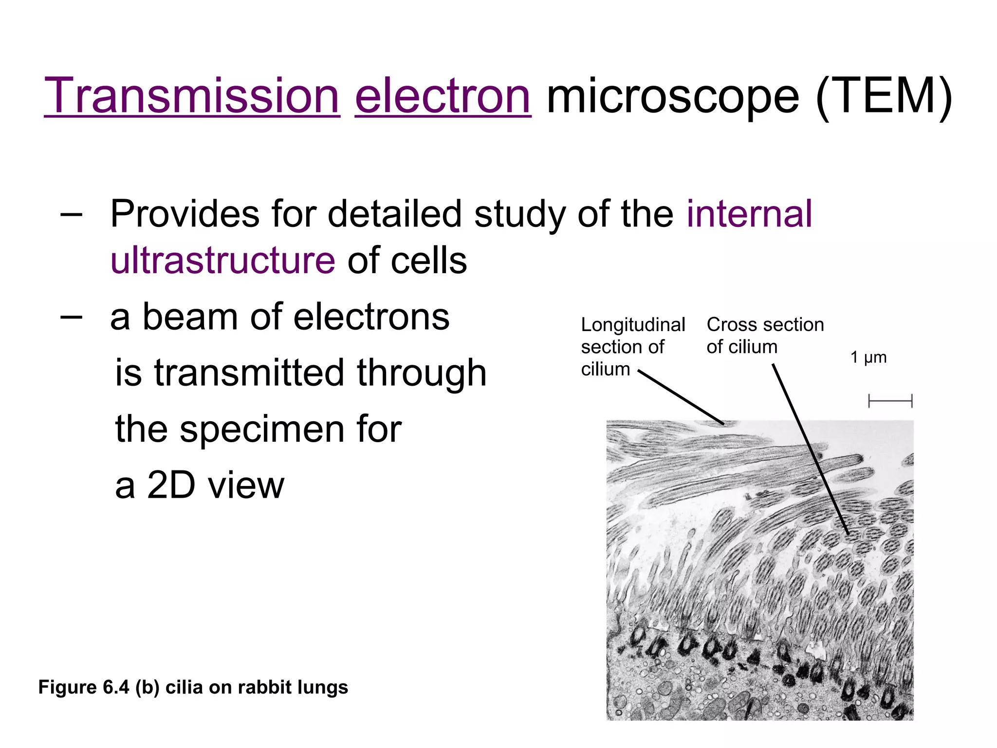
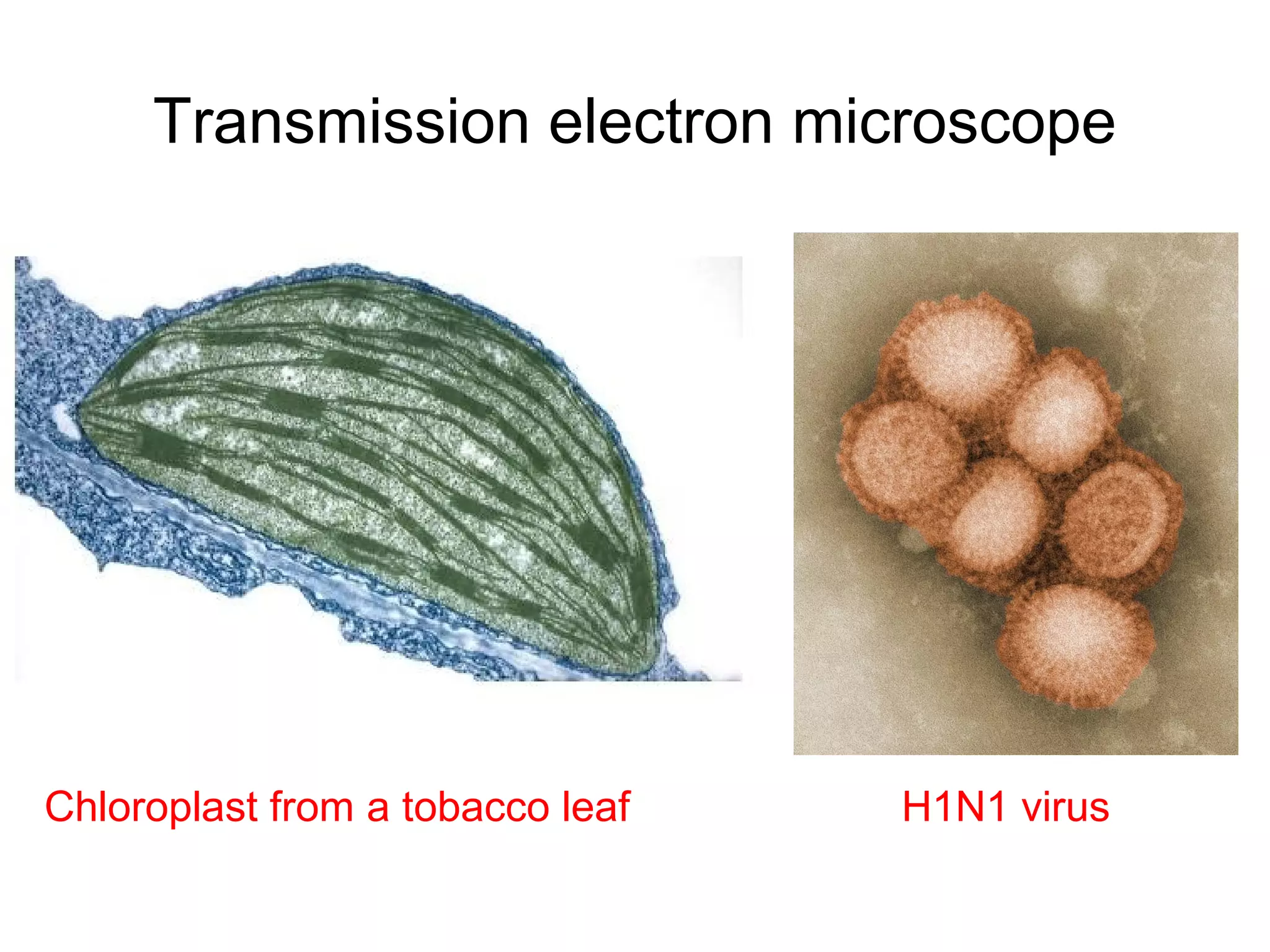
Transmission electron microscope(TEM)
– Provides for detailed study of the internal
ultrastructure of cells
– a beam of electrons Longitudinal Cross section
section of of cilium 1 µm
is transmitted through cilium
the specimen for
a 2D view
Figure 6.4 (b) cilia on rabbit lungs
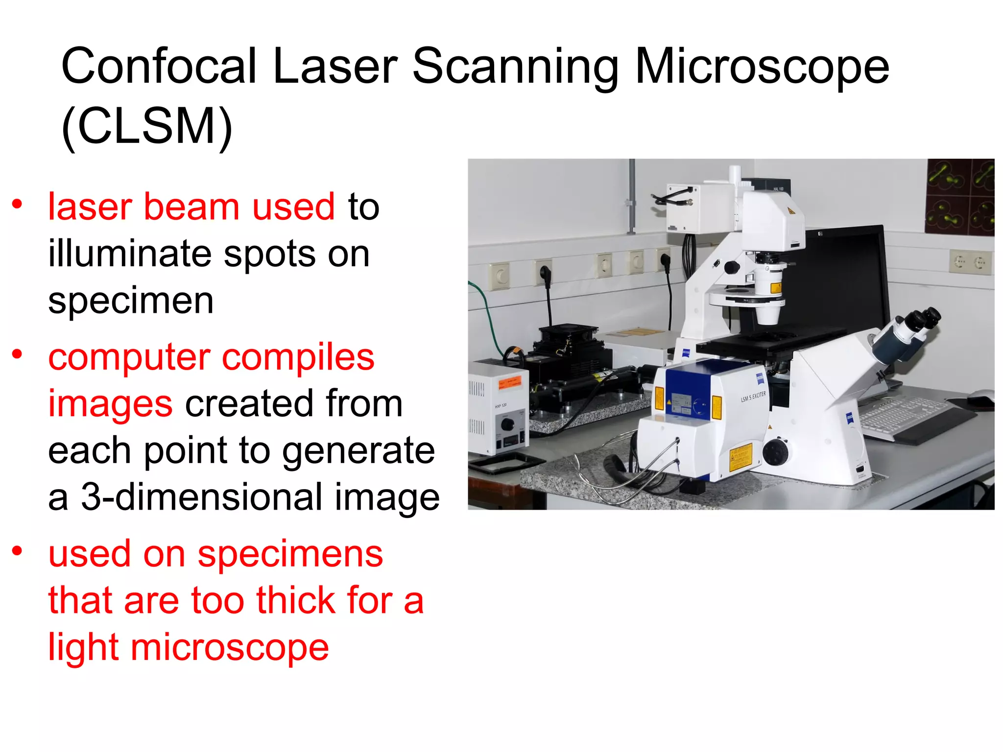
Confocal Laser ScanningMicroscope
(CLSM)
• laser beam used to
illuminate spots on
specimen
• computer compiles
images created from
each point to generate
a 3-dimensional image
• used on specimens
that are too thick for a
light microscope
16.
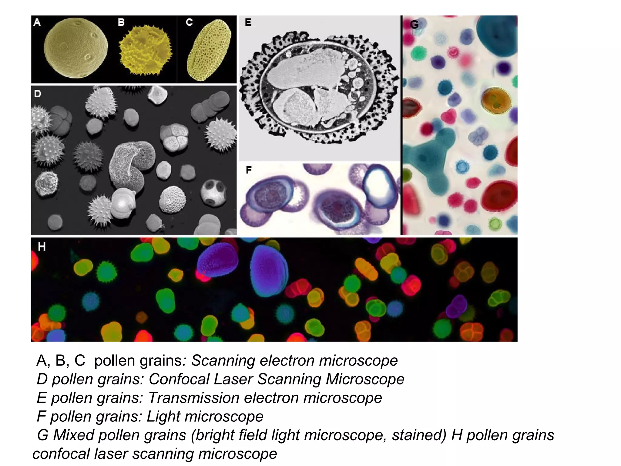
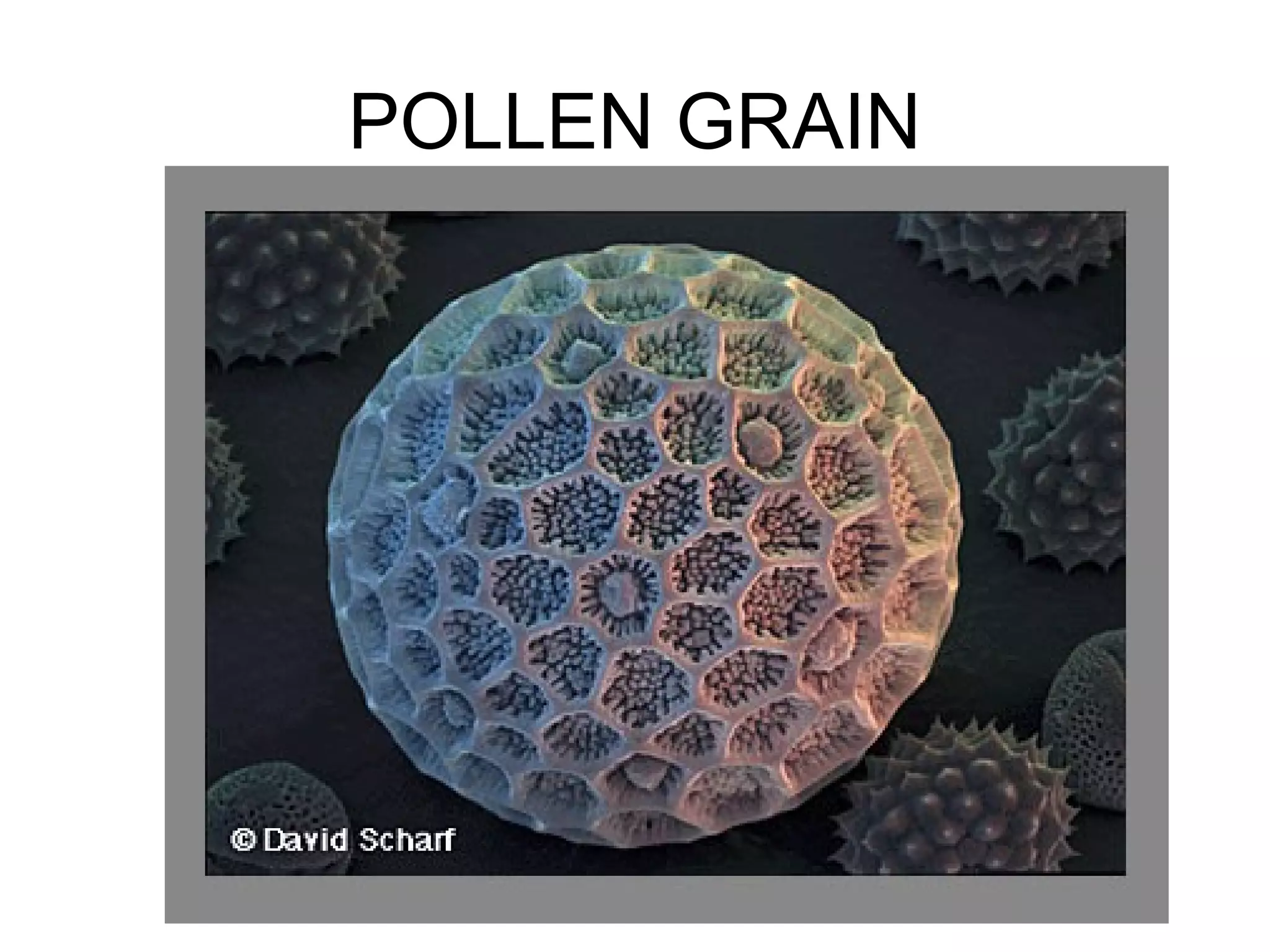
A, B, Cpollen grains: Scanning electron microscope
D pollen grains: Confocal Laser Scanning Microscope
E pollen grains: Transmission electron microscope
F pollen grains: Light microscope
G Mixed pollen grains (bright field light microscope, stained) H pollen grains
confocal laser scanning microscope
What is thedifference between a…
VIRUS and CELL?
E.coli bacterial cells
38.
VIRUS BACTERIA
- can’t live on its own- must - can exist on its own
live inside another cell
- much smaller (20 – 400nm) - larger (1000 nm = 1μm)
- none are beneficial - some can be beneficial
(bacteria in gut)
- no cell wall, only a protein - outer cell wall
coat
- cannot be killed by antibiotics - are killed by antibiotics


![Vibe Coding vs. Spec-Driven Development [Free Meetup]](https://cdn.slidesharecdn.com/ss_thumbnails/vibecodingvsspecdrivendevelopment-251209105622-43f455e7-thumbnail.jpg?width=640&height=640&fit=bounds)


