Downloaded 307 times

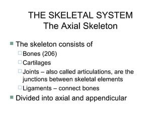
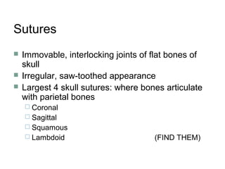
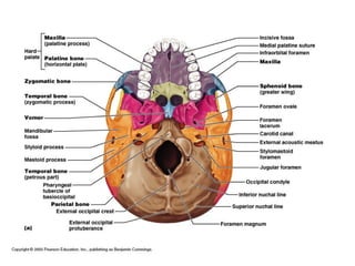
![Remember, the skull is composed of:
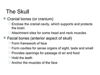
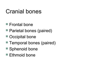
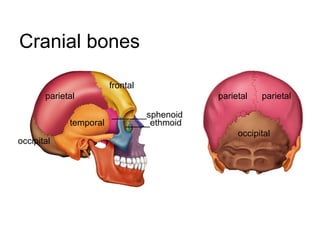
1. Cranial bones (or cranium)
[these were just reviewed]
and
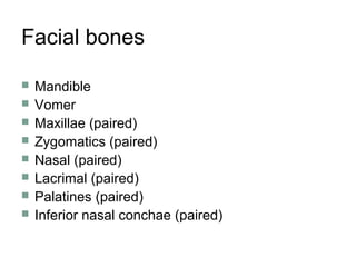
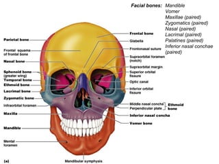
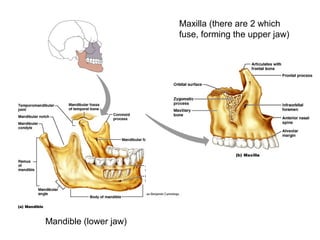
2. Facial bones (anterior aspect of skull)
Form framework of face
Form cavities for sense organs of sight, taste
and smell
Provides openings for passage of air and food
Hold the teeth
Anchor the muscles of the face](https://image.slidesharecdn.com/axialskeleton-150302023925-conversion-gate02/85/Axial-skeleton-17-320.jpg)


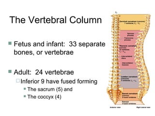
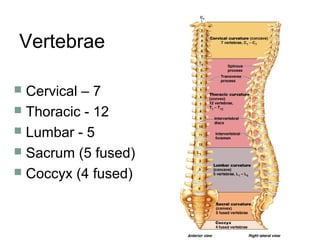
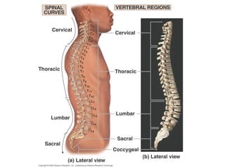

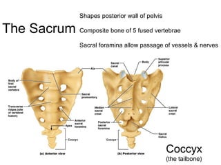
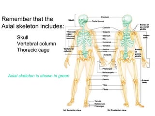
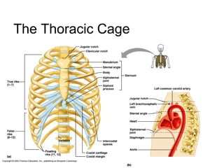

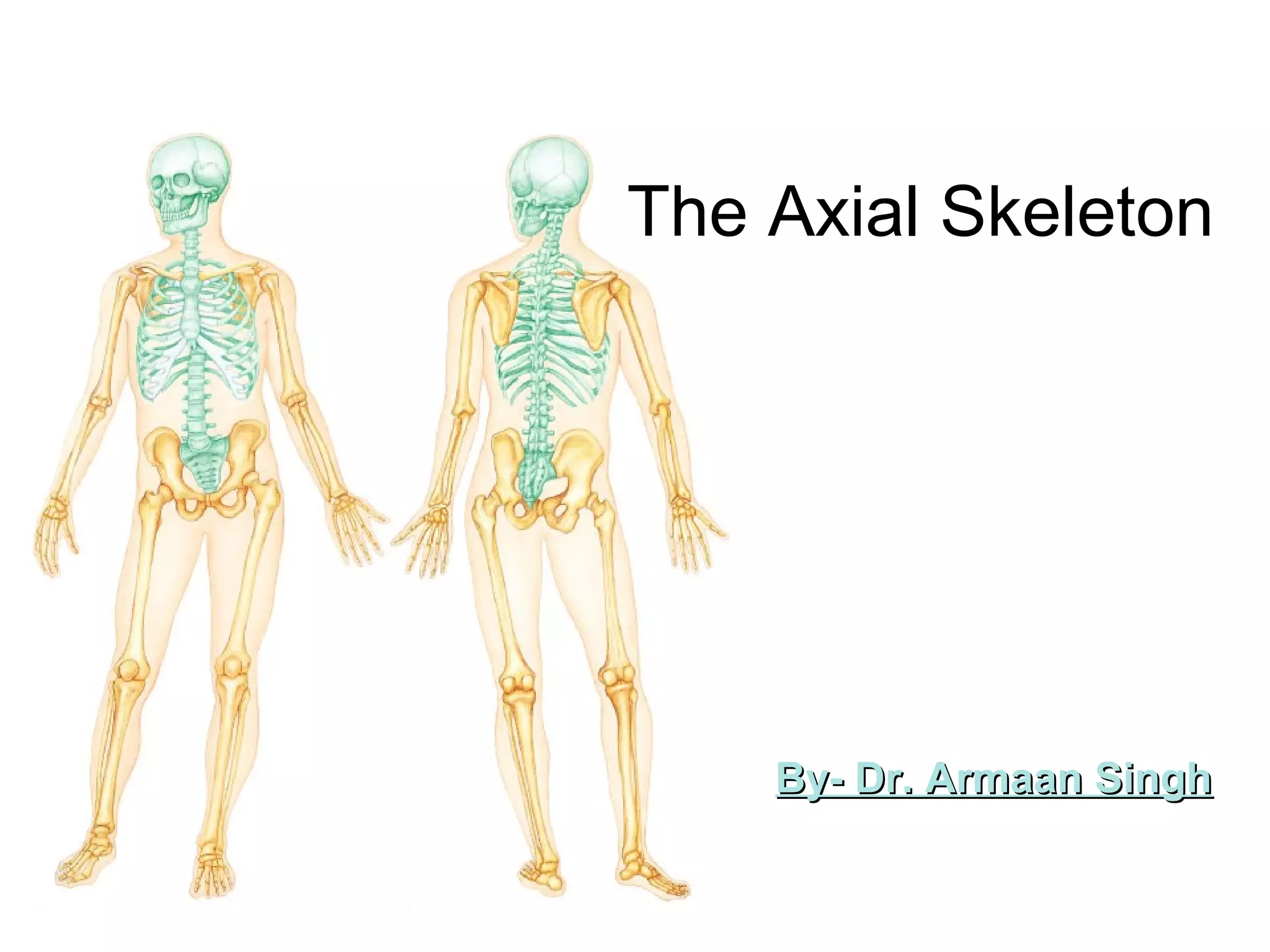
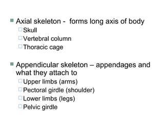
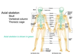
The document summarizes key aspects of the axial skeleton, including: 1. The axial skeleton consists of the skull, vertebral column, and thoracic cage. It forms the central axis of the body. 2. The skull is made up of multiple cranial and facial bones that protect the brain and house sensory organs. It includes prominent features like the foramen magnum, cranial fossae, and sutures. 3. The vertebral column consists of 33 vertebrae that fuse into 24 bones in adulthood. It includes cervical, thoracic, lumbar, sacral and coccygeal regions with characteristic features. 4. The thoracic cage is formed from ribs, sternum and


![CTEV [ clubfoot] DR ARUN LAL ,DR MOHAMED ASHRAF travancore medical college k...](https://cdn.slidesharecdn.com/ss_thumbnails/ctevclubfootdrarunlaldrmohamedashraftravancoremedicalcollegekollamkeralaindia-260208063247-18fc466c-thumbnail.jpg?width=640&height=640&fit=bounds)


