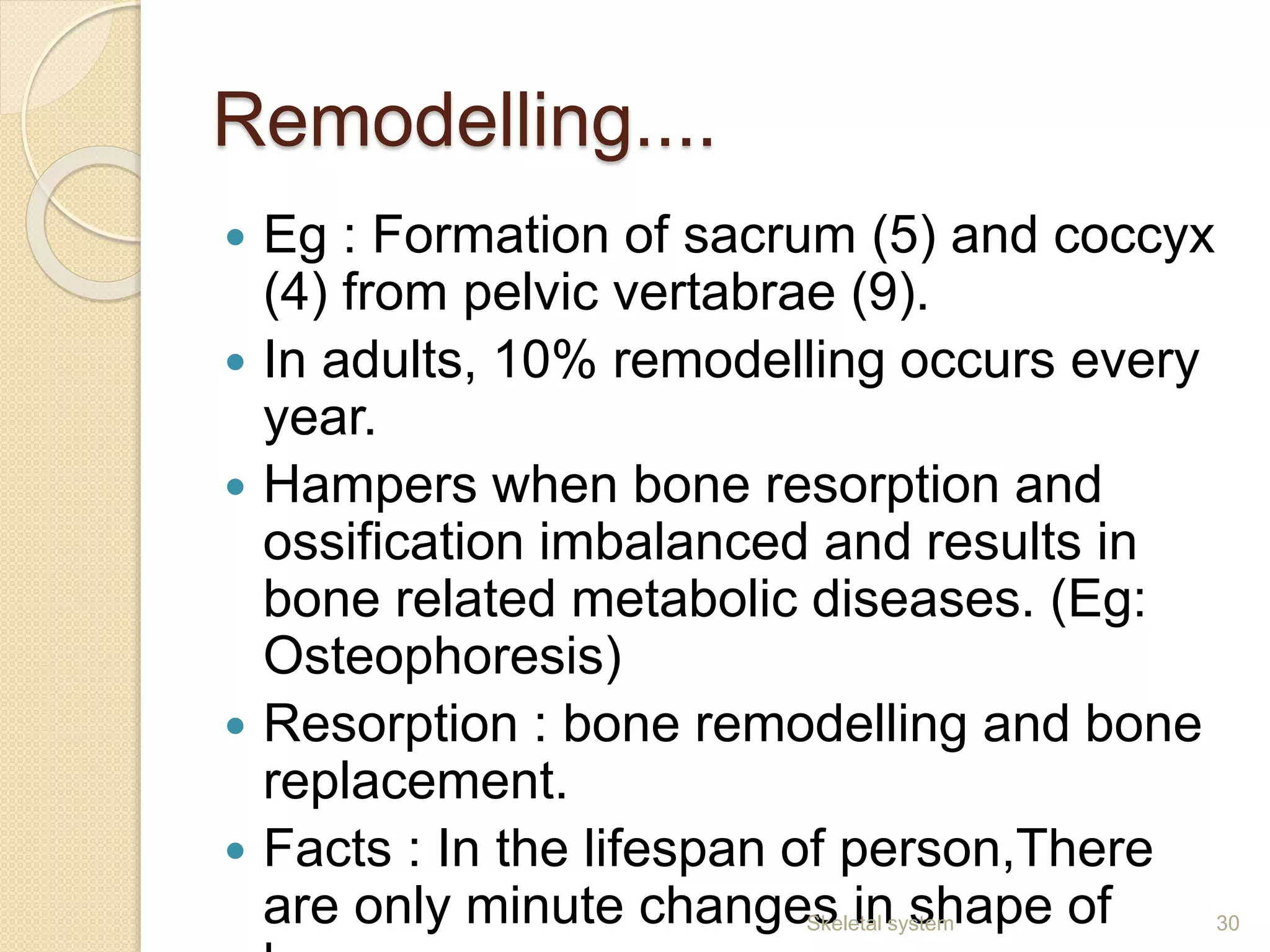
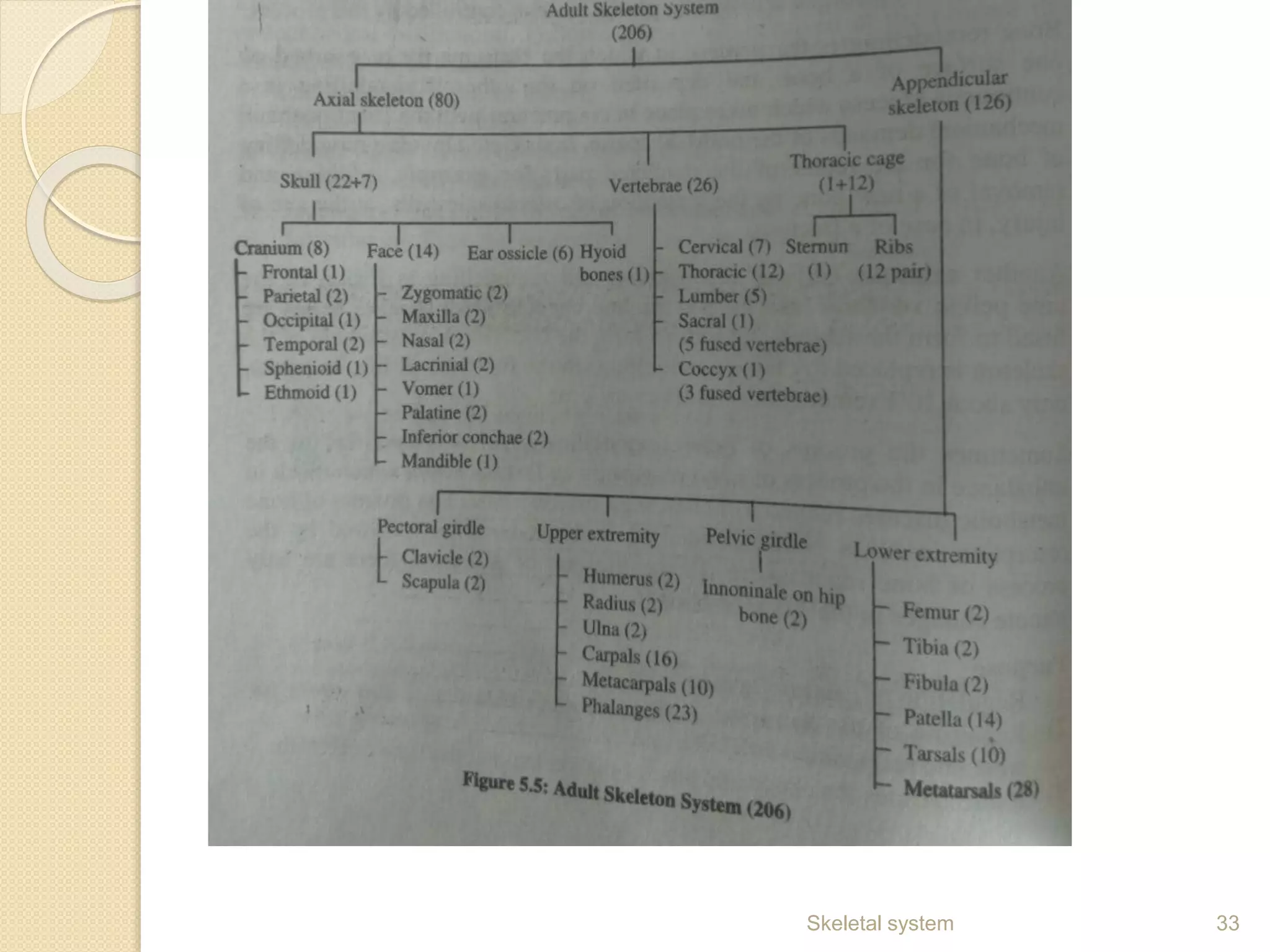
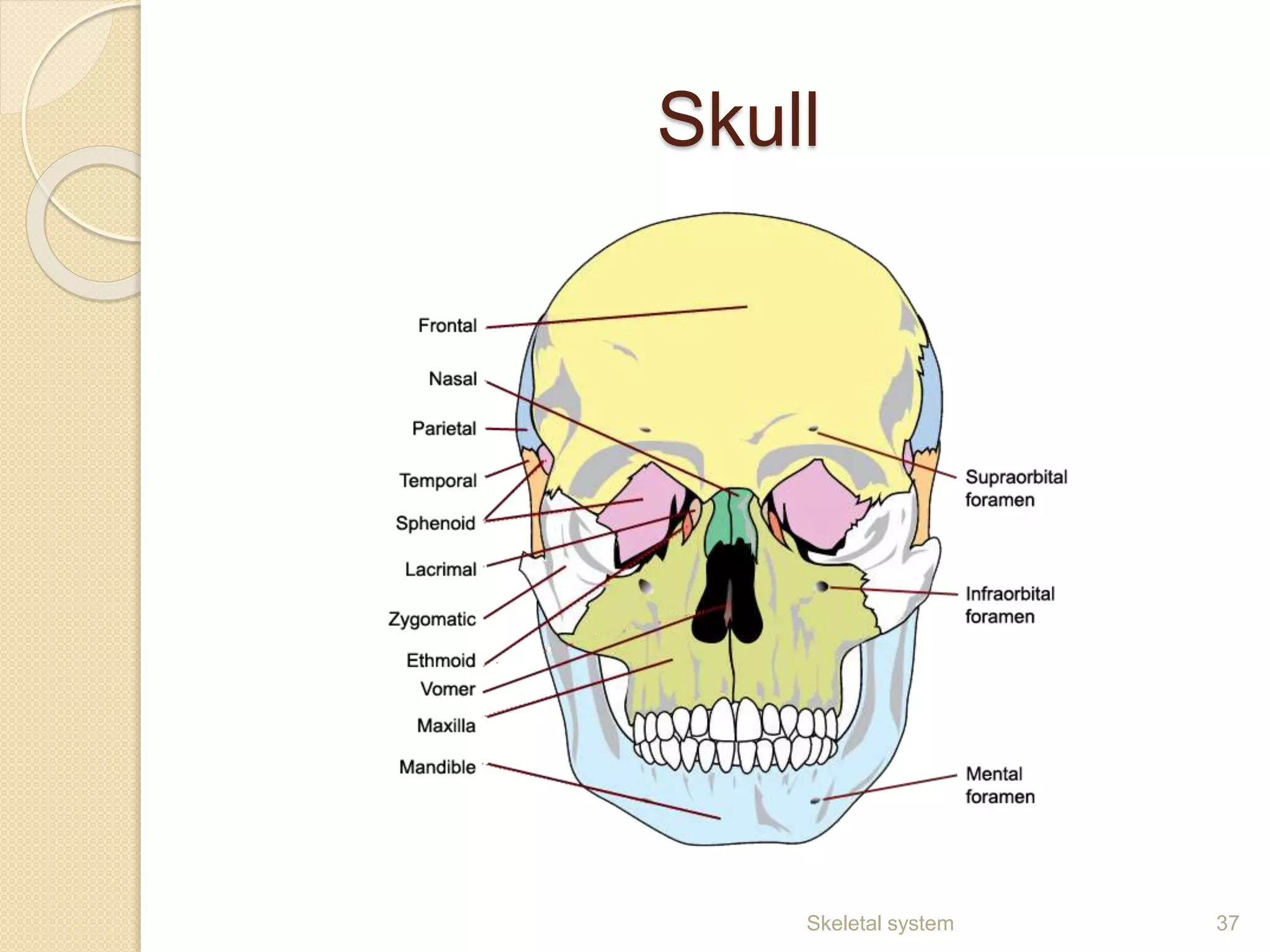
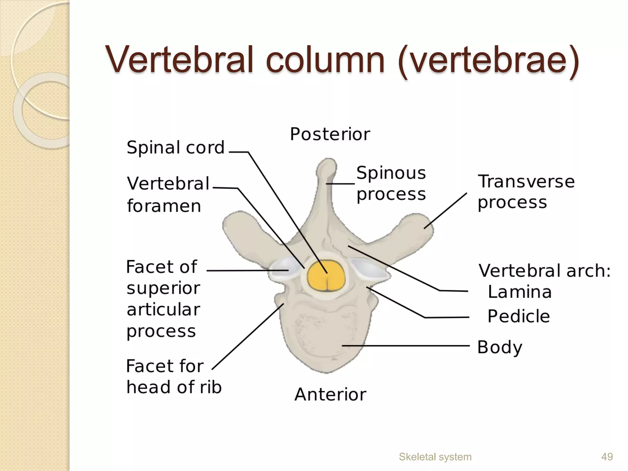
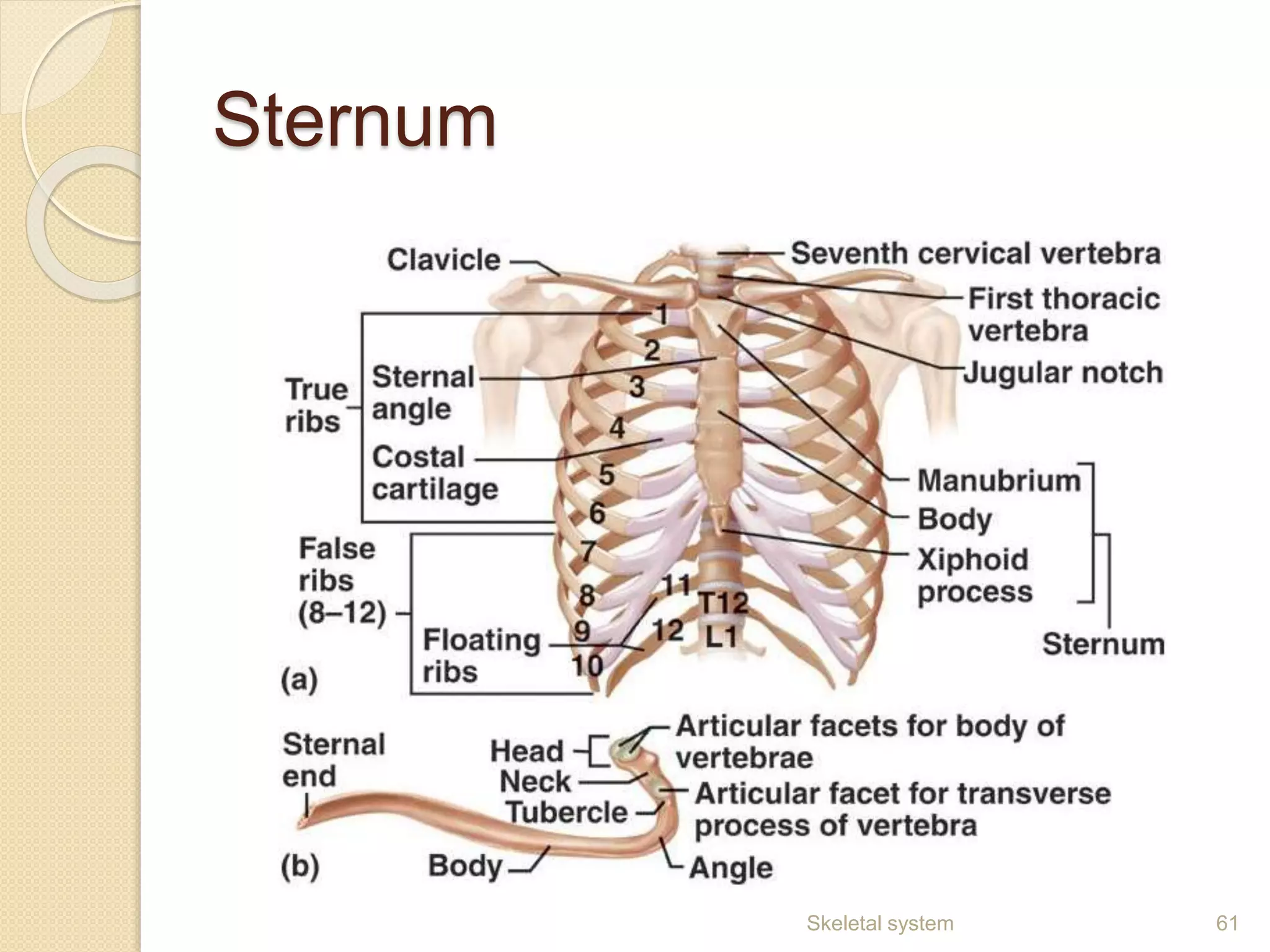
SKELETAL SYSTEM
M.ASIYABI. M.pharm.,
ASSISTANT PROFESSOR
DEPT OF PHARMACOLOGY
1Skeletal system
Introduction
•Framework of joined bones.
•Helps to stand straight, to move, to perform any
physical activity
•Aids in breathing.
•It comprises of : Bones, cartilages, tendons,
and liagments.
•Bones comprise of salts like calcium phosphate
– gives stone like appearance.
Bones
 Osteology – study of bone and teeth.
 Applied in anthropology, forensic
science and archaeology.
 To determine nutritional value, health,
the position of bone.
3Skeletal system
Types of bones
1.) Long bones :
Shaft present. Length greater than
width.
Usually curved, imparts high strength
Eg : femur, tibia, ulna, humerus.
4Skeletal system
2.) Short bones : Equal length and
width. Eg : Ankle and wrist. Cubical
in shape.
5Skeletal system
3.) Flat bones: Thin, offer wide surface for
muscle attachment, provides mechanical
protection. Eg : Cranial bones (Protects brain)
6Skeletal system
4.) Irregular bones : Assymetrical,
complicated shape. Shaped to fulfill certain
functions in body. Eg : Vertebrae and facial
bones.
7Skeletal system
5.) Sesamoid bones: Present in Palms
of hand and soles of feet (Knee cap) .
Develop in some tendons under
stresses.
8Skeletal system
 6.) Sutural bones: Very small bones
found within sutural joints of cranium.
Categorised on basis of location not
shape.
9Skeletal system
Salient features
 Hard in consistency
 Light weighed tissue made of calcium
phosphate provides rigidity to bone.
 High compressive strength
 Lacks tensile strength.
 Brittle, but significant elastic due to the
presence of collagen.
 Made of living cells.
 Non-uniform structures.
10Skeletal system
Macroscopic structure of
bones
C.S of Compact and cancellous bone.
11Skeletal system
Compact / Cortical / Dense bone
 Hard outermost layer of bone.
 Minimum voids/spaces.
 Gives white , smooth and sturdy
outlook to bones.
 Harvesian system/osteons:
Cylindrical structure.
Has Concentric collagen layers called
Harvesian lamellae (Within this
Harvesian canal present which carry
blood vessels, lymph vessels and
nerve fibres). 12Skeletal system
Compact bone
 Volkmann’s canals : Connects
Harvesian systems.
 Lacuane – Small tiny spaces and has
tissue fluid and spider shaped
osteocytes (Has canaliculi, which joins
together to form network).
 Periosteum : Covers outer surface of
bone. Does not cover joints. Made of
vascularised connective tissue.
 Two layers : Internal (osteogenic) and
outer (fibrous).
13Skeletal system
Trabecular / cancellous /
Spongy bone
 Filled with trabecular bone tissue
which contains network of spongy
bones.
 Contains rod and plate like elements
which provides space for blood
vessels and bone marrow.
 20% lighter and 10 times greater
surface area than compact bone.
14Skeletal system
Microscopic structure
1.) Osteoblasts : Mononucleate cells
formed from osteoprogenitor cells.
Leads to formation of proteinaceous
mixture (osteoid, which gets
mineralised to form bone).
2.) Osteocytes : Mature bone cells
formed from osteoblasts.
Occupies specific space called lacuane.
Mechno-sensory receptor : Regulate
the response of bone to stress.
15Skeletal system
Microscopic structure
3.) Osteoclasts : Large multinucleated
cells that involved in re-modelling of
bone (Bone resorption).
Located on small depression on bone’s
surface called Howship’s lacunae/
resorption pits (Appear as scalloped
surfaces in case of bone fracture).
16Skeletal system
Molecular structure
INORGANIC CONSTITUENTS :
The organic constisuents of matrix are
usually mineralised salt and calcium in
crystaline forms ,that occurs as
hydroxypatite.
The oesteoblast get mineralised by
alkaine phosphate ,released secreting
vesicles.
17Skeletal system
Organic constituents:
Type I collagen - main part of organic
matrix .
its made up of tropocollagen
Glycosaminoglycans, bone
sialoproteins ,osteocalcin, oesteonectin
and cell attachment factor were also
present in organic matrix.
Based on pattern of collagen
formed by osteoid
1.) Woven bone: Weak, Little amount of
collagen fibres oriented randomly. It forms
quickly independent of previous structure at
the time of growth and repair.
2.) Lamellar bone : Layered and very strong.
Several layers are piled up on one another. It
consist of collagen fibres lying parallel to
other fibres. They withstand torsion forces
due to the lamellar bone arrangement (Opp in
each alternate layer)
Bony substitution : When fracture, lamellar
bone replaced by woven bone (at hyaline
cartilage). 19Skeletal system
Formation of bones :
During prenatal stage, bones developed
by two methods:
1.) Intramembranous ossification
2.) Endochondral ossification
20Skeletal system
Intramembranous ossification
 Ossification –formation of bones.
 Flat bones of cranium formed from
mesenchyme tissue by a process
called Intramembranous ossification.
 Steps :
1.) Ossification centre development
2.) Process of calcification
3.) Trabeculae formation
4.) Periosteum development.
21Skeletal system
Intramembranous ossification
showing :
22Skeletal system
 Membranous ossification : At
specific region, direct mineralisation of
vascularised connective tissue takes
place.
 At that centre, Osteoprogenitor cells
multiply and accumulate. Around
vessels, collagen fibres are present.
 Osteoprogenitor cells osteoblasts
osteoid Osteoblasts (bony matrix)
osteocytes (lined by active osteoblasts)
 This depostion, Primarily has
mesenchymal stem cells, later forms
the basic bone tissue. 23Skeletal system
 Thickness of trabeculae increases ad
spongy layer becomes narrowed.
Cancellous bone slows down the
process and occupied by hemopoitic
tissue.
 Mesenchyme become denser to form
fibrovascular periosteum. Peiosteum
grows and form woven bone.
 Stem cells from periosteum further
extends and is called ossification.
24Skeletal system
Endochondral ossification
 Long bones from limbs mainly formed from
cartlage by the process of endochondral
ossification.
Steps:
1.) Cartilage development model.
2.) cartilage growth model.
3.) Primary ossification development centre.
4.) Medullary cavity development.
5.) Secondary ossification development centre,
and
6.) Articular cartilage and epiphyseal plate
formation.
25Skeletal system
Endochondreal ossification
26Skeletal system
Endochondrial....
 Starting point – Primary ossification centre
formed (during fetal development and post
natal phase) – responsible for the formation
of short bones, diaphysis of long bones and
irregular bones.
 Secondary ossification : In post natal period,
epiphysis of long bones, extremities of flat
bones and other parts of irregular bones were
formed.
 Epiphyseal plate – zone for cartilage
formation. At age 18 to 25, all cartilages
ossified and converted to bones (Epiphysis
and diaphysis fuse together by epiphyseal
closure). 27Skeletal system
Regeneration and repair of
bones
28Skeletal system
Remodelling or bone turnover
 Removal of old bone cells and addition
of new bone cells.
 Lifelong process (if fractured, it will
reshape or replace the bone).
 The bone matrix is resorbed on one
surface of abone and deposited on other.
 It is continuous process.
 Takes place in conjuction with functional
and mechanical demands of body.
 Exercise , injury also involved in
remodelling.
Eg: Formation of new bone in at injured 29Skeletal system
Remodelling....
 Eg : Formation of sacrum (5) and coccyx
(4) from pelvic vertabrae (9).
 In adults, 10% remodelling occurs every
year.
 Hampers when bone resorption and
ossification imbalanced and results in
bone related metabolic diseases. (Eg:
Osteophoresis)
 Resorption : bone remodelling and bone
replacement.
 Facts : In the lifespan of person,There
are only minute changes in shape of 30Skeletal system
Calcium balance
 Osteoclast release stored Ca2+ into
systemic circulation and thus
helps in regulating Ca2+ .
 From bloodstream Ca2+ used to
fix damaged bone simultaneously
Ca2+ released by resorption.
 Both resorption and remodelling
interlinked and occurs at specific
site only. Eg: Fractured site .
31Skeletal system
Divisions of skeletal system
1.)Axial skeleton
2.) Appendicular skeleton
32Skeletal system
33Skeletal system
Functions of bones:
 Shape and support.
 Protection : Protects internal organs (Eg :Skull
protects brain)
 Body Movement : Aids in physical movement of
body.
 Acid-Base balance : Alkaline salt Ca2+ either
released or absorbed and helps to maintain PH of
blood.
 Mineral storage : Eg - Ca2+ and P.
 Blood production: blood cells produced from
bone marrow.
 Detoxification : Toxin absorbed from blood and
excreted gradually.
 Sound transduction : Ear bones involved in
hearing. 34Skeletal system
35Skeletal system
Axial skeleton
 Helps in the formation of body axis.
 Supports and protects skull, neck and
trunk.
 Adult : 80 bones ; Children : 87 bones
Bones Number
Skull
Cranium 8
Facial skeleton 14
Ear ossicles 6
Hyoid bone 1
Vertebral column 26 (33 in children)
Thorax
cage
Ribs 24 (12 pair)
Sterum 1
36Skeletal system
Skull
37Skeletal system
Skull
 Bones of head region forms skull.
 Located over vertebral column along vertical
axis.
 Skull of adult comprise of 28 bones
(Immovable , except mandible).
 Two regions : Cranium and facial bones
 Various cavities are formed by bones of skull
and face. Eg : Cranial cavity
 3 parts of sutures (Synarthroidal or
immovable joints) : Coronal suture,
Squamous suture and Lambdoid suture.
 Neurocranium (8 bones) supports brain
 Splanchocranium (14 bones) supports face. 38Skeletal system
Cranium
 Spherical box like structure –
Protective covering of brain.
 Basal region – numerous blood
vessels and nerves enter and exit the
cranial cavity.
39Skeletal system
Bones of cranium :
1.) Frontal bones :
i.) Frontal squama : thick plate like
structure that forms forehead
ii.) Supraorbital margin : Thick
region located above eyes
iii.) Frontal sinuses : Cavities in
frontal bone
2.) Parietal bones : Form the major
portion of both sides of the skull.
40Skeletal system
Cranium...
3.) Temporal bones : Present along the ear.
i) Temporal squama : temple of skull
ii) Zygomatic arch : part of jaw
iii) Carotid canal : Passage for carotid artery
and causes pulsation.
iv) Jugular foramen : passage for jugular vein
along with 3 cranial nerves.
v) Mandibular fossa : Forms Tempero-maibular
joint (TMJ).
vi) TMJ : articulates mandible with temporal
bone.
vii) Mastoid process : behind ear to which neck
muscles connects
viii) Styloid process : Keeps hyoid bone in its
place with the help of neck, muscles of tongue
and ligaments. 41Skeletal system
Cranium...
4.) Occipital bones : Back of skull
i) Foramen magnum : Site where
Spinal cord attached to brain.
ii) Occipital condyles : Articulates with
vertebrae and helps in nodding of head.
5.) Sphenoid : Bat like structure present in
the centre of skull.
i) Sella turcica : holds pituitary gland
ii) Greater (2 outer) and lesser (2
inner) wings
iii) Pterygoid process : Between
greater wings and body.
42Skeletal system
Cranium...
6.) Ethmoid : Anterior part of skull and
seperates nasal cavity from brain. It forms the
upper part of nasal cavity. Act as pivot
(center) for almost all the skull bones.
i) Cribriform plate : Perforate with
olfactory foramina through which 1st cranial
nerves passes. Forms roof of nasal cavity.
ii) Labyrinth or lateral mass : Contains
Thin walled cellular cavities, including
ethmoidal cells (sequenced in 3 groups :
Anterior, middle, posterior)
iii) Perpendicular plate : Consists of thin
flattened lamina and polygonal shaped
perpendicular plate. Forms nasal septum (via
cribriform plate)
43Skeletal system
Facial bones
Facial skeleton, also called as visceral skull,
attaches anterior and inferior surfaces of
cranium.
Different bones of face includes:
1.) Nasal bones : cartilaginous and provide
flexibility. Helps in formation of nasal
bridge.
2.) Nasal septum : seperates right and left
part of nose, composed of ethmoid, vomer
and cartilages. Three concho of nasal
cavity : middle concho, inferior concho,
superior concho.
3.)Maxillae : Paired bones of upper jaw.
Holds upper set of teeth and act as roof
for mouth and floor of nose and eye cavity.44Skeletal system
Facial bones..
4.) Cleft : Seperates oral and nasal cavity.
Roof of mouth.
5.) Zygomatic bones : Cheek
bones(articulate with maxilla, sphenoid
and temporal bones)
6.) Mandible : Strongest and largest bone.
Only movable bone of face. Forms lower
jaw and holds lower teeth.
7.) Vomer : Only unpaired bone of face
which forms nasal septum.
8.) Lacrimal bones : Smallest bone.
Lacrimal ducts present and responsible
for lacrimation.
9.) Orbit : The socket or cavity holding the 45Skeletal system
Hyoid bone • Horse shoe shaped bone
• Also known as lingual bone
• Located middle of neck, between
thyroid cartilage and chin
• Floor of oral cavity
46Skeletal system
Hyoid bone ..
 Thyroid ligament holds this bone.
 Only skeletal bone not attached to any
other bone.
 Act as point of attachment for : Muscles of
oral cavity and tongue (ventrally) , The
larynx (Dorsally).
 Posterior surface attached to epiglottis and
pharynx.
 Two parts :
- centrally loacted part (Body)
- Two pairs of cornu: Greater cornu (2) ,
lesser cornu
(2) 47Skeletal system
Functions of hyoid bone
 Anchoring structure for tongue and
larynx, anterior neck muscles.
 Aids in tongue movement.
 Helps in swallowing.
 Helps in production of sound and its
variations.
48Skeletal system
Vertebral column (vertebrae)
49Skeletal system
Vertebrae
 The backbone of human being
comprises of 33 vertebrae.
 Also known as spine
 Provides protection to spinal cord and
its membranes.
50Skeletal system
Basic structure of a vertebra
1.) Body : Anterior portion of vertebra
which is joined to the intervertebral disc.
2.) Vertebral arch : Arch of posterior part
of body and protects spinal cord. It has
pedicles and laminae.
3.) Vertebral foramen : Opening through
arch. Spinal cord passes through this
foramen.
4.) Processes : Projection or outgrowth of
tissue from vertebrae. Three types :
transverse process, spinous process and
superior articular process. Attachment 51Skeletal system
Parts of vertebrae
52Skeletal system
Parts...
1.) Cervical (7) : form neck region.
Presence of foramen in transverse
process.
2.) Thoracic (12) : Externally visible as
chest or thorax. Presence of facets (ribs)
3.) Lumbar (5) : Absence of facet and
foramen. Belove thoracic region and
above pelvic region. Larger and stronger
than other vertebrae.
4.)Sacral (5) : Formation of sacral region
or upper pelvis.
5.) Coccyx (1) : Fusion of 4 coccygeal
vertebrae, in lower part of pelvis.
53Skeletal system
Curvatures of vetebral column
 4 types :
1. The cervical curvature
2. The thoracic curvature
3. The lumbar curvature
4. The pelvic curvature.
54Skeletal system
Functions of vertebral column
 Maintain erect body posture
 Maintain body balance.
 Vertebral architecture : supporting framework
for the trunk of body.
 Support system : Supports upper limb and
head to make them move freely. Point of
attachment for various muscles, ribs and
organs.
 Protection : protects spinal cord, kidney and
liver.
 Other functions : Maintain and balance
distribution of weight. Haemopoiesis in foetus
or infants. Forms site of attachment for 55Skeletal system
Thoracic cage / thorax
 Lies between head and abdomen,
excluding upper limbs.
 It constitute of sternum, thoracic
vertebrae and ribs.
 Encloses and protects heart and lungs
as well as blood vessels and nerves.
 Three parts : Ribs, sternum, thoracic
vertebrae.
 Out of 12 pair of ribs, upper 10 pairs
attached to sternum via cartilage from
anterior end. From posterior end, all the
12 pairs are attached to thoracic 56Skeletal system
Functions of thoracic cage
 Motion : Mobility to thorax region.
 Costal breathing :Attached to costal
cartilages and helps in costal
breathing.
 Protection : Encapsulates several
organs like heart and lungs, and
protects from damage.
57Skeletal system
Ribs:
 Long semi- circular bones forming rib cage.
Encapsulate chest forming periphery.
 They also allow the expansion of lungs for
respiration by expanding the chest (thorax).
 Organs like heart and lungs prtoected by the
ribs.
Basic structure of ribs : 12 pairs of ribs
3 parts : 1. Head : Attaches rib to the vertebrae.
Neck lies adjacent to neck.
2. Shaft : Length of rib. It has costal
where the blood vessels and nerves pass.
3. Tubercle : Terminal parts of rib and
attached to vertebrae of thorax. 58Skeletal system
Central rib of the left side
59Skeletal system
Types of ribs
 Sub divided into 3 categories :
1. True ribs : Directly attached to
sternum (via cartilages)
2. False ribs : Not directly attached
to the sternum.
3. Floating ribs : Not attached to
the sternum.
60Skeletal system
Sternum
61Skeletal system
Sternum
 Sternum or breastbone is a T shaped
or dagger shaped long flat bone
located at the centre of thorax.
 Lies in the centre of thoracic cavity.
 Joins to the ribs with the help of
cartilages and forms the frontal part of
the rib cage.
 The rib cage protects the heart, lungs
and major blood vessels from physical
injury or damage.
62Skeletal system
Three main regions of
sternum
1. Manubrium : Uppermost part of the
sternum consisting of two notches. i.e.,
Jugular notch and clavicular notch.
Clavicle (collar bone) attached at
clavicular notch.
2. Body : Rins (12) gets attached to the
sternum.
3. Xiphoid process : At the time of birth,
xiphoid process occurs as a cartilage,
which hardens due to ossification.
i.) It act as the point of attachment of
muscles of abdomen
ii.) Main point for cardiopulmonary
resuscitation 63Skeletal system
Functions of sternum
 Protecting vital organs
 Providing flexibility (costal cartilages)
 Protects aorta, vena cava and thymus.
 Fills the space in the mid region of the
chest just above the lungs and the
heart.
64Skeletal system
Appendicular skeleton
 Bony framework of upper and lower
limbs and their supporting girdles
including 126 bones that anchor the
appendages to the axial skeleton.
65Skeletal system
Pectoral girdle
Divisions : Left and right halves
Each half contains : Scapula and
clavicle
66Skeletal system
Scapula : Large, irregular and triangular flat
bone. Attaches muscles with trunk and the
upper limb.
3 processes :
i.) Spinous process : Posterior surface of the
upper broader part of each scapula. Distinct
ridges or spines are present.
ii.) Acromian process : The lateral end
articlated with acromion process at acromio-
clavicular joint.
iii.) Coracoid process : Small projection which
extends out of the scapula. Tip of this
process forms the point of attachment of
biceps and coracobronchialis muscle.
Just benead coracoid, glenoid cavity (cup like
cavity) is present. 67Skeletal system
Clavicle (collar bone)
 S shaped subcutaneous bone.
 Present on both sides of neck and
connects upper limbs with trunk.
 Inner and outer end articulates with
sternum and acromian process (at
clavicular facet )
 No medullary cavity.
 Inner surface of lateral end shows
elevation called Conoid tubercle and
ridge known as trapezoid ridge.
68Skeletal system
Functions of pectoral girdle
 Support and suspension : for upper
extremities.
 Shock absorber : After the act of
jumping or leaping.
 Maintanence : Maintains shape of
thorax and provides space for
insertion of muscle of the back.
 Locomotor movement : It forms the
site of attachment for muscles of
upper limb and helps in movement.
69Skeletal system
Upper limb : 30 bones and divided into 3
parts (Humerus, forearm and carpals)
1.) Humerus or arm bone:
70Skeletal system
• Longest bone of fore limb
• Articulates with glenoid cavity
of scapula
• Joined with radius and ulna at
elbow joint
• To perform pull, push, lift and
rotation
Radius and ulna
Skeletal system 71
Ulna and Radius
Radius and ulna together forms forearm
Ulna : Forms medial bone in the fore limb.
It consist of following notches and
process:
i. Olecranon process
ii. Coronoid process
iii. Radial notch and
iv. Trochlear notch
Radius : Lateral bone of forearm of the
body. The upper surface of the radius
articulates with capitulum of the
humerus, distal end articulates with ulnar
Skeletal system 72
Carpals, metacarpals and
phalanges
Skeletal system 73
 Carpals: 8 irregularly shaped bones,
arranged in proximal (scaphoid,
lunate,triquetral and pisiform) and
distal (Trapezium, trapezoid, capitate
and hamate) rows.
 Metacarpals: 5, which are numbered
from lateral to medial side and have
the head, the shaft and base.
 Phalanges: There are 14 phalanges
present in single hand (2 in thumb, 12
in other four fingers 3 each).
Skeletal system 74
Pelvic girdle
 Irregular, arch shaped structure made
up of coxae or innominate bones.
 The arch of pelvic girdle is articulated
by highly flexible structure called
pubic symphysis.
 Pelvis : Ring like structure formed by
sacrum and coccyx.
 Pelvic girdle made of 3 distinct bones
(Ilium, Ischium and pubis), fused
completely in adult.
Skeletal system 75
Pelvic girdle
Skeletal system 76
Bones of pelvic girdle
1.) Ilium : Plate like structure Upper
expanded part of the hip bone. External
surface acts as an attachment site for
gluteus muscles such as Guteus
maximus, medius and minimus.
3 parts :
i.) Sacroiliac joint : Attach sacrum and
ilium
ii.) Greater sciatic notch : Pathway for
sciatic nerve passes towards the leg.
iii.) Iliac fossa : Flat surface of ilium
Skeletal system 77
2.) Ischium : Posterior inferior region of hip
bone. It consists of body and Ramus along
with ischial spine and ischial tuberosity.
3 Parts:
i.) Obturator foramen : Large hole through
which blood vessel and nerve passes,
covered by fibrous network.
ii.) Ischial tuberosity : Rough and thicck
surface of ischial body. Strongest part of hip.
Supports whole weight of body, while sitting.
iii.) Acetabulum : Wide socket like cavity
that holds the head of femur (thigh).
3.)Pubis : Anterior-inferior part of the hip bone.
Three parts : Superior ramus, inferior ramus
and body. (together forms pubis). Meets to
form small area, called Acetabulum (like
deep socket).
Skeletal system 78
Difference
Skeletal system 79
Male pelvis Female pelvis
Massive size Comparatively
smaller
Iliac crests are
placed close
together
Placed farther apart
Less flexible More flexible
Variations in structure proves that the pelvis of female plays a vital
role in
pregnancy and delivery of child. During parturition, child passes
through
mother’s pelvis. If it is small, then caesarean is done
Functions of pelvic girdle
 Support : Weight of body from
vertebral column is supported by
pelvis
 Protection : Supports and protects the
organs related to reproduction and
urinary system as well as developing
fetus in pregnant females.
Skeletal system 80
Lower limb
Skeletal system 81
Lower limb comprises of 32
bones as given below :
1. Femur: Strongest and longest bone of
human body. Present at upper end of
femur. The lower condylar end present
two articular surfaces : Patellar surface
(patella) and tibial surface (tibia).
2. Patella : Sesamoid bone located right in
front of distal end of femur.
3. Tibia : larger bone of lower leg and
located medially.
4. Fibula : Lateral bone present in lower
limb , articulate with lateral condyle of
tibia. Skeletal system 82
Tibio-
fibula
Skeletal system 83
Femur
Skeletal system 84
Skeletal system 85
5. )Tarsal : Proximal (Talus and calcaneus) and distal (medial cuneiform,
internal
cuneiform, lateral cuneiform, cuboid and navicular bones) rows.
6.) Metatarsal : articulate proximally with uniform and cuboid bones, and
distally with phalanges.
7.) Phalanges : 14 phalanges in single foot (2 in great toe remaining in
other
fingers)
Foot contain two additional bones called sesamoid bones. Muscle of big
Skeletal system 86

Skeletal system

  • 1.
    SKELETAL SYSTEM M.ASIYABI. M.pharm., ASSISTANTPROFESSOR DEPT OF PHARMACOLOGY 1Skeletal system
  • 2.
    Introduction •Framework of joinedbones. •Helps to stand straight, to move, to perform any physical activity •Aids in breathing. •It comprises of : Bones, cartilages, tendons, and liagments. •Bones comprise of salts like calcium phosphate – gives stone like appearance.
  • 3.
    Bones  Osteology –study of bone and teeth.  Applied in anthropology, forensic science and archaeology.  To determine nutritional value, health, the position of bone. 3Skeletal system
  • 4.
    Types of bones 1.)Long bones : Shaft present. Length greater than width. Usually curved, imparts high strength Eg : femur, tibia, ulna, humerus. 4Skeletal system
  • 5.
    2.) Short bones: Equal length and width. Eg : Ankle and wrist. Cubical in shape. 5Skeletal system
  • 6.
    3.) Flat bones:Thin, offer wide surface for muscle attachment, provides mechanical protection. Eg : Cranial bones (Protects brain) 6Skeletal system
  • 7.
    4.) Irregular bones: Assymetrical, complicated shape. Shaped to fulfill certain functions in body. Eg : Vertebrae and facial bones. 7Skeletal system
  • 8.
    5.) Sesamoid bones:Present in Palms of hand and soles of feet (Knee cap) . Develop in some tendons under stresses. 8Skeletal system
  • 9.
     6.) Suturalbones: Very small bones found within sutural joints of cranium. Categorised on basis of location not shape. 9Skeletal system
  • 10.
    Salient features  Hardin consistency  Light weighed tissue made of calcium phosphate provides rigidity to bone.  High compressive strength  Lacks tensile strength.  Brittle, but significant elastic due to the presence of collagen.  Made of living cells.  Non-uniform structures. 10Skeletal system
  • 11.
    Macroscopic structure of bones C.Sof Compact and cancellous bone. 11Skeletal system
  • 12.
    Compact / Cortical/ Dense bone  Hard outermost layer of bone.  Minimum voids/spaces.  Gives white , smooth and sturdy outlook to bones.  Harvesian system/osteons: Cylindrical structure. Has Concentric collagen layers called Harvesian lamellae (Within this Harvesian canal present which carry blood vessels, lymph vessels and nerve fibres). 12Skeletal system
  • 13.
    Compact bone  Volkmann’scanals : Connects Harvesian systems.  Lacuane – Small tiny spaces and has tissue fluid and spider shaped osteocytes (Has canaliculi, which joins together to form network).  Periosteum : Covers outer surface of bone. Does not cover joints. Made of vascularised connective tissue.  Two layers : Internal (osteogenic) and outer (fibrous). 13Skeletal system
  • 14.
    Trabecular / cancellous/ Spongy bone  Filled with trabecular bone tissue which contains network of spongy bones.  Contains rod and plate like elements which provides space for blood vessels and bone marrow.  20% lighter and 10 times greater surface area than compact bone. 14Skeletal system
  • 15.
    Microscopic structure 1.) Osteoblasts: Mononucleate cells formed from osteoprogenitor cells. Leads to formation of proteinaceous mixture (osteoid, which gets mineralised to form bone). 2.) Osteocytes : Mature bone cells formed from osteoblasts. Occupies specific space called lacuane. Mechno-sensory receptor : Regulate the response of bone to stress. 15Skeletal system
  • 16.
    Microscopic structure 3.) Osteoclasts: Large multinucleated cells that involved in re-modelling of bone (Bone resorption). Located on small depression on bone’s surface called Howship’s lacunae/ resorption pits (Appear as scalloped surfaces in case of bone fracture). 16Skeletal system
  • 17.
    Molecular structure INORGANIC CONSTITUENTS: The organic constisuents of matrix are usually mineralised salt and calcium in crystaline forms ,that occurs as hydroxypatite. The oesteoblast get mineralised by alkaine phosphate ,released secreting vesicles. 17Skeletal system
  • 18.
    Organic constituents: Type Icollagen - main part of organic matrix . its made up of tropocollagen Glycosaminoglycans, bone sialoproteins ,osteocalcin, oesteonectin and cell attachment factor were also present in organic matrix.
  • 19.
    Based on patternof collagen formed by osteoid 1.) Woven bone: Weak, Little amount of collagen fibres oriented randomly. It forms quickly independent of previous structure at the time of growth and repair. 2.) Lamellar bone : Layered and very strong. Several layers are piled up on one another. It consist of collagen fibres lying parallel to other fibres. They withstand torsion forces due to the lamellar bone arrangement (Opp in each alternate layer) Bony substitution : When fracture, lamellar bone replaced by woven bone (at hyaline cartilage). 19Skeletal system
  • 20.
    Formation of bones: During prenatal stage, bones developed by two methods: 1.) Intramembranous ossification 2.) Endochondral ossification 20Skeletal system
  • 21.
    Intramembranous ossification  Ossification–formation of bones.  Flat bones of cranium formed from mesenchyme tissue by a process called Intramembranous ossification.  Steps : 1.) Ossification centre development 2.) Process of calcification 3.) Trabeculae formation 4.) Periosteum development. 21Skeletal system
  • 22.
  • 23.
     Membranous ossification: At specific region, direct mineralisation of vascularised connective tissue takes place.  At that centre, Osteoprogenitor cells multiply and accumulate. Around vessels, collagen fibres are present.  Osteoprogenitor cells osteoblasts osteoid Osteoblasts (bony matrix) osteocytes (lined by active osteoblasts)  This depostion, Primarily has mesenchymal stem cells, later forms the basic bone tissue. 23Skeletal system
  • 24.
     Thickness oftrabeculae increases ad spongy layer becomes narrowed. Cancellous bone slows down the process and occupied by hemopoitic tissue.  Mesenchyme become denser to form fibrovascular periosteum. Peiosteum grows and form woven bone.  Stem cells from periosteum further extends and is called ossification. 24Skeletal system
  • 25.
    Endochondral ossification  Longbones from limbs mainly formed from cartlage by the process of endochondral ossification. Steps: 1.) Cartilage development model. 2.) cartilage growth model. 3.) Primary ossification development centre. 4.) Medullary cavity development. 5.) Secondary ossification development centre, and 6.) Articular cartilage and epiphyseal plate formation. 25Skeletal system
  • 26.
  • 27.
    Endochondrial....  Starting point– Primary ossification centre formed (during fetal development and post natal phase) – responsible for the formation of short bones, diaphysis of long bones and irregular bones.  Secondary ossification : In post natal period, epiphysis of long bones, extremities of flat bones and other parts of irregular bones were formed.  Epiphyseal plate – zone for cartilage formation. At age 18 to 25, all cartilages ossified and converted to bones (Epiphysis and diaphysis fuse together by epiphyseal closure). 27Skeletal system
  • 28.
    Regeneration and repairof bones 28Skeletal system
  • 29.
    Remodelling or boneturnover  Removal of old bone cells and addition of new bone cells.  Lifelong process (if fractured, it will reshape or replace the bone).  The bone matrix is resorbed on one surface of abone and deposited on other.  It is continuous process.  Takes place in conjuction with functional and mechanical demands of body.  Exercise , injury also involved in remodelling. Eg: Formation of new bone in at injured 29Skeletal system
  • 30.
    Remodelling....  Eg :Formation of sacrum (5) and coccyx (4) from pelvic vertabrae (9).  In adults, 10% remodelling occurs every year.  Hampers when bone resorption and ossification imbalanced and results in bone related metabolic diseases. (Eg: Osteophoresis)  Resorption : bone remodelling and bone replacement.  Facts : In the lifespan of person,There are only minute changes in shape of 30Skeletal system
  • 31.
    Calcium balance  Osteoclastrelease stored Ca2+ into systemic circulation and thus helps in regulating Ca2+ .  From bloodstream Ca2+ used to fix damaged bone simultaneously Ca2+ released by resorption.  Both resorption and remodelling interlinked and occurs at specific site only. Eg: Fractured site . 31Skeletal system
  • 32.
    Divisions of skeletalsystem 1.)Axial skeleton 2.) Appendicular skeleton 32Skeletal system
  • 33.
  • 34.
    Functions of bones: Shape and support.  Protection : Protects internal organs (Eg :Skull protects brain)  Body Movement : Aids in physical movement of body.  Acid-Base balance : Alkaline salt Ca2+ either released or absorbed and helps to maintain PH of blood.  Mineral storage : Eg - Ca2+ and P.  Blood production: blood cells produced from bone marrow.  Detoxification : Toxin absorbed from blood and excreted gradually.  Sound transduction : Ear bones involved in hearing. 34Skeletal system
  • 35.
  • 36.
    Axial skeleton  Helpsin the formation of body axis.  Supports and protects skull, neck and trunk.  Adult : 80 bones ; Children : 87 bones Bones Number Skull Cranium 8 Facial skeleton 14 Ear ossicles 6 Hyoid bone 1 Vertebral column 26 (33 in children) Thorax cage Ribs 24 (12 pair) Sterum 1 36Skeletal system
  • 37.
  • 38.
    Skull  Bones ofhead region forms skull.  Located over vertebral column along vertical axis.  Skull of adult comprise of 28 bones (Immovable , except mandible).  Two regions : Cranium and facial bones  Various cavities are formed by bones of skull and face. Eg : Cranial cavity  3 parts of sutures (Synarthroidal or immovable joints) : Coronal suture, Squamous suture and Lambdoid suture.  Neurocranium (8 bones) supports brain  Splanchocranium (14 bones) supports face. 38Skeletal system
  • 39.
    Cranium  Spherical boxlike structure – Protective covering of brain.  Basal region – numerous blood vessels and nerves enter and exit the cranial cavity. 39Skeletal system
  • 40.
    Bones of cranium: 1.) Frontal bones : i.) Frontal squama : thick plate like structure that forms forehead ii.) Supraorbital margin : Thick region located above eyes iii.) Frontal sinuses : Cavities in frontal bone 2.) Parietal bones : Form the major portion of both sides of the skull. 40Skeletal system
  • 41.
    Cranium... 3.) Temporal bones: Present along the ear. i) Temporal squama : temple of skull ii) Zygomatic arch : part of jaw iii) Carotid canal : Passage for carotid artery and causes pulsation. iv) Jugular foramen : passage for jugular vein along with 3 cranial nerves. v) Mandibular fossa : Forms Tempero-maibular joint (TMJ). vi) TMJ : articulates mandible with temporal bone. vii) Mastoid process : behind ear to which neck muscles connects viii) Styloid process : Keeps hyoid bone in its place with the help of neck, muscles of tongue and ligaments. 41Skeletal system
  • 42.
    Cranium... 4.) Occipital bones: Back of skull i) Foramen magnum : Site where Spinal cord attached to brain. ii) Occipital condyles : Articulates with vertebrae and helps in nodding of head. 5.) Sphenoid : Bat like structure present in the centre of skull. i) Sella turcica : holds pituitary gland ii) Greater (2 outer) and lesser (2 inner) wings iii) Pterygoid process : Between greater wings and body. 42Skeletal system
  • 43.
    Cranium... 6.) Ethmoid :Anterior part of skull and seperates nasal cavity from brain. It forms the upper part of nasal cavity. Act as pivot (center) for almost all the skull bones. i) Cribriform plate : Perforate with olfactory foramina through which 1st cranial nerves passes. Forms roof of nasal cavity. ii) Labyrinth or lateral mass : Contains Thin walled cellular cavities, including ethmoidal cells (sequenced in 3 groups : Anterior, middle, posterior) iii) Perpendicular plate : Consists of thin flattened lamina and polygonal shaped perpendicular plate. Forms nasal septum (via cribriform plate) 43Skeletal system
  • 44.
    Facial bones Facial skeleton,also called as visceral skull, attaches anterior and inferior surfaces of cranium. Different bones of face includes: 1.) Nasal bones : cartilaginous and provide flexibility. Helps in formation of nasal bridge. 2.) Nasal septum : seperates right and left part of nose, composed of ethmoid, vomer and cartilages. Three concho of nasal cavity : middle concho, inferior concho, superior concho. 3.)Maxillae : Paired bones of upper jaw. Holds upper set of teeth and act as roof for mouth and floor of nose and eye cavity.44Skeletal system
  • 45.
    Facial bones.. 4.) Cleft: Seperates oral and nasal cavity. Roof of mouth. 5.) Zygomatic bones : Cheek bones(articulate with maxilla, sphenoid and temporal bones) 6.) Mandible : Strongest and largest bone. Only movable bone of face. Forms lower jaw and holds lower teeth. 7.) Vomer : Only unpaired bone of face which forms nasal septum. 8.) Lacrimal bones : Smallest bone. Lacrimal ducts present and responsible for lacrimation. 9.) Orbit : The socket or cavity holding the 45Skeletal system
  • 46.
    Hyoid bone •Horse shoe shaped bone • Also known as lingual bone • Located middle of neck, between thyroid cartilage and chin • Floor of oral cavity 46Skeletal system
  • 47.
    Hyoid bone .. Thyroid ligament holds this bone.  Only skeletal bone not attached to any other bone.  Act as point of attachment for : Muscles of oral cavity and tongue (ventrally) , The larynx (Dorsally).  Posterior surface attached to epiglottis and pharynx.  Two parts : - centrally loacted part (Body) - Two pairs of cornu: Greater cornu (2) , lesser cornu (2) 47Skeletal system
  • 48.
    Functions of hyoidbone  Anchoring structure for tongue and larynx, anterior neck muscles.  Aids in tongue movement.  Helps in swallowing.  Helps in production of sound and its variations. 48Skeletal system
  • 49.
  • 50.
    Vertebrae  The backboneof human being comprises of 33 vertebrae.  Also known as spine  Provides protection to spinal cord and its membranes. 50Skeletal system
  • 51.
    Basic structure ofa vertebra 1.) Body : Anterior portion of vertebra which is joined to the intervertebral disc. 2.) Vertebral arch : Arch of posterior part of body and protects spinal cord. It has pedicles and laminae. 3.) Vertebral foramen : Opening through arch. Spinal cord passes through this foramen. 4.) Processes : Projection or outgrowth of tissue from vertebrae. Three types : transverse process, spinous process and superior articular process. Attachment 51Skeletal system
  • 52.
  • 53.
    Parts... 1.) Cervical (7): form neck region. Presence of foramen in transverse process. 2.) Thoracic (12) : Externally visible as chest or thorax. Presence of facets (ribs) 3.) Lumbar (5) : Absence of facet and foramen. Belove thoracic region and above pelvic region. Larger and stronger than other vertebrae. 4.)Sacral (5) : Formation of sacral region or upper pelvis. 5.) Coccyx (1) : Fusion of 4 coccygeal vertebrae, in lower part of pelvis. 53Skeletal system
  • 54.
    Curvatures of vetebralcolumn  4 types : 1. The cervical curvature 2. The thoracic curvature 3. The lumbar curvature 4. The pelvic curvature. 54Skeletal system
  • 55.
    Functions of vertebralcolumn  Maintain erect body posture  Maintain body balance.  Vertebral architecture : supporting framework for the trunk of body.  Support system : Supports upper limb and head to make them move freely. Point of attachment for various muscles, ribs and organs.  Protection : protects spinal cord, kidney and liver.  Other functions : Maintain and balance distribution of weight. Haemopoiesis in foetus or infants. Forms site of attachment for 55Skeletal system
  • 56.
    Thoracic cage /thorax  Lies between head and abdomen, excluding upper limbs.  It constitute of sternum, thoracic vertebrae and ribs.  Encloses and protects heart and lungs as well as blood vessels and nerves.  Three parts : Ribs, sternum, thoracic vertebrae.  Out of 12 pair of ribs, upper 10 pairs attached to sternum via cartilage from anterior end. From posterior end, all the 12 pairs are attached to thoracic 56Skeletal system
  • 57.
    Functions of thoraciccage  Motion : Mobility to thorax region.  Costal breathing :Attached to costal cartilages and helps in costal breathing.  Protection : Encapsulates several organs like heart and lungs, and protects from damage. 57Skeletal system
  • 58.
    Ribs:  Long semi-circular bones forming rib cage. Encapsulate chest forming periphery.  They also allow the expansion of lungs for respiration by expanding the chest (thorax).  Organs like heart and lungs prtoected by the ribs. Basic structure of ribs : 12 pairs of ribs 3 parts : 1. Head : Attaches rib to the vertebrae. Neck lies adjacent to neck. 2. Shaft : Length of rib. It has costal where the blood vessels and nerves pass. 3. Tubercle : Terminal parts of rib and attached to vertebrae of thorax. 58Skeletal system
  • 59.
    Central rib ofthe left side 59Skeletal system
  • 60.
    Types of ribs Sub divided into 3 categories : 1. True ribs : Directly attached to sternum (via cartilages) 2. False ribs : Not directly attached to the sternum. 3. Floating ribs : Not attached to the sternum. 60Skeletal system
  • 61.
  • 62.
    Sternum  Sternum orbreastbone is a T shaped or dagger shaped long flat bone located at the centre of thorax.  Lies in the centre of thoracic cavity.  Joins to the ribs with the help of cartilages and forms the frontal part of the rib cage.  The rib cage protects the heart, lungs and major blood vessels from physical injury or damage. 62Skeletal system
  • 63.
    Three main regionsof sternum 1. Manubrium : Uppermost part of the sternum consisting of two notches. i.e., Jugular notch and clavicular notch. Clavicle (collar bone) attached at clavicular notch. 2. Body : Rins (12) gets attached to the sternum. 3. Xiphoid process : At the time of birth, xiphoid process occurs as a cartilage, which hardens due to ossification. i.) It act as the point of attachment of muscles of abdomen ii.) Main point for cardiopulmonary resuscitation 63Skeletal system
  • 64.
    Functions of sternum Protecting vital organs  Providing flexibility (costal cartilages)  Protects aorta, vena cava and thymus.  Fills the space in the mid region of the chest just above the lungs and the heart. 64Skeletal system
  • 65.
    Appendicular skeleton  Bonyframework of upper and lower limbs and their supporting girdles including 126 bones that anchor the appendages to the axial skeleton. 65Skeletal system
  • 66.
    Pectoral girdle Divisions :Left and right halves Each half contains : Scapula and clavicle 66Skeletal system
  • 67.
    Scapula : Large,irregular and triangular flat bone. Attaches muscles with trunk and the upper limb. 3 processes : i.) Spinous process : Posterior surface of the upper broader part of each scapula. Distinct ridges or spines are present. ii.) Acromian process : The lateral end articlated with acromion process at acromio- clavicular joint. iii.) Coracoid process : Small projection which extends out of the scapula. Tip of this process forms the point of attachment of biceps and coracobronchialis muscle. Just benead coracoid, glenoid cavity (cup like cavity) is present. 67Skeletal system
  • 68.
    Clavicle (collar bone) S shaped subcutaneous bone.  Present on both sides of neck and connects upper limbs with trunk.  Inner and outer end articulates with sternum and acromian process (at clavicular facet )  No medullary cavity.  Inner surface of lateral end shows elevation called Conoid tubercle and ridge known as trapezoid ridge. 68Skeletal system
  • 69.
    Functions of pectoralgirdle  Support and suspension : for upper extremities.  Shock absorber : After the act of jumping or leaping.  Maintanence : Maintains shape of thorax and provides space for insertion of muscle of the back.  Locomotor movement : It forms the site of attachment for muscles of upper limb and helps in movement. 69Skeletal system
  • 70.
    Upper limb :30 bones and divided into 3 parts (Humerus, forearm and carpals) 1.) Humerus or arm bone: 70Skeletal system • Longest bone of fore limb • Articulates with glenoid cavity of scapula • Joined with radius and ulna at elbow joint • To perform pull, push, lift and rotation
  • 71.
  • 72.
    Ulna and Radius Radiusand ulna together forms forearm Ulna : Forms medial bone in the fore limb. It consist of following notches and process: i. Olecranon process ii. Coronoid process iii. Radial notch and iv. Trochlear notch Radius : Lateral bone of forearm of the body. The upper surface of the radius articulates with capitulum of the humerus, distal end articulates with ulnar Skeletal system 72
  • 73.
  • 74.
     Carpals: 8irregularly shaped bones, arranged in proximal (scaphoid, lunate,triquetral and pisiform) and distal (Trapezium, trapezoid, capitate and hamate) rows.  Metacarpals: 5, which are numbered from lateral to medial side and have the head, the shaft and base.  Phalanges: There are 14 phalanges present in single hand (2 in thumb, 12 in other four fingers 3 each). Skeletal system 74
  • 75.
    Pelvic girdle  Irregular,arch shaped structure made up of coxae or innominate bones.  The arch of pelvic girdle is articulated by highly flexible structure called pubic symphysis.  Pelvis : Ring like structure formed by sacrum and coccyx.  Pelvic girdle made of 3 distinct bones (Ilium, Ischium and pubis), fused completely in adult. Skeletal system 75
  • 76.
  • 77.
    Bones of pelvicgirdle 1.) Ilium : Plate like structure Upper expanded part of the hip bone. External surface acts as an attachment site for gluteus muscles such as Guteus maximus, medius and minimus. 3 parts : i.) Sacroiliac joint : Attach sacrum and ilium ii.) Greater sciatic notch : Pathway for sciatic nerve passes towards the leg. iii.) Iliac fossa : Flat surface of ilium Skeletal system 77
  • 78.
    2.) Ischium :Posterior inferior region of hip bone. It consists of body and Ramus along with ischial spine and ischial tuberosity. 3 Parts: i.) Obturator foramen : Large hole through which blood vessel and nerve passes, covered by fibrous network. ii.) Ischial tuberosity : Rough and thicck surface of ischial body. Strongest part of hip. Supports whole weight of body, while sitting. iii.) Acetabulum : Wide socket like cavity that holds the head of femur (thigh). 3.)Pubis : Anterior-inferior part of the hip bone. Three parts : Superior ramus, inferior ramus and body. (together forms pubis). Meets to form small area, called Acetabulum (like deep socket). Skeletal system 78
  • 79.
    Difference Skeletal system 79 Malepelvis Female pelvis Massive size Comparatively smaller Iliac crests are placed close together Placed farther apart Less flexible More flexible Variations in structure proves that the pelvis of female plays a vital role in pregnancy and delivery of child. During parturition, child passes through mother’s pelvis. If it is small, then caesarean is done
  • 80.
    Functions of pelvicgirdle  Support : Weight of body from vertebral column is supported by pelvis  Protection : Supports and protects the organs related to reproduction and urinary system as well as developing fetus in pregnant females. Skeletal system 80
  • 81.
  • 82.
    Lower limb comprisesof 32 bones as given below : 1. Femur: Strongest and longest bone of human body. Present at upper end of femur. The lower condylar end present two articular surfaces : Patellar surface (patella) and tibial surface (tibia). 2. Patella : Sesamoid bone located right in front of distal end of femur. 3. Tibia : larger bone of lower leg and located medially. 4. Fibula : Lateral bone present in lower limb , articulate with lateral condyle of tibia. Skeletal system 82
  • 83.
  • 84.
  • 85.
    Skeletal system 85 5.)Tarsal : Proximal (Talus and calcaneus) and distal (medial cuneiform, internal cuneiform, lateral cuneiform, cuboid and navicular bones) rows. 6.) Metatarsal : articulate proximally with uniform and cuboid bones, and distally with phalanges. 7.) Phalanges : 14 phalanges in single foot (2 in great toe remaining in other fingers) Foot contain two additional bones called sesamoid bones. Muscle of big
  • 86.