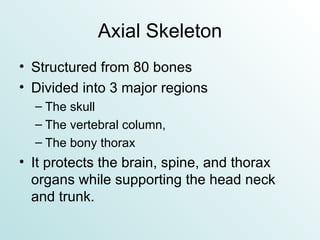
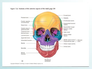
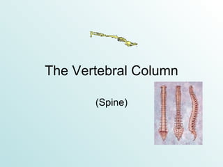
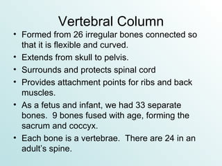
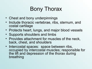
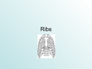
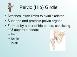
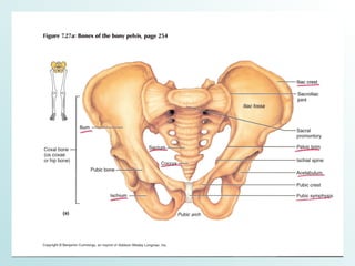
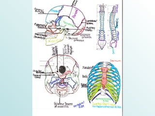
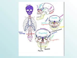
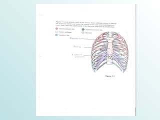
The Skeleton Chapter 7
Axial Skeleton Structured from 80 bones  Divided into 3 major regions The skull The vertebral column,  The bony thorax It protects the brain, spine, and thorax organs while supporting the head neck and trunk.
The Skull    
Skull Most complex bony structure Formed by cranial and facial bones (22 in all) All skull bones are united by joints called sutures, which have a saw-tooth or serrated appearance and are named for the bones they connect.
Facial Bones Form the framework of the face Contain cavities for sense organs (sight, taste, and smell) Provide openings for air passages Secure the teeth Anchor facial muscles
Parts of the Skull
The Cranium The cranium is made of eight cranial bones. It protects the brain, serving as a “helmet.”
Frontal Bone Anterior portion of the cranium Most anterior part is the frontal squama (forehead). The forehead ends at the supraorbital margins (thick part under the eyebrows). It forms the upper part of the orbits (eye sockets). The glabella is the smooth portion between the orbits. Frontonasal suture is where the frontal bone meets the nasal bone.
Parietal Bones and Major Sutures 2 curved, rectangular bones that form the superior and lateral aspects of the skull. They are the bulk of the cranium. 4 of the largest sutures occur at parietal joints: Coronal suture – parietal meets frontal  Sagittal suture – right and left parietals meet at midline Lambdoidal suture – parietal meets occipital Squamous suture – parietal meets temporal
Occipital Bone Forms most of the skull’s posterior wall and base Internally, it forms the walls of the posterior cranial fossa, which supports the cerebellum. At the bottom is the foramen magnum (“big hole”) where the brain connects with the spinal cord.
Occipital (con’t) Occipital condyles allow the head to nod. The external occipital protuberance is the knob-like projection on the posterior of the skull.
Temporal Bones Temporal bones (2) lie inferior to the parietal bones. They are called the temples (Latin:  temporous = time) because of gray hair forming signified time passing. They have a complicated shape and are described in terms of regions.
Temporal Regions  Squamous region:  cheekbone and condyle (“ball”) of the lower jawbone Tympanic region:  external ear canal and styloid process; serves as an attachment point for several muscles of the tongue and neck. Mastoid region:  facial nerves, neck muscles, and mastoid sinuses Petrous region:  middle and inner ear cavities, jugular vein, carotid arteries (supply 80% of the blood to the brain)
Sphenoid Bone Butterfly-shaped Wedged in the other cranial bones Consists of a central body and 3 processes.
Ethmoid Bone Between the sphenoid and nasal bones Forms the roof of the nasal cavities Punctured by olfactory foramina (holes), which allow nerves to pass through that aid in smelling. Forms the upper part of the nasal septum
Sutural Bones Also, sutural (AKA Wormian) bones are found in the cranium. These bones are tiny, irregularly shaped bones or bone clusters within the sutures.


Facial Bones There are 14 facial bones. The facial skeleton for men is more elongated than for women.  Women’s facial skeleton is rounder and less angular.
Mandible Lower jawbone U-shaped Largest and strongest bone in the face Consists of a chin and two rami (branches) A large muscle (temporalis) elevates the jaw. The lower teeth are anchored into the mandible
Mandible (con’t) The mandibular symphysis is the line of fusion of the mandible in infancy. The mandibular foramina allows the lower teeth to have sensation.
Maxilla Upper jaw and central portion of the face Anchors the upper teeth The palatine processes of the maxilla form 2/3 of the hard palate of the mouth. The frontal processes form part of the bridge of the nose
Other facial bones… Zygomatic bones – cheek bones Nasal bones – part of the bridge of the nose Lacrimal bones – house lacrimal sac (tear duct) Palatine bones – palates Vomer – nasal septum
Orbits Bony cavities within which the eyes are encases and cushioned by fatty tissue. Also, houses muscles that move eyes and the glands that produce tears. Formed by parts of frontal, sphenoid, zygomatic, maxilla, palatine, lacrimal and ethmoid bones.
Nasal Cavity Constructed by bone and hyaline cartilage Divided in ½ by nasal septum Covered in mucus-secreting mucosa, which moisten and warm the air.
 
The Vertebral Column (Spine)
Vertebral Column Formed from 26 irregular bones connected so that it is flexible and curved. Extends from skull to pelvis. Surrounds and protects spinal cord Provides attachment points for ribs and back muscles. As a fetus and infant, we had 33 separate bones.  9 bones fused with age, forming the sacrum and coccyx. Each bone is a vertebrae.  There are 24 in an adult’s spine.
Division & Curvature of Spine ~28 inches long in an average adult Has 5 major divisions All humans have the same number of cervical vertebrae.  However, there is a variation in the number for the rest of the vertebrae in 5% of humans in other regions. There are 4 curvatures, giving it a sinusoid (“S”) shape, which increases flexibility.
Divisions of the Spine  Cervical vertebrae – 7 vertebrae of the neck Thoracic vertebrae – next 12 vertebrae Lumbar vertebrae – 5 vertebrae supporting the lower back Sacrum – hip bones and pelvis Coccyx – tiny end of spine
 
Homeostatic Imbalances of the Spine  Scoliosis:  abnormal curvature of the spine usually in thoracic region, commonly in females
Kyphosis:  hunchback found in older adults Lordosis:  swayback; exaggerated lumbar curvature
Ligaments of the Spine Cables that cause the spine to be upright Major supporting ligaments are anterior and posterior, running down the front and back of the spine.  They prevent hyperextension and hyperflexion. Short ligaments connect each vertebrae to those above and below.
Intervertebral disks Cushion like pads Composed of 2 parts: Nucleus pulposus: “rubber ball;” elastic and compressible Annulus fibrosus: girdle for the nuclus pulposus, holds vertebrae together and resists tention Act as shock absorbers Account for ~25% of the spine’s length
Discs (con’t) Flatten during the day    taller in the morning Herniated (prolapsed) disc:  annulus fibrosus ruptures, allowing the nucleus pulposus to protrude
General Structure of Vertebrae Each vertebrae consists of a body (centrum) anteriorly and a vertebral arch posteriorly Body and arch enclose an opening called verebral foramen. Stacked foramen form a canal for the spinal cord to pass.
 
Vertebral Column Characteristics
Cervical Vertebrae  C 1  – C 7 Smallest and lightest C 1  & C 2 Have no discs in between them Allow the head to rotate
Thoracic Vertebrae All articulate with ribs T 1  – T 12 Have a long spinous process that points down
Lumbar Vertebrae “ Small of the Back” Receives the most stress L 1  – L 5 Have short, flat spinous processes that are hatchet-shaped – can be seen when bent over
Sacrum Five vertebrae that are fused S 1  – S 5 The remainder of the vertebral column continues as the sacral column Sacral hiatus:  enlarged opening where the 5 th  vertebrae fails to fuse completely
Coccyx “Tailbone” Small triangular bone 3-5 vertebrae fused together Nearly useless – gives slight support for the pelvic organs
The Bony Thorax
 
Bony Thorax Chest and bony underpinnings Include thoracic vertebrae, ribs, sternum, and costal cartilage Protects heart, lungs, and major blood vessels Supports shoulders and limbs Provides attachment for muscles of the neck, back, chest, and shoulders Intercostal spaces:  space between ribs, occupied by intercostal muscles; responsible for the lift and depression of the thorax during breathing
Sternum “Breastbone” Anterior midline of thorax Resembles a dagger Flat bone ~6 inches long
3 Parts that make up the Sternum  Manubrium:  superior portion; shaped like a necktie knot Body:  midportion; forms the bulk and the 2 nd  – 7 th  ribs attach Xiphoid process:  forms inferior end; it is hyaline cartilage of youth, but bone in adult (attachment point of abdominal muscles)
Ribs
Ribs 12 pair of ribs All ribs attach to the thoracic vertebrae Divided into two types of ribs True False Ribs increase in length from pair 1 – pair 7; then, they decrease from pair 8 – pair 12 Typical ribs are bowed flat bones.
True Ribs The superior 7 pairs of ribs are true ribs. They are known as vertebrosternal ribs, because they attach both to the spinal column and the sternum.
False Ribs The remaining 5 pairs of ribs are false ribs. Pairs 8-10 attach indirectly to the sternum – vertebrochondral ribs Pairs 11 & 12 do not attach at all to the sternum – floating ribs
 
Appendicular Skeleton Bones of the limbs and their girdles Enable us to carry out movement
Pectoral (Shoulder) Girdle Consists of the clavicle and scapula Attach the upper limbs to the axial skeleton Provide attachment for many muscles Very light and because the scapula is unattached and the socket is shallow, it allows a lot of mobility.
Clavicle (Collar Bone) Slender, curved long bone Cone shaped at the end and attaches to sternum Flat at the end that attaches to the shoulder blades Act as braces, holding arms and shoulder blades out laterally Not very strong; easily fractured and sensitive
Scapula Shoulder blade Thin and triangular flat bones Dorsal side of the ribcage
The Upper Limb Consists of 30 bones.
Arm Humerus:  sole bone of the arm Long bone Largest and longest of upper limb Connects with scapulae and forearm Only part of the upper limb between the shoulder and elbow
Forearm Ulna Longer bone of forearm Found medial when standing anatomically correct Radius Anchor bicep muscle When radius moves, the hand moves.
Hand
Carpus Wrist Consists of 8 marble-sized short bones (carpals) United by ligaments Carpal Tunnel Syndrome Overuse and inflammation of tendons, causes swelling Compresses nerves Causes numbness
Metacarpus Palm Bones radiate from wrist (metacarpals) Numbered 1-5 Heads of bones are knuckles Metacarpal 1 is the thumb and is the shortest and most mobile
Phalanges Fingers, AKA digits Numbered 1-5 Each hand has 14 mini long bones Each finger has 3 phalanges: distal, middle, and proximal Thumb has only 2 phalanges (no middle)
 
Pelvic (Hip) Girdle Attaches lower limbs to axial skeleton Supports and protects pelvic organs Formed by a pair of hip bones, consisting of 3 separate bones: Ilium Ischium Pubis
Ilium Large, flaring bone Consists of a body and winglike portion called ala (singular) or alae (plural) When resting your hands on your hips, you are resting on the thickened superior margins of alae.
Ischium Posteroinferior part of the hip L-shaped When we sit, the weight is entirely on the ischium.
Pubis Anterior portion of the hip Bladder rests on it V-shaped The pubic arch helps to differentiate males from females.
 
The Lower Limb The thicker and stronger bones due to the weight and stress on them.
Thigh Femur:  single bone of the thigh Largest and longest bone Strongest bone ~ ¼ of a person’s height
Leg Tibia Shin bone 2 nd  largest and strongest bone Medial surface not covered by muscles Fibula Sticklike bone with expanded ends Forms lateral ankle bulge Doesn’t bear weight, but it is attachment point for many muscles.
Foot Supports body weight Propels us forward when we walk.
Tarsus 7 Tarsal bones Weight is carried by the talus (ankle) and the calcaneus (heel). Achilles (calcaneal) tendon attaches to calcaneus.
Metatarsus 5 Small long (metatarsal) bones Numbered 1-5 Enlarged head of 1 st  metatarsal forms “ball” of foot
Phalanges (Toes) 14 phalanges Smaller than fingers Less maneuverable
 
Arches Distribute weight evenly between ball and heel of foot Gives foot great strength and mobility


The Skeleton Chapter 7

  • 1.
  • 2.
    Axial Skeleton Structuredfrom 80 bones Divided into 3 major regions The skull The vertebral column, The bony thorax It protects the brain, spine, and thorax organs while supporting the head neck and trunk.
  • 3.
  • 4.
    Skull Most complexbony structure Formed by cranial and facial bones (22 in all) All skull bones are united by joints called sutures, which have a saw-tooth or serrated appearance and are named for the bones they connect.
  • 5.
    Facial Bones Formthe framework of the face Contain cavities for sense organs (sight, taste, and smell) Provide openings for air passages Secure the teeth Anchor facial muscles
  • 6.
  • 7.
    The Cranium Thecranium is made of eight cranial bones. It protects the brain, serving as a “helmet.”
  • 8.
    Frontal Bone Anteriorportion of the cranium Most anterior part is the frontal squama (forehead). The forehead ends at the supraorbital margins (thick part under the eyebrows). It forms the upper part of the orbits (eye sockets). The glabella is the smooth portion between the orbits. Frontonasal suture is where the frontal bone meets the nasal bone.
  • 9.
    Parietal Bones andMajor Sutures 2 curved, rectangular bones that form the superior and lateral aspects of the skull. They are the bulk of the cranium. 4 of the largest sutures occur at parietal joints: Coronal suture – parietal meets frontal Sagittal suture – right and left parietals meet at midline Lambdoidal suture – parietal meets occipital Squamous suture – parietal meets temporal
  • 10.
    Occipital Bone Formsmost of the skull’s posterior wall and base Internally, it forms the walls of the posterior cranial fossa, which supports the cerebellum. At the bottom is the foramen magnum (“big hole”) where the brain connects with the spinal cord.
  • 11.
    Occipital (con’t) Occipitalcondyles allow the head to nod. The external occipital protuberance is the knob-like projection on the posterior of the skull.
  • 12.
    Temporal Bones Temporalbones (2) lie inferior to the parietal bones. They are called the temples (Latin: temporous = time) because of gray hair forming signified time passing. They have a complicated shape and are described in terms of regions.
  • 13.
    Temporal Regions Squamous region: cheekbone and condyle (“ball”) of the lower jawbone Tympanic region: external ear canal and styloid process; serves as an attachment point for several muscles of the tongue and neck. Mastoid region: facial nerves, neck muscles, and mastoid sinuses Petrous region: middle and inner ear cavities, jugular vein, carotid arteries (supply 80% of the blood to the brain)
  • 14.
    Sphenoid Bone Butterfly-shapedWedged in the other cranial bones Consists of a central body and 3 processes.
  • 15.
    Ethmoid Bone Betweenthe sphenoid and nasal bones Forms the roof of the nasal cavities Punctured by olfactory foramina (holes), which allow nerves to pass through that aid in smelling. Forms the upper part of the nasal septum
  • 16.
    Sutural Bones Also,sutural (AKA Wormian) bones are found in the cranium. These bones are tiny, irregularly shaped bones or bone clusters within the sutures.
  • 17.
  • 18.
  • 19.
  • 20.
    Facial Bones Thereare 14 facial bones. The facial skeleton for men is more elongated than for women. Women’s facial skeleton is rounder and less angular.
  • 21.
    Mandible Lower jawboneU-shaped Largest and strongest bone in the face Consists of a chin and two rami (branches) A large muscle (temporalis) elevates the jaw. The lower teeth are anchored into the mandible
  • 22.
    Mandible (con’t) Themandibular symphysis is the line of fusion of the mandible in infancy. The mandibular foramina allows the lower teeth to have sensation.
  • 23.
    Maxilla Upper jawand central portion of the face Anchors the upper teeth The palatine processes of the maxilla form 2/3 of the hard palate of the mouth. The frontal processes form part of the bridge of the nose
  • 24.
    Other facial bones…Zygomatic bones – cheek bones Nasal bones – part of the bridge of the nose Lacrimal bones – house lacrimal sac (tear duct) Palatine bones – palates Vomer – nasal septum
  • 25.
    Orbits Bony cavitieswithin which the eyes are encases and cushioned by fatty tissue. Also, houses muscles that move eyes and the glands that produce tears. Formed by parts of frontal, sphenoid, zygomatic, maxilla, palatine, lacrimal and ethmoid bones.
  • 26.
    Nasal Cavity Constructedby bone and hyaline cartilage Divided in ½ by nasal septum Covered in mucus-secreting mucosa, which moisten and warm the air.
  • 27.
  • 28.
  • 29.
    Vertebral Column Formedfrom 26 irregular bones connected so that it is flexible and curved. Extends from skull to pelvis. Surrounds and protects spinal cord Provides attachment points for ribs and back muscles. As a fetus and infant, we had 33 separate bones. 9 bones fused with age, forming the sacrum and coccyx. Each bone is a vertebrae. There are 24 in an adult’s spine.
  • 30.
    Division & Curvatureof Spine ~28 inches long in an average adult Has 5 major divisions All humans have the same number of cervical vertebrae. However, there is a variation in the number for the rest of the vertebrae in 5% of humans in other regions. There are 4 curvatures, giving it a sinusoid (“S”) shape, which increases flexibility.
  • 31.
    Divisions of theSpine Cervical vertebrae – 7 vertebrae of the neck Thoracic vertebrae – next 12 vertebrae Lumbar vertebrae – 5 vertebrae supporting the lower back Sacrum – hip bones and pelvis Coccyx – tiny end of spine
  • 32.
  • 33.
    Homeostatic Imbalances ofthe Spine Scoliosis: abnormal curvature of the spine usually in thoracic region, commonly in females
  • 34.
    Kyphosis: hunchbackfound in older adults Lordosis: swayback; exaggerated lumbar curvature
  • 35.
    Ligaments of theSpine Cables that cause the spine to be upright Major supporting ligaments are anterior and posterior, running down the front and back of the spine. They prevent hyperextension and hyperflexion. Short ligaments connect each vertebrae to those above and below.
  • 36.
    Intervertebral disks Cushionlike pads Composed of 2 parts: Nucleus pulposus: “rubber ball;” elastic and compressible Annulus fibrosus: girdle for the nuclus pulposus, holds vertebrae together and resists tention Act as shock absorbers Account for ~25% of the spine’s length
  • 37.
    Discs (con’t) Flattenduring the day  taller in the morning Herniated (prolapsed) disc: annulus fibrosus ruptures, allowing the nucleus pulposus to protrude
  • 38.
    General Structure ofVertebrae Each vertebrae consists of a body (centrum) anteriorly and a vertebral arch posteriorly Body and arch enclose an opening called verebral foramen. Stacked foramen form a canal for the spinal cord to pass.
  • 39.
  • 40.
  • 41.
    Cervical Vertebrae C 1 – C 7 Smallest and lightest C 1 & C 2 Have no discs in between them Allow the head to rotate
  • 42.
    Thoracic Vertebrae Allarticulate with ribs T 1 – T 12 Have a long spinous process that points down
  • 43.
    Lumbar Vertebrae “Small of the Back” Receives the most stress L 1 – L 5 Have short, flat spinous processes that are hatchet-shaped – can be seen when bent over
  • 44.
    Sacrum Five vertebraethat are fused S 1 – S 5 The remainder of the vertebral column continues as the sacral column Sacral hiatus: enlarged opening where the 5 th vertebrae fails to fuse completely
  • 45.
    Coccyx “Tailbone” Smalltriangular bone 3-5 vertebrae fused together Nearly useless – gives slight support for the pelvic organs
  • 46.
  • 47.
  • 48.
    Bony Thorax Chestand bony underpinnings Include thoracic vertebrae, ribs, sternum, and costal cartilage Protects heart, lungs, and major blood vessels Supports shoulders and limbs Provides attachment for muscles of the neck, back, chest, and shoulders Intercostal spaces: space between ribs, occupied by intercostal muscles; responsible for the lift and depression of the thorax during breathing
  • 49.
    Sternum “Breastbone” Anteriormidline of thorax Resembles a dagger Flat bone ~6 inches long
  • 50.
    3 Parts thatmake up the Sternum Manubrium: superior portion; shaped like a necktie knot Body: midportion; forms the bulk and the 2 nd – 7 th ribs attach Xiphoid process: forms inferior end; it is hyaline cartilage of youth, but bone in adult (attachment point of abdominal muscles)
  • 51.
  • 52.
    Ribs 12 pairof ribs All ribs attach to the thoracic vertebrae Divided into two types of ribs True False Ribs increase in length from pair 1 – pair 7; then, they decrease from pair 8 – pair 12 Typical ribs are bowed flat bones.
  • 53.
    True Ribs Thesuperior 7 pairs of ribs are true ribs. They are known as vertebrosternal ribs, because they attach both to the spinal column and the sternum.
  • 54.
    False Ribs Theremaining 5 pairs of ribs are false ribs. Pairs 8-10 attach indirectly to the sternum – vertebrochondral ribs Pairs 11 & 12 do not attach at all to the sternum – floating ribs
  • 55.
  • 56.
    Appendicular Skeleton Bonesof the limbs and their girdles Enable us to carry out movement
  • 57.
    Pectoral (Shoulder) GirdleConsists of the clavicle and scapula Attach the upper limbs to the axial skeleton Provide attachment for many muscles Very light and because the scapula is unattached and the socket is shallow, it allows a lot of mobility.
  • 58.
    Clavicle (Collar Bone)Slender, curved long bone Cone shaped at the end and attaches to sternum Flat at the end that attaches to the shoulder blades Act as braces, holding arms and shoulder blades out laterally Not very strong; easily fractured and sensitive
  • 59.
    Scapula Shoulder bladeThin and triangular flat bones Dorsal side of the ribcage
  • 60.
    The Upper LimbConsists of 30 bones.
  • 61.
    Arm Humerus: sole bone of the arm Long bone Largest and longest of upper limb Connects with scapulae and forearm Only part of the upper limb between the shoulder and elbow
  • 62.
    Forearm Ulna Longerbone of forearm Found medial when standing anatomically correct Radius Anchor bicep muscle When radius moves, the hand moves.
  • 63.
  • 64.
    Carpus Wrist Consistsof 8 marble-sized short bones (carpals) United by ligaments Carpal Tunnel Syndrome Overuse and inflammation of tendons, causes swelling Compresses nerves Causes numbness
  • 65.
    Metacarpus Palm Bonesradiate from wrist (metacarpals) Numbered 1-5 Heads of bones are knuckles Metacarpal 1 is the thumb and is the shortest and most mobile
  • 66.
    Phalanges Fingers, AKAdigits Numbered 1-5 Each hand has 14 mini long bones Each finger has 3 phalanges: distal, middle, and proximal Thumb has only 2 phalanges (no middle)
  • 67.
  • 68.
    Pelvic (Hip) GirdleAttaches lower limbs to axial skeleton Supports and protects pelvic organs Formed by a pair of hip bones, consisting of 3 separate bones: Ilium Ischium Pubis
  • 69.
    Ilium Large, flaringbone Consists of a body and winglike portion called ala (singular) or alae (plural) When resting your hands on your hips, you are resting on the thickened superior margins of alae.
  • 70.
    Ischium Posteroinferior partof the hip L-shaped When we sit, the weight is entirely on the ischium.
  • 71.
    Pubis Anterior portionof the hip Bladder rests on it V-shaped The pubic arch helps to differentiate males from females.
  • 72.
  • 73.
    The Lower LimbThe thicker and stronger bones due to the weight and stress on them.
  • 74.
    Thigh Femur: single bone of the thigh Largest and longest bone Strongest bone ~ ¼ of a person’s height
  • 75.
    Leg Tibia Shinbone 2 nd largest and strongest bone Medial surface not covered by muscles Fibula Sticklike bone with expanded ends Forms lateral ankle bulge Doesn’t bear weight, but it is attachment point for many muscles.
  • 76.
    Foot Supports bodyweight Propels us forward when we walk.
  • 77.
    Tarsus 7 Tarsalbones Weight is carried by the talus (ankle) and the calcaneus (heel). Achilles (calcaneal) tendon attaches to calcaneus.
  • 78.
    Metatarsus 5 Smalllong (metatarsal) bones Numbered 1-5 Enlarged head of 1 st metatarsal forms “ball” of foot
  • 79.
    Phalanges (Toes) 14phalanges Smaller than fingers Less maneuverable
  • 80.
  • 81.
    Arches Distribute weightevenly between ball and heel of foot Gives foot great strength and mobility
  • 82.
  • 83.
  • 84.