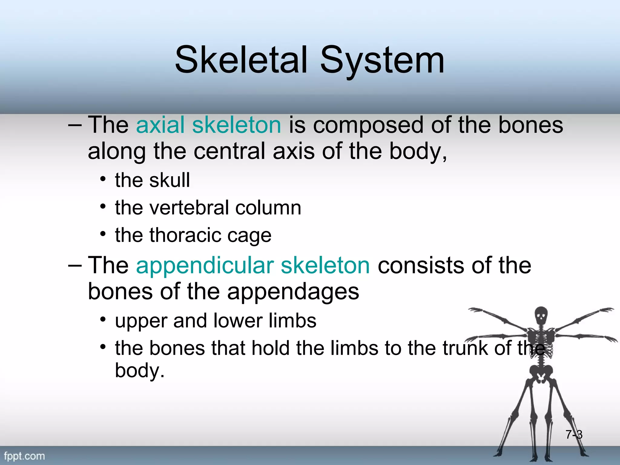
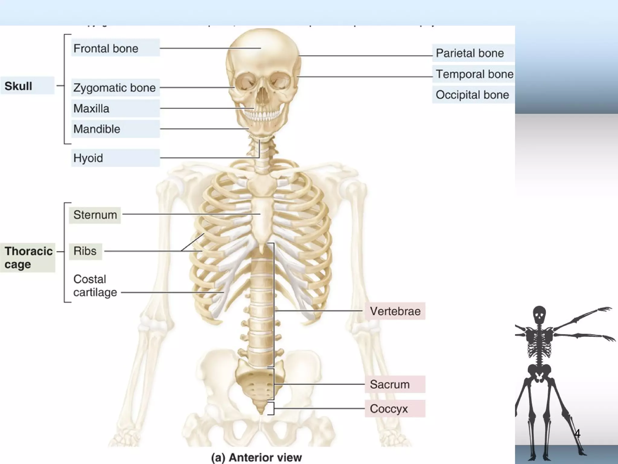
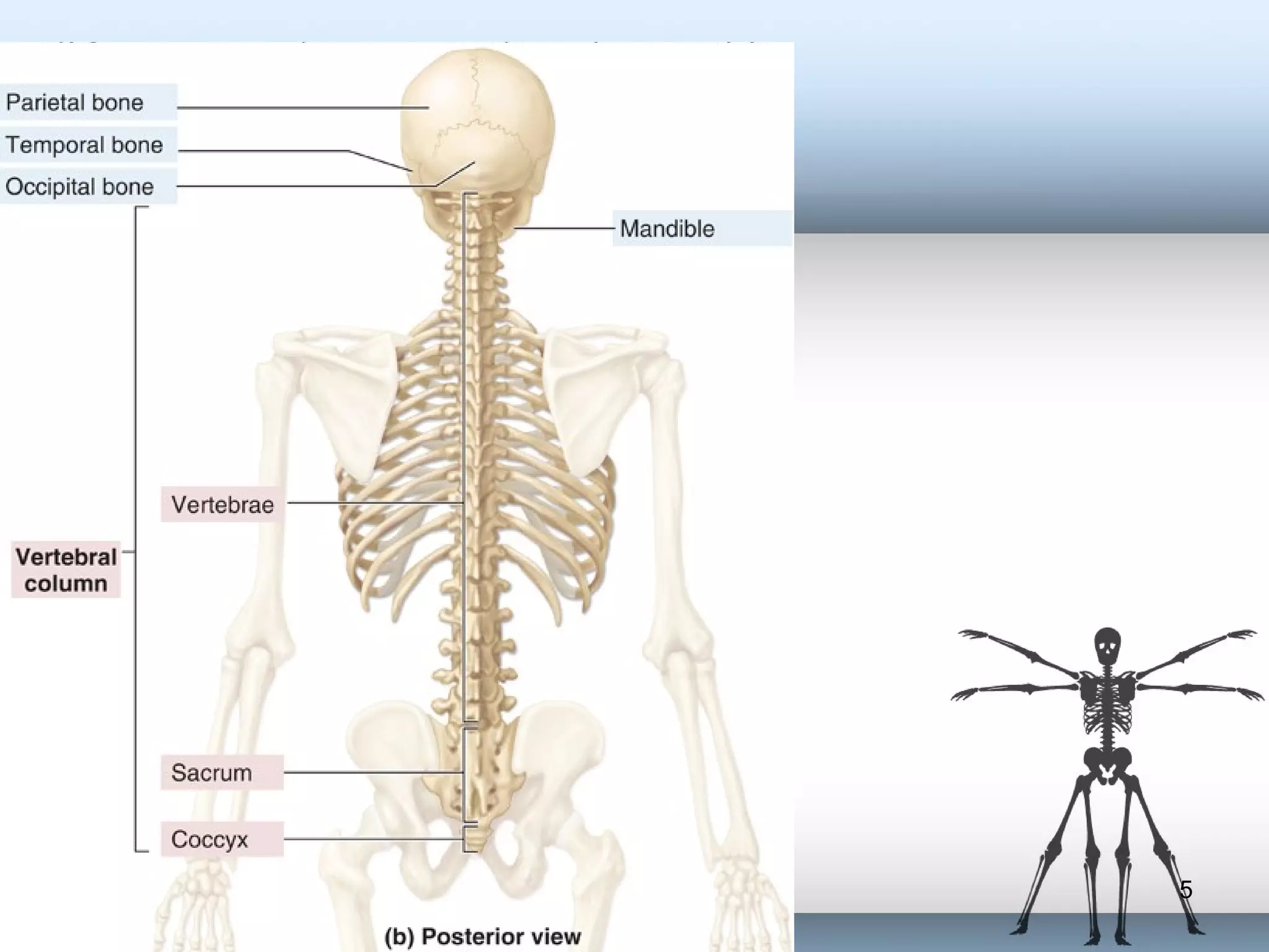
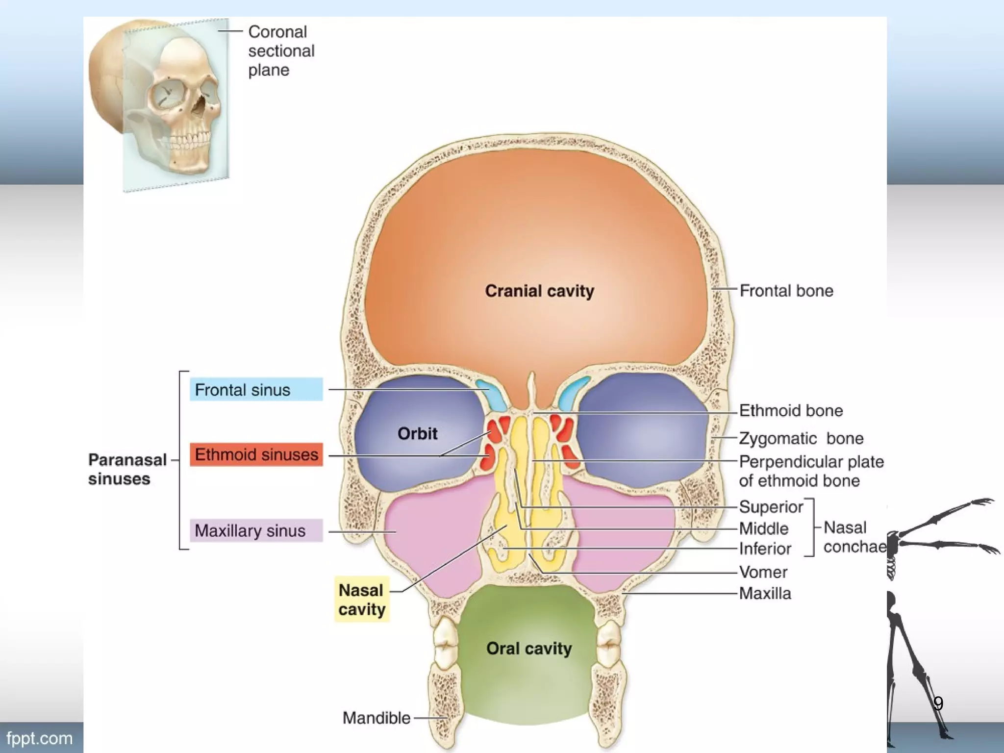
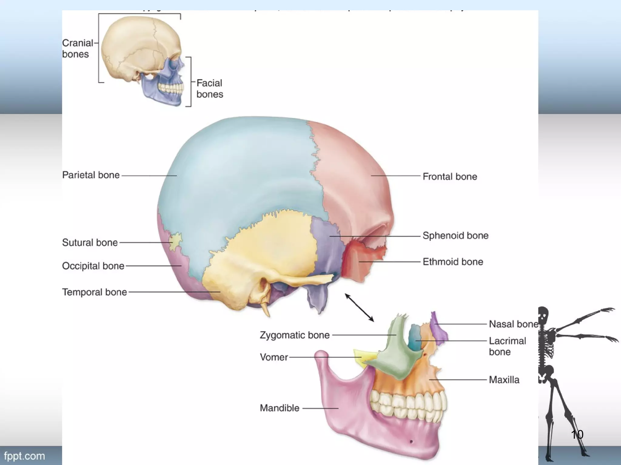
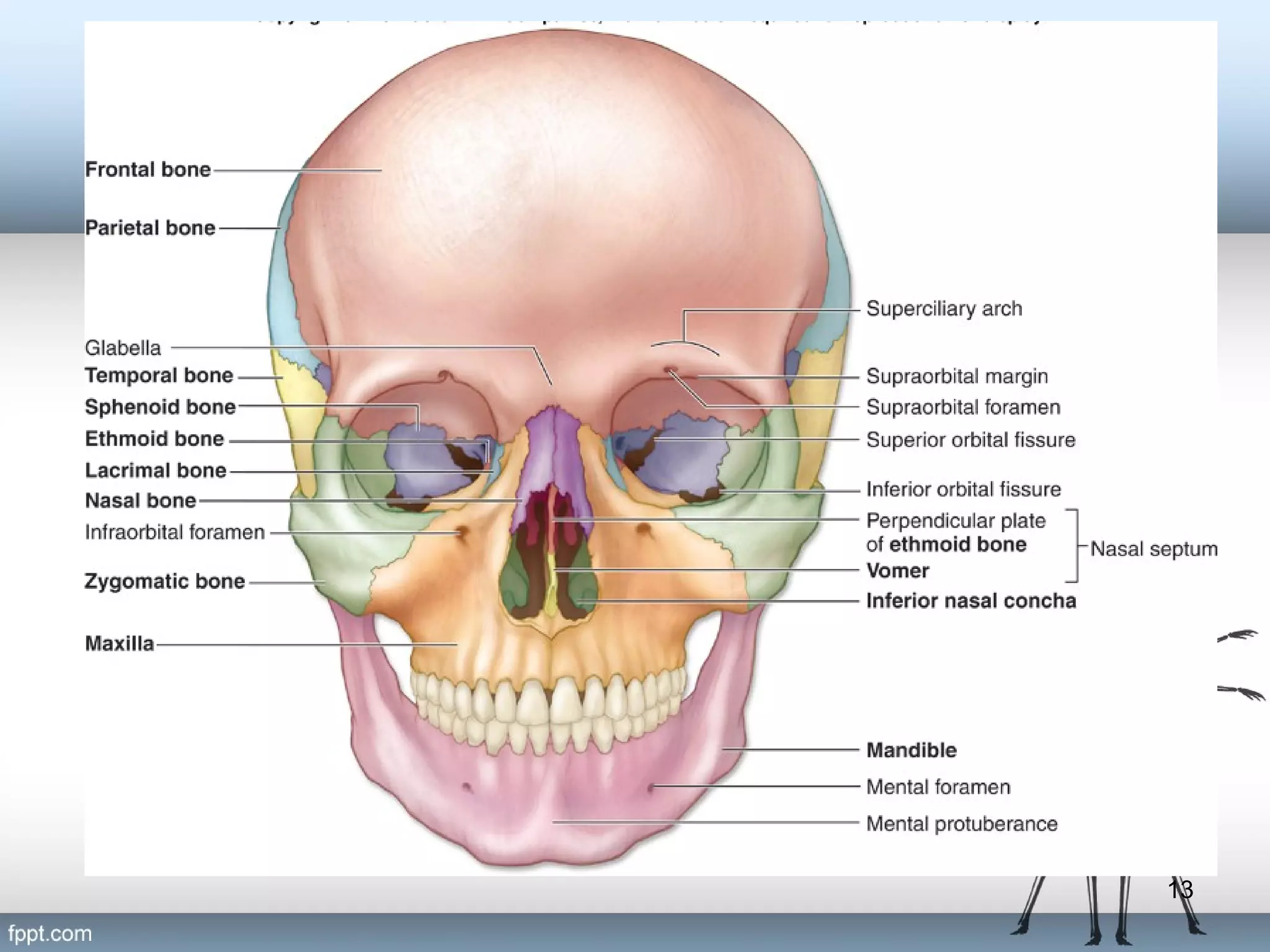
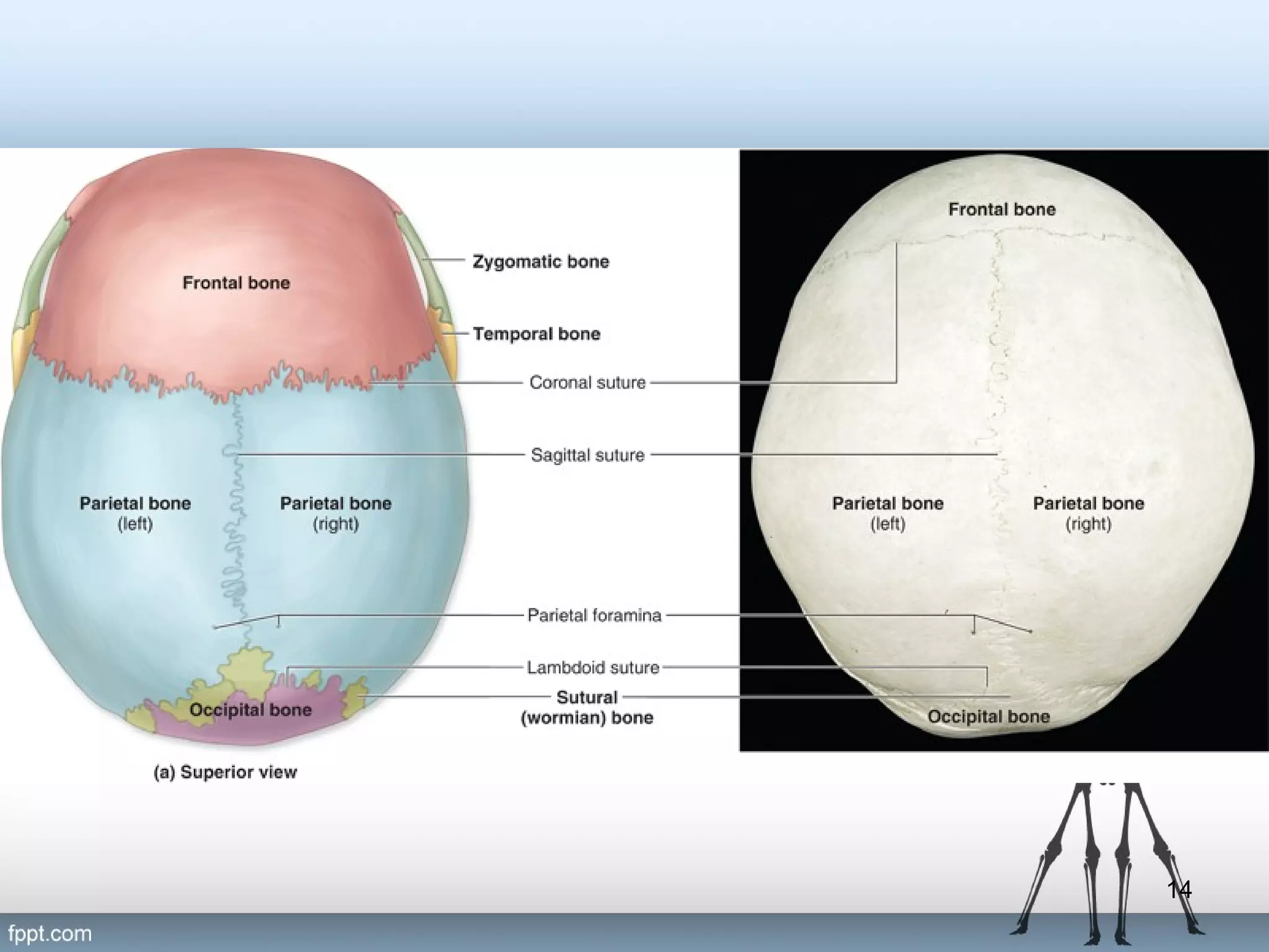
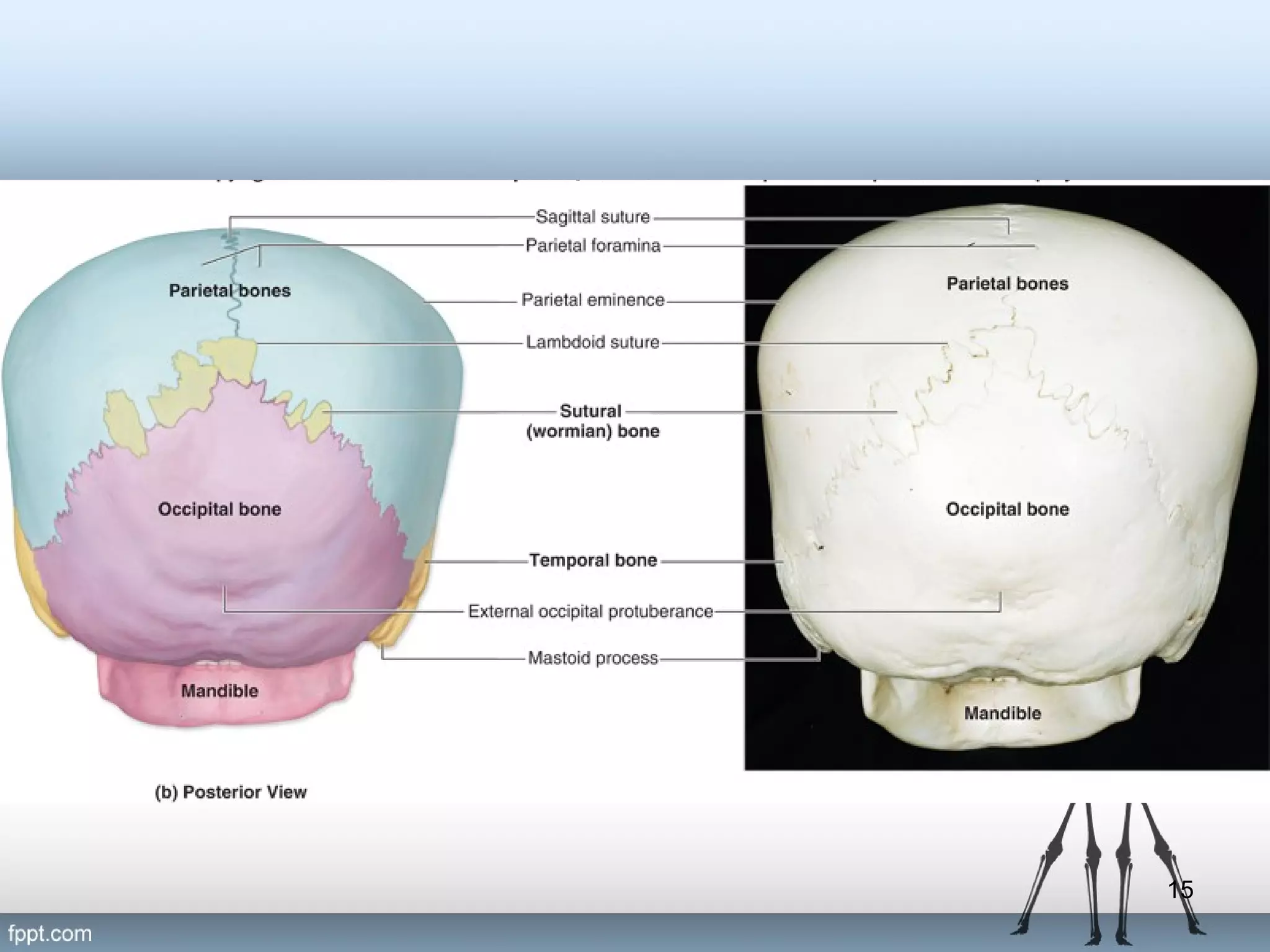
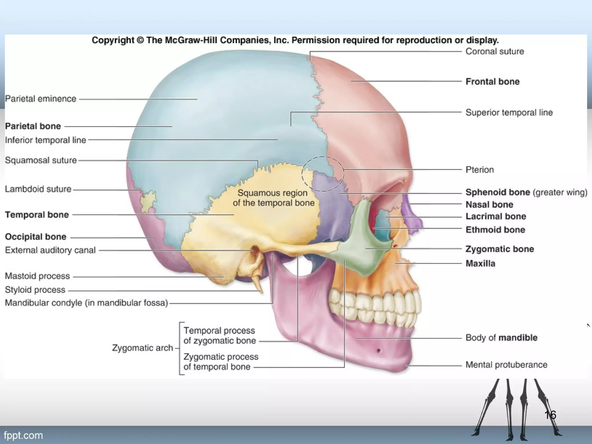
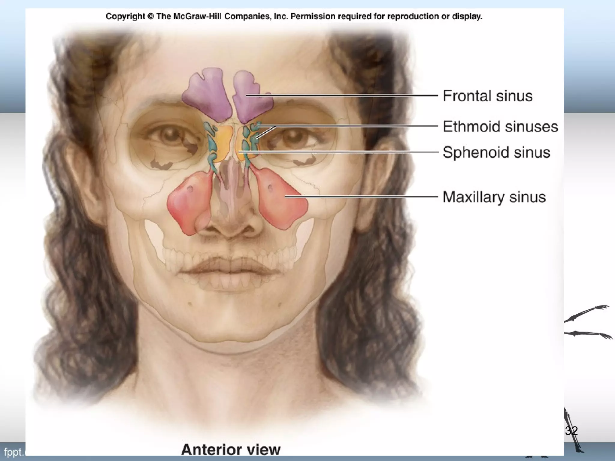
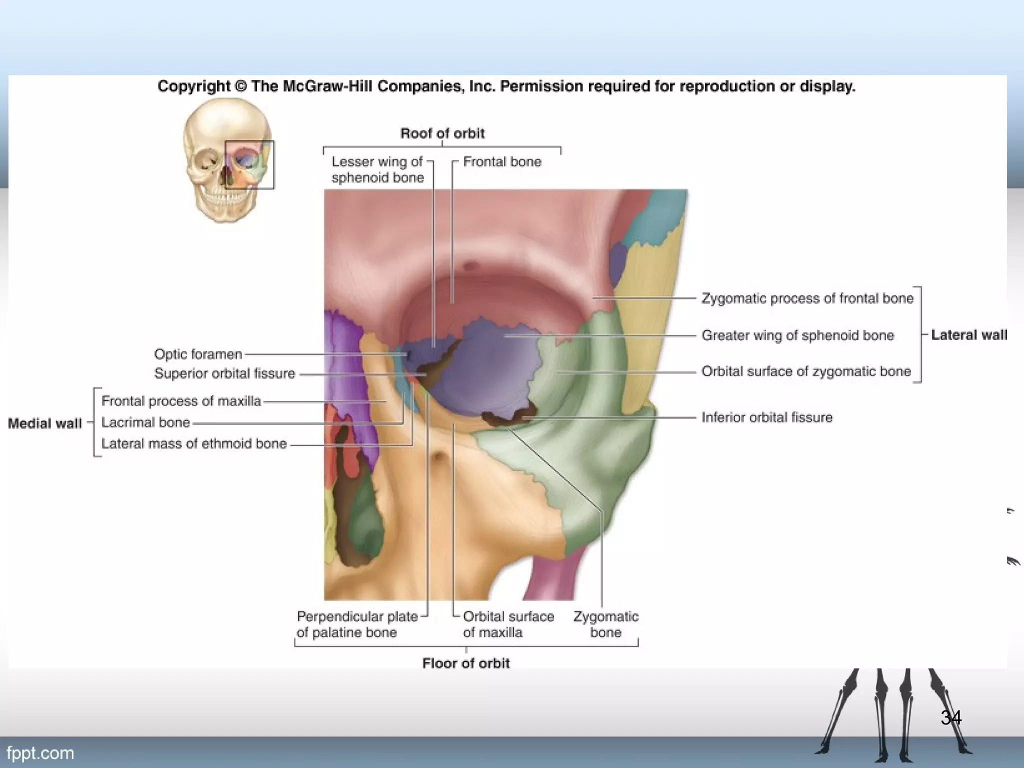
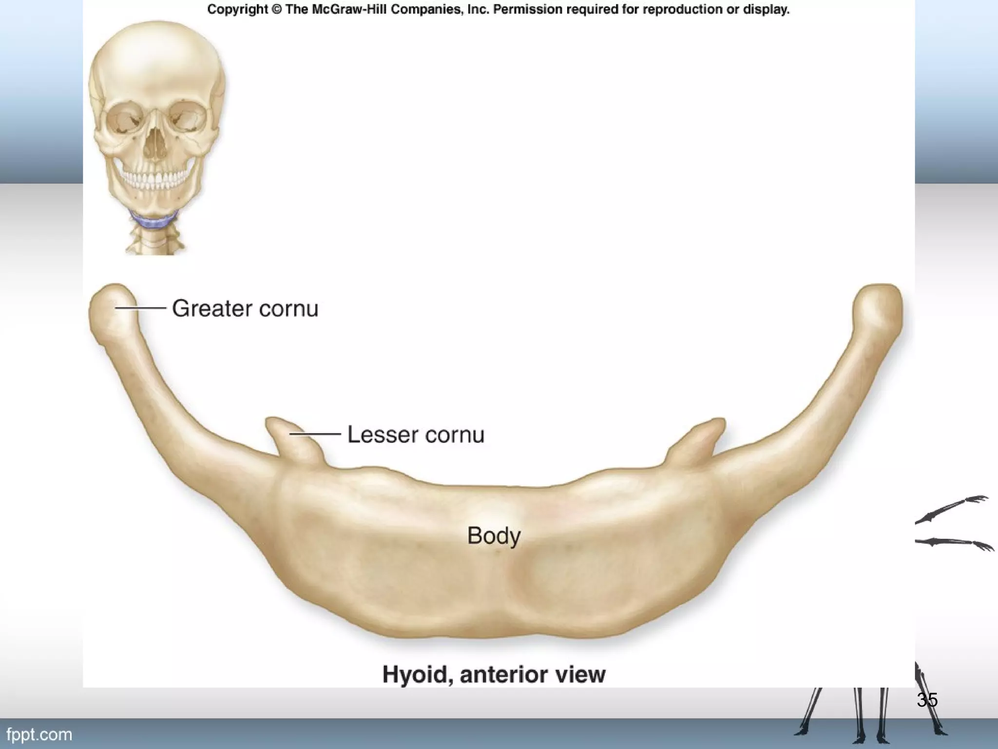
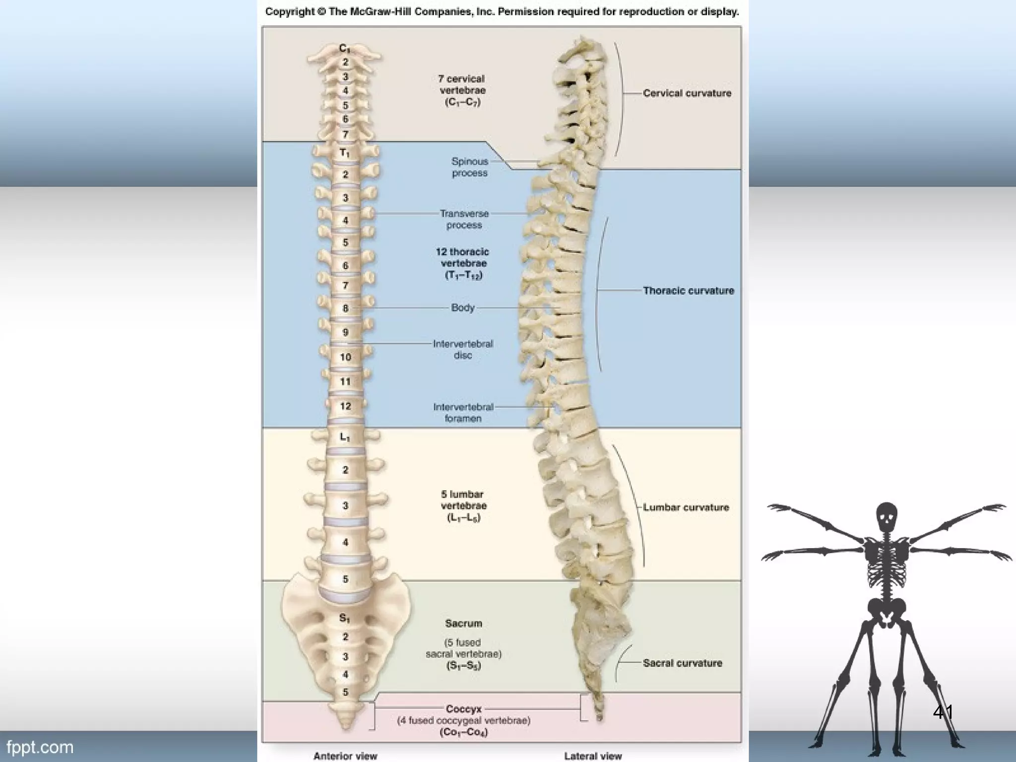
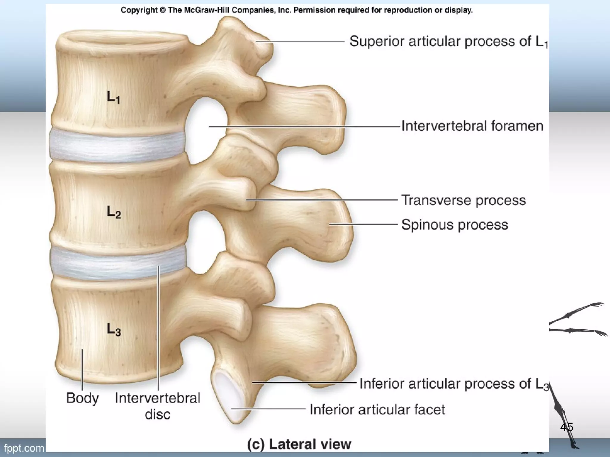
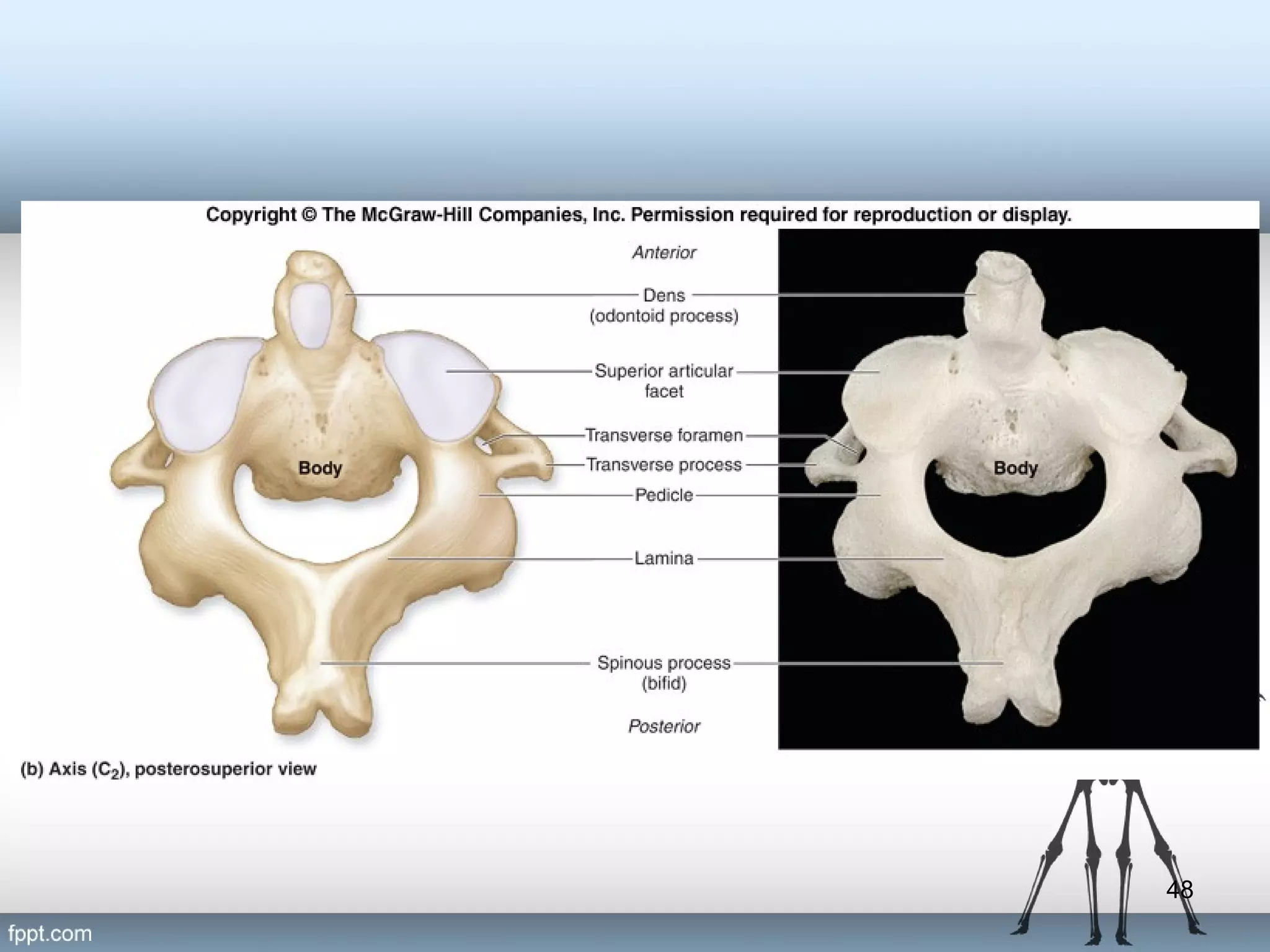
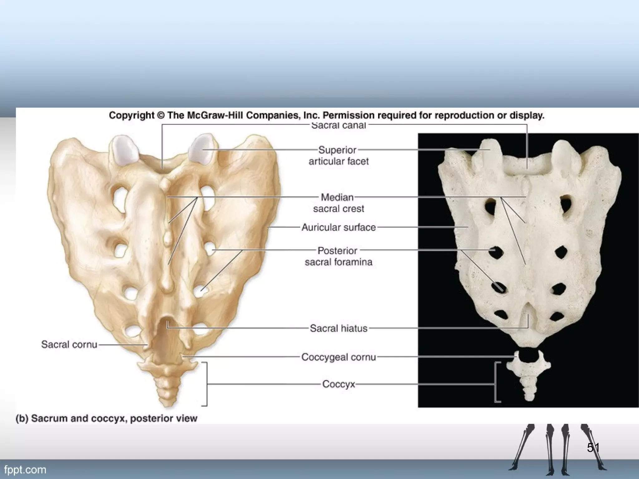
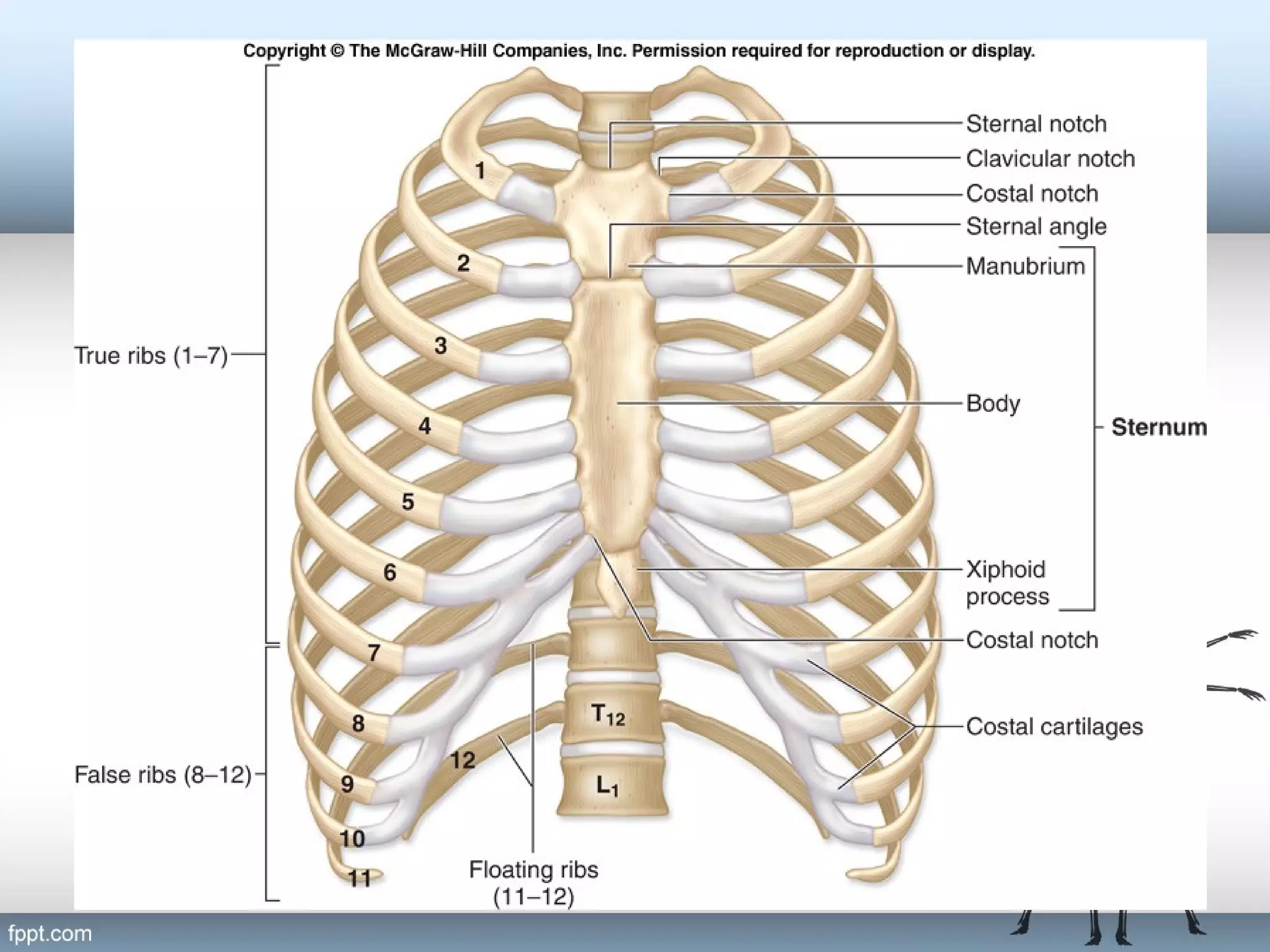
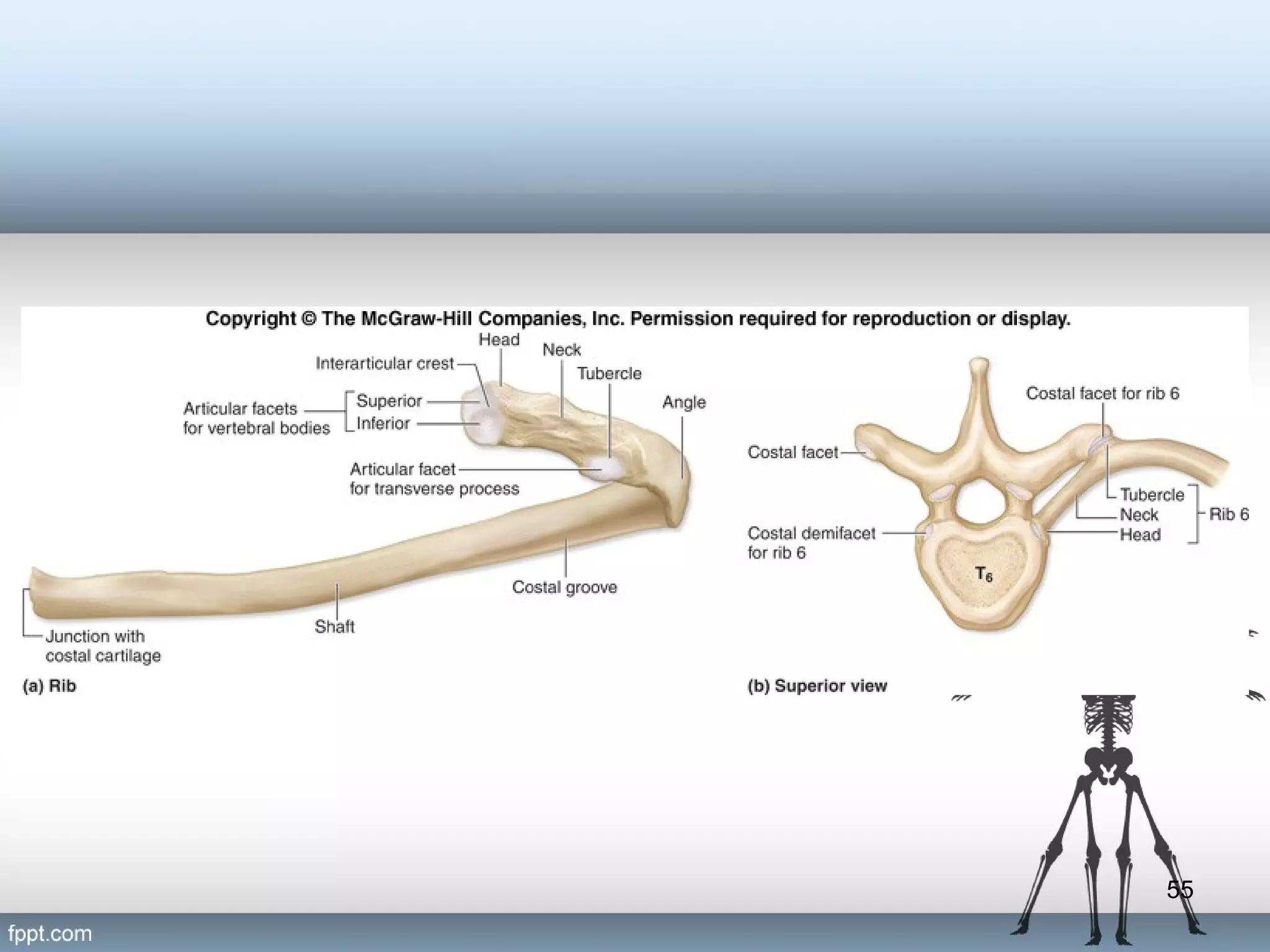
Human Anatomy
(Axial Skeleton)
By:
Yogananta Ramadhan,
MD
1
Skeletal System
• The bones of the skeleton form an internal framework to
support soft tissues, protect vital organs, bear the body’s
weight, and help us move.
• Typically, there are 206 bones in an adult skeleton,
although this number varies in some individuals.
• A larger number of bones are present at birth, but the
total number decreases with growth and maturity as
some separate bones fuse.

7-2
Skeletal System
– The axial skeleton is composed of the bones
along the central axis of the body,
• the skull
• the vertebral column
• the thoracic cage

– The appendicular skeleton consists of the
bones of the appendages
• upper and lower limbs
• the bones that hold the limbs to the trunk of the
body.
7-3
4
5
The Skull
– Cranial bones form the rounded cranium,
which completely surrounds and encloses the
brain.
– Facial bones form the bones of the face. They
also
• protect the entrances to the digestive and
respiratory systems as well as
• provide attachment sites for facial muscles

7-6
The Mandible
• The lower jaw is formed by the mandible.
• The prominent “chin” of the mandible is
called the mental protuberance.

7-7
Cavities of The Skull
– The largest cavity is the cranial cavity, which
encloses, cushions, and supports the brain.
– The skull also has several smaller cavities,
including the orbits (eye sockets), the oral
cavity (mouth), the nasal cavity, and the
paranasal sinuses.

7-8
9
10
Markings of the Skull
• Numerous bone markings
– canals
– fissures
– foramina
• passageways for blood vessels and nerves

7-11
Sutures of the Skull
• Sutures are immovable fibrous joints that form
the boundaries between the cranial bones.
• Dense regular connective tissue seals cranial
bones firmly together at a suture.
• Allow the cranium to grow and expand during
childhood.
• In adulthood, when cranial growth has stopped,
the sutures fuse and are obliterated.

7-12
13
14
15
16
17
Insert Fig. 7.9 superior view of skull

18
19
20
21
22
23
24
25
26
27
28
29
30
Sinuses
• Have a mucous lining that helps to
humidify and warm inhaled air.
• Cause these skull bones to be lighter.
• Provide resonance to the voice.

7-31
32
33
34
35
Auditory Ossicles
• Three tiny ear bones called auditory
ossicles are housed within the petrous
region of each temporal bone.
• the malleus
• the incus
• the stapes

7-36
Hyoid Bone
– Slender, curved bone located inferior to the
skull between the mandible and the larynx
(voice box).
– Does not articulate with any other bone in the
skeleton.
– Serves as sites for attachment for tongue and
larynx muscles and ligaments.

7-37
38
39
Fontanels
• The regions between the cranial bones are thickened,
fibrous membrane remnants that are not yet ossified.
• Sometimes referred to as the “soft spots” on a baby’s
head.
• They close by 15 months of age.
• When a baby travels through the birth canal, the cranial
bones overlap at these fontanels, in order to ease the
baby’s passage.
• Newborns frequently have a “cone-shaped” head due to
this temporary deformation.
7-40
41
The Vertebral Column
•

Composed of 26 bones, including
– 24 individual vertebrae and the
– fused vertebrae that form both the sacrum and the coccyx

•

The vertebral column has several functions:
– providing vertical support for the body
– supporting the weight of the head
– helping to maintain upright body position
– helping to transfer axial skeletal weight to the appendicular
skeleton of the lower limbs
– housing and protecting the delicate spinal cord and providing a
passageway for spinal nerves connecting to the spinal cord

7-42
43
44
45
Three Main Spinal Curvature
Deformities
– Kyphosis is an exaggerated thoracic curvature that is directed
posteriorly, producing a “hunchback” look.
– Lordosis is an exaggerated lumbar curvature, often called
“swayback,” that is observed as a protrusion of the abdomen
and buttocks.
– Scoliosis is an abnormal lateral curvature that sometimes results
during development when both the vertebral arch and body fail
to form, or form incompletely, on one side of a vertebra.
• scoliosis is the most common spinal curvature deformity.

7-46
47
48
49
50
51
Thoracic Cage
• Consists of the thoracic vertebrae posteriorly,
the ribs laterally, and the sternum anteriorly.
• Acts as a protective cage around vital organs,
such as the heart, lungs, trachea, and
esophagus.
• Provides attachment points for many muscles
supporting the pectoral girdles, the chest, the
neck, the shoulders, the back, and the muscles
involved in respiration.

7-52
53
Ribs
• Both males and females 12 pairs
– Ribs 1–7 are called true ribs. At the anterior body wall, the true
ribs connect individually to the sternum by separate cartilaginous
extensions called costal cartilages.
– Ribs 8–12 are called false ribs because their costal cartilages do
not attach directly to the sternum. The costal cartilages of ribs 8–
10 fuse to the costal cartilage of rib 7 and thus indirectly
articulate with the sternum.
– The last two pairs of false ribs (ribs 11 and 12) are called floating
ribs because they have no connection with the sternum.

7-54
55

Axial Skeleton

  • 1.
  • 2.
    Skeletal System • Thebones of the skeleton form an internal framework to support soft tissues, protect vital organs, bear the body’s weight, and help us move. • Typically, there are 206 bones in an adult skeleton, although this number varies in some individuals. • A larger number of bones are present at birth, but the total number decreases with growth and maturity as some separate bones fuse. 7-2
  • 3.
    Skeletal System – Theaxial skeleton is composed of the bones along the central axis of the body, • the skull • the vertebral column • the thoracic cage – The appendicular skeleton consists of the bones of the appendages • upper and lower limbs • the bones that hold the limbs to the trunk of the body. 7-3
  • 4.
  • 5.
  • 6.
    The Skull – Cranialbones form the rounded cranium, which completely surrounds and encloses the brain. – Facial bones form the bones of the face. They also • protect the entrances to the digestive and respiratory systems as well as • provide attachment sites for facial muscles 7-6
  • 7.
    The Mandible • Thelower jaw is formed by the mandible. • The prominent “chin” of the mandible is called the mental protuberance. 7-7
  • 8.
    Cavities of TheSkull – The largest cavity is the cranial cavity, which encloses, cushions, and supports the brain. – The skull also has several smaller cavities, including the orbits (eye sockets), the oral cavity (mouth), the nasal cavity, and the paranasal sinuses. 7-8
  • 9.
  • 10.
  • 11.
    Markings of theSkull • Numerous bone markings – canals – fissures – foramina • passageways for blood vessels and nerves 7-11
  • 12.
    Sutures of theSkull • Sutures are immovable fibrous joints that form the boundaries between the cranial bones. • Dense regular connective tissue seals cranial bones firmly together at a suture. • Allow the cranium to grow and expand during childhood. • In adulthood, when cranial growth has stopped, the sutures fuse and are obliterated. 7-12
  • 13.
  • 14.
  • 15.
  • 16.
  • 17.
  • 18.
    Insert Fig. 7.9superior view of skull 18
  • 19.
  • 20.
  • 21.
  • 22.
  • 23.
  • 24.
  • 25.
  • 26.
  • 27.
  • 28.
  • 29.
  • 30.
  • 31.
    Sinuses • Have amucous lining that helps to humidify and warm inhaled air. • Cause these skull bones to be lighter. • Provide resonance to the voice. 7-31
  • 32.
  • 33.
  • 34.
  • 35.
  • 36.
    Auditory Ossicles • Threetiny ear bones called auditory ossicles are housed within the petrous region of each temporal bone. • the malleus • the incus • the stapes 7-36
  • 37.
    Hyoid Bone – Slender,curved bone located inferior to the skull between the mandible and the larynx (voice box). – Does not articulate with any other bone in the skeleton. – Serves as sites for attachment for tongue and larynx muscles and ligaments. 7-37
  • 38.
  • 39.
  • 40.
    Fontanels • The regionsbetween the cranial bones are thickened, fibrous membrane remnants that are not yet ossified. • Sometimes referred to as the “soft spots” on a baby’s head. • They close by 15 months of age. • When a baby travels through the birth canal, the cranial bones overlap at these fontanels, in order to ease the baby’s passage. • Newborns frequently have a “cone-shaped” head due to this temporary deformation. 7-40
  • 41.
  • 42.
    The Vertebral Column • Composedof 26 bones, including – 24 individual vertebrae and the – fused vertebrae that form both the sacrum and the coccyx • The vertebral column has several functions: – providing vertical support for the body – supporting the weight of the head – helping to maintain upright body position – helping to transfer axial skeletal weight to the appendicular skeleton of the lower limbs – housing and protecting the delicate spinal cord and providing a passageway for spinal nerves connecting to the spinal cord 7-42
  • 43.
  • 44.
  • 45.
  • 46.
    Three Main SpinalCurvature Deformities – Kyphosis is an exaggerated thoracic curvature that is directed posteriorly, producing a “hunchback” look. – Lordosis is an exaggerated lumbar curvature, often called “swayback,” that is observed as a protrusion of the abdomen and buttocks. – Scoliosis is an abnormal lateral curvature that sometimes results during development when both the vertebral arch and body fail to form, or form incompletely, on one side of a vertebra. • scoliosis is the most common spinal curvature deformity. 7-46
  • 47.
  • 48.
  • 49.
  • 50.
  • 51.
  • 52.
    Thoracic Cage • Consistsof the thoracic vertebrae posteriorly, the ribs laterally, and the sternum anteriorly. • Acts as a protective cage around vital organs, such as the heart, lungs, trachea, and esophagus. • Provides attachment points for many muscles supporting the pectoral girdles, the chest, the neck, the shoulders, the back, and the muscles involved in respiration. 7-52
  • 53.
  • 54.
    Ribs • Both malesand females 12 pairs – Ribs 1–7 are called true ribs. At the anterior body wall, the true ribs connect individually to the sternum by separate cartilaginous extensions called costal cartilages. – Ribs 8–12 are called false ribs because their costal cartilages do not attach directly to the sternum. The costal cartilages of ribs 8– 10 fuse to the costal cartilage of rib 7 and thus indirectly articulate with the sternum. – The last two pairs of false ribs (ribs 11 and 12) are called floating ribs because they have no connection with the sternum. 7-54
  • 55.