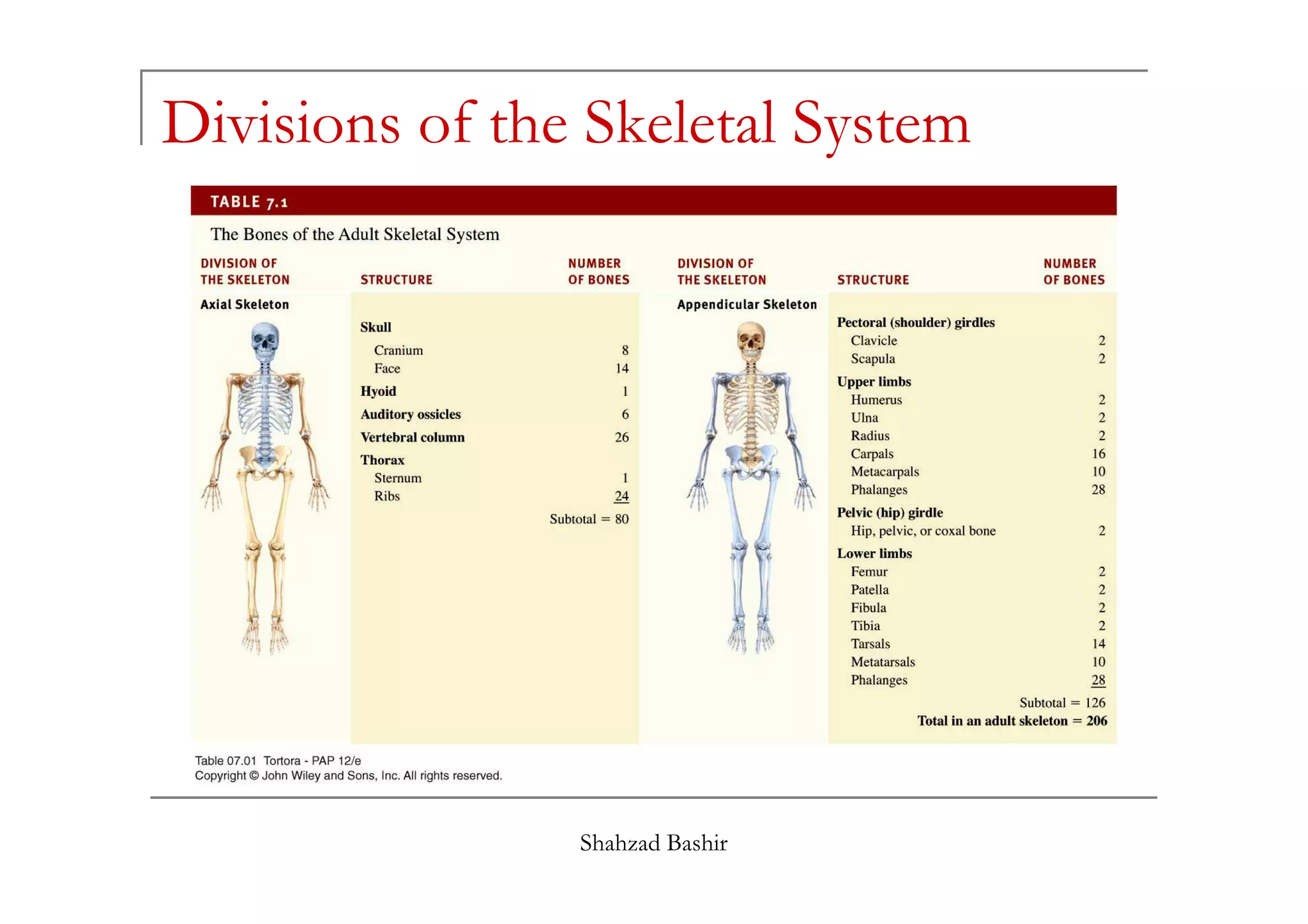
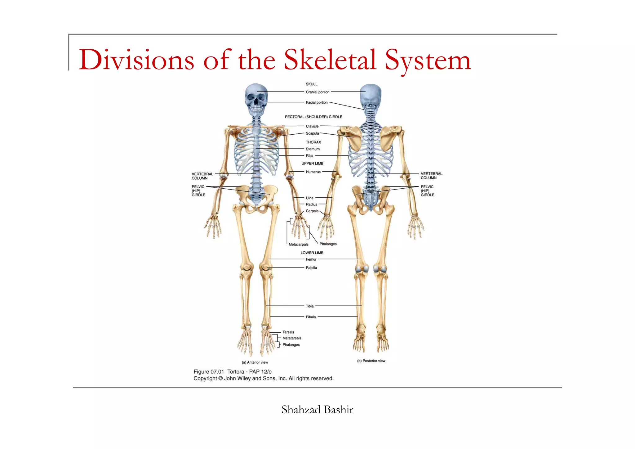
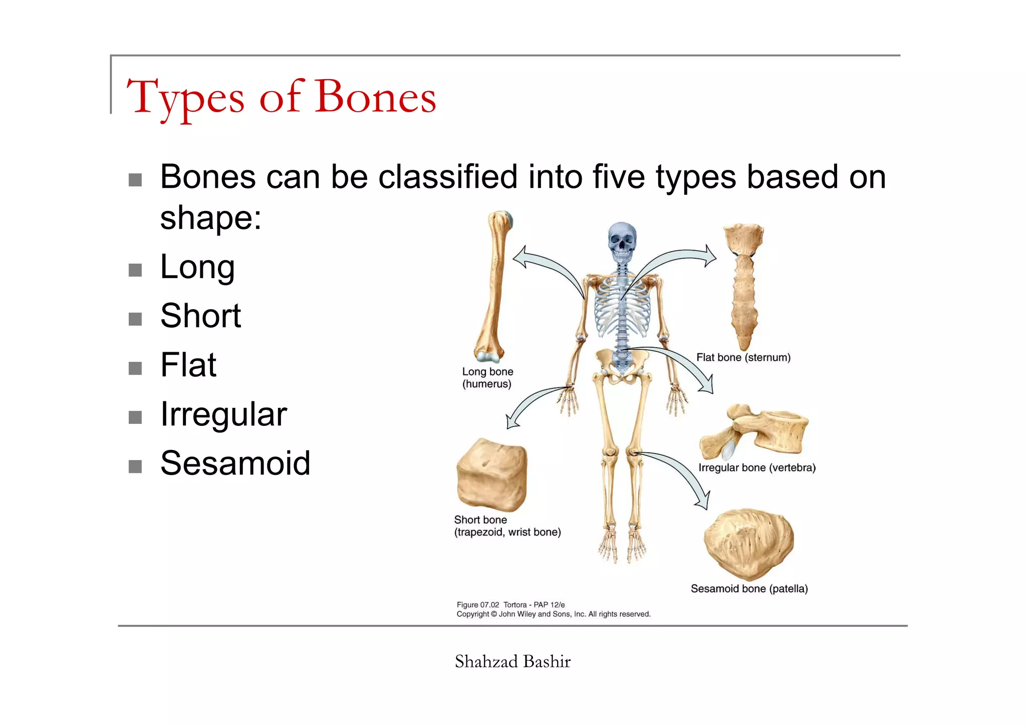
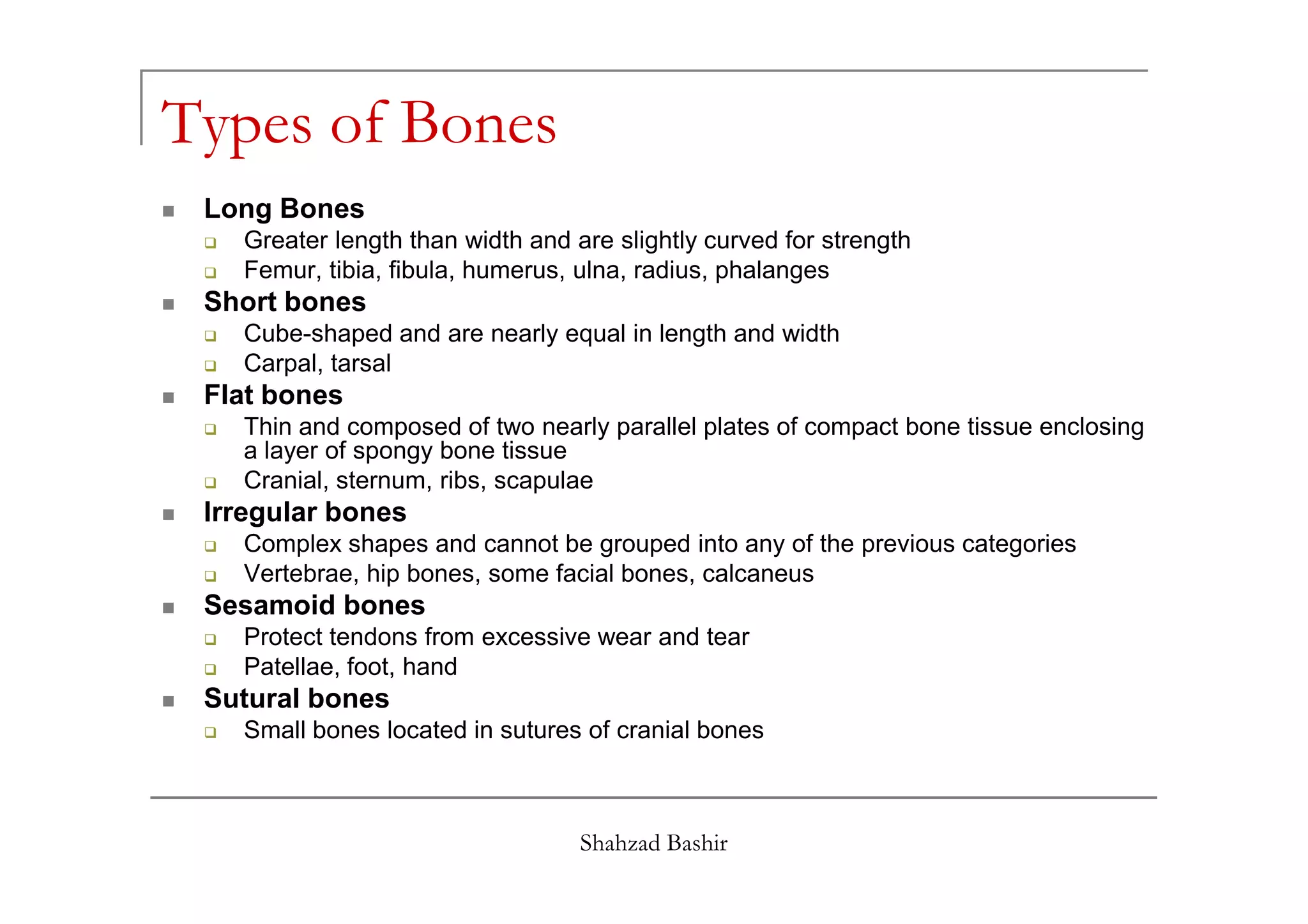
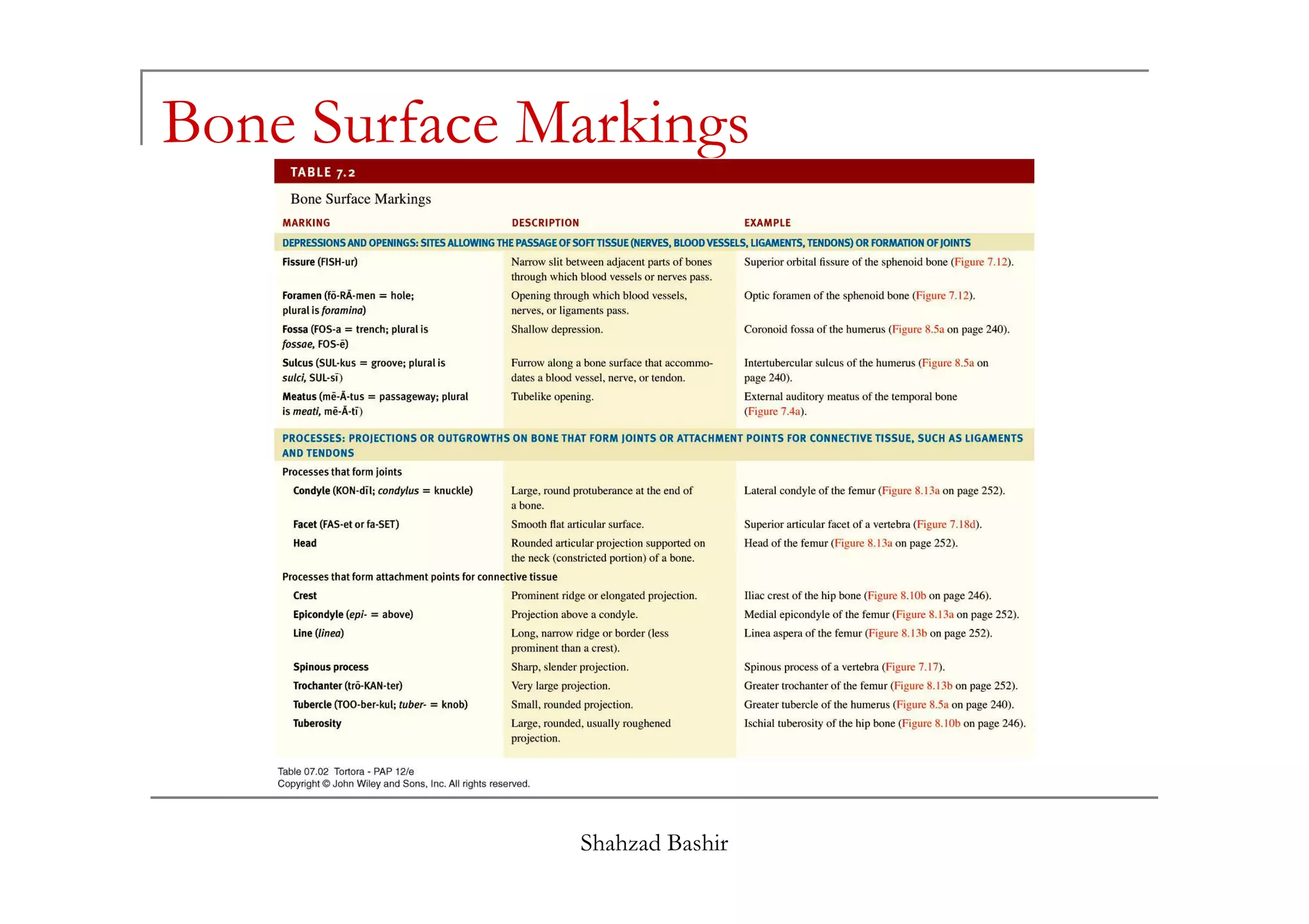
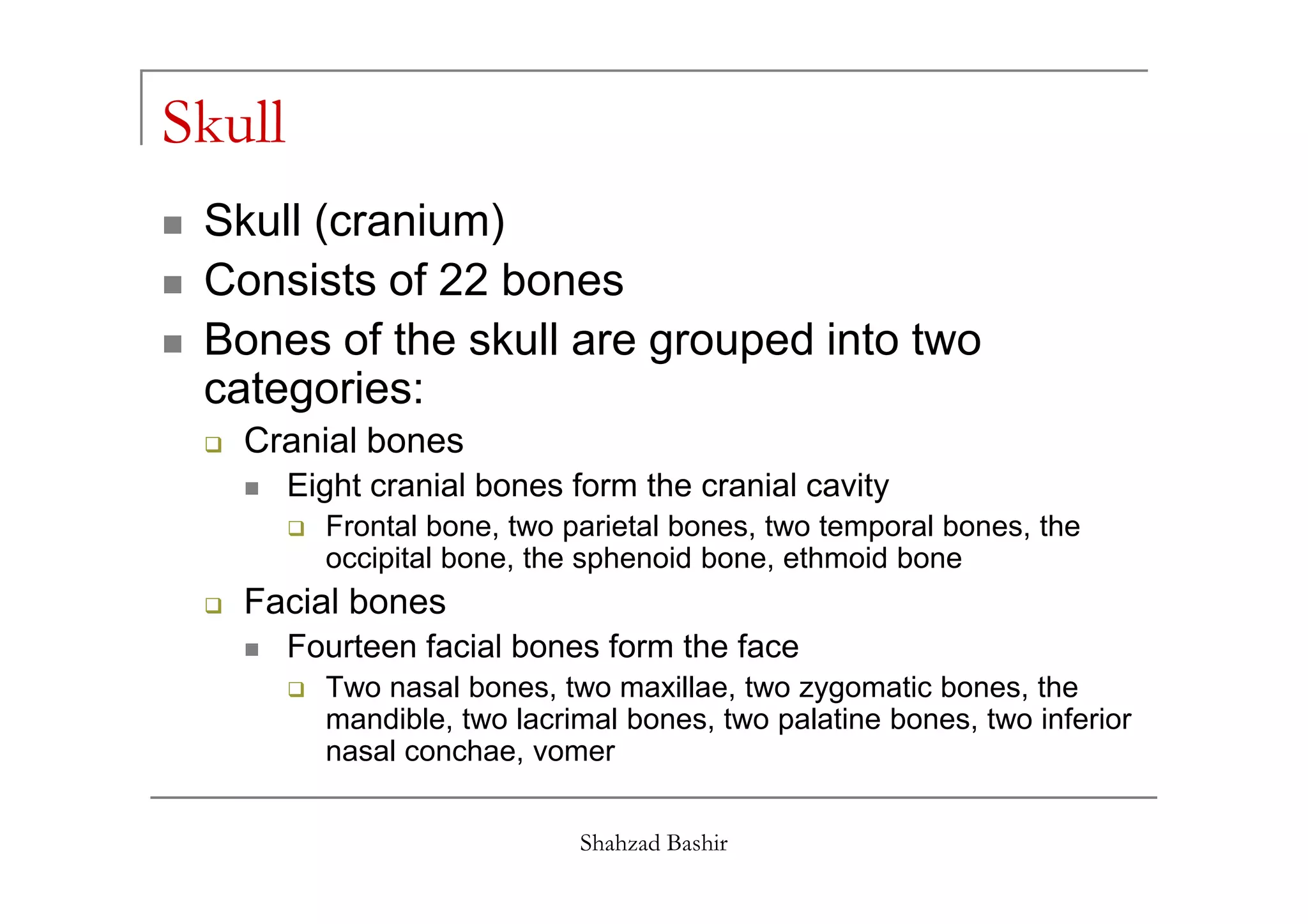
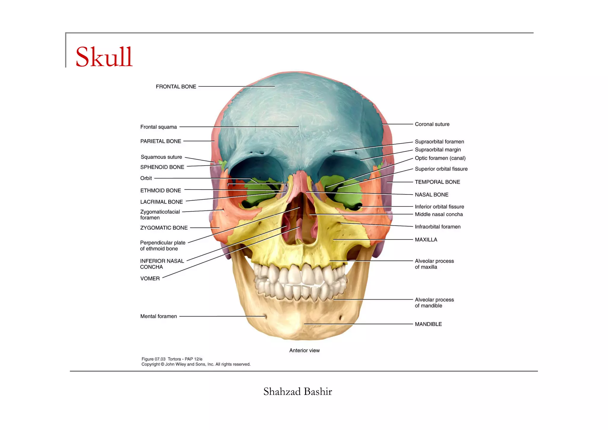
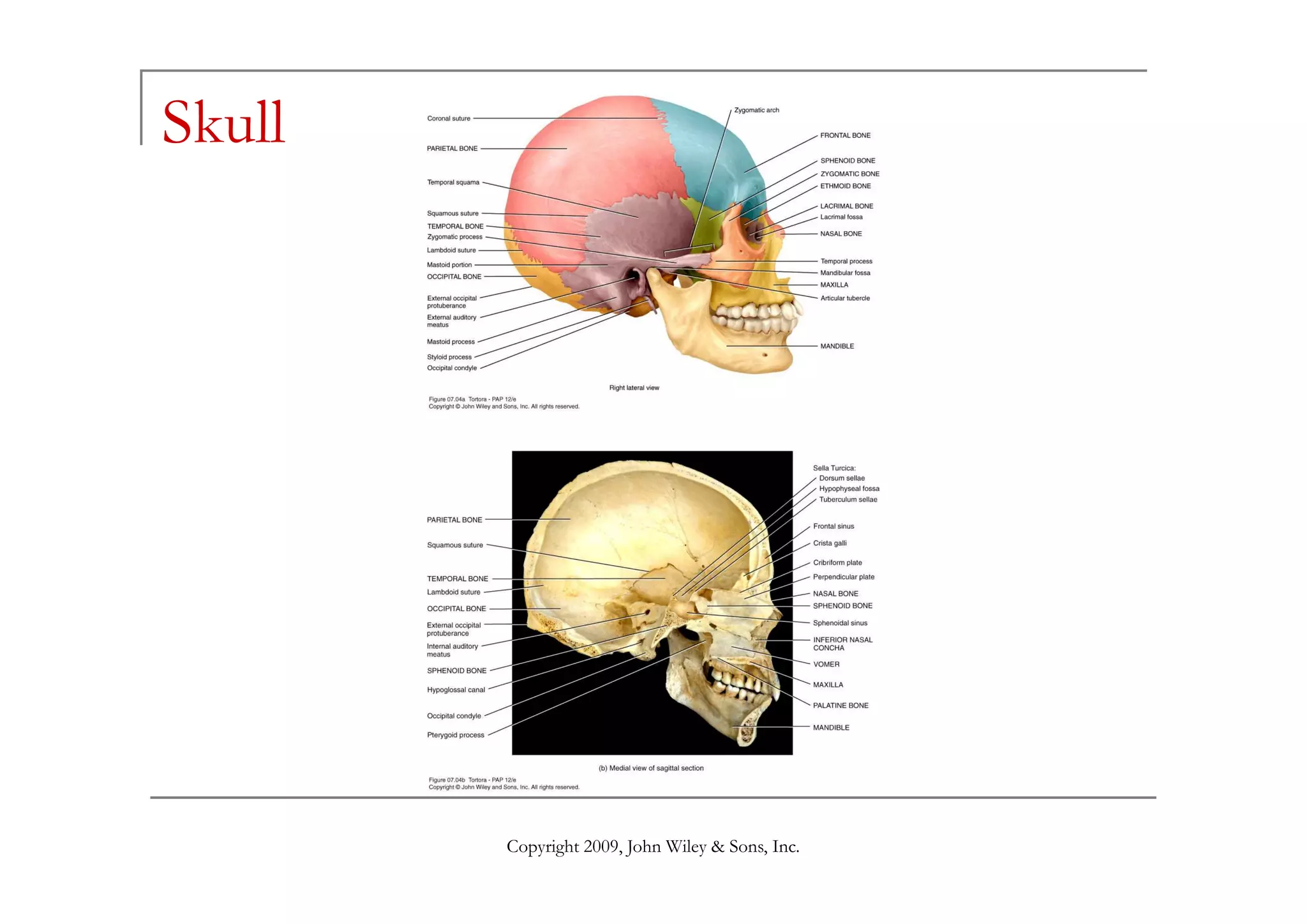
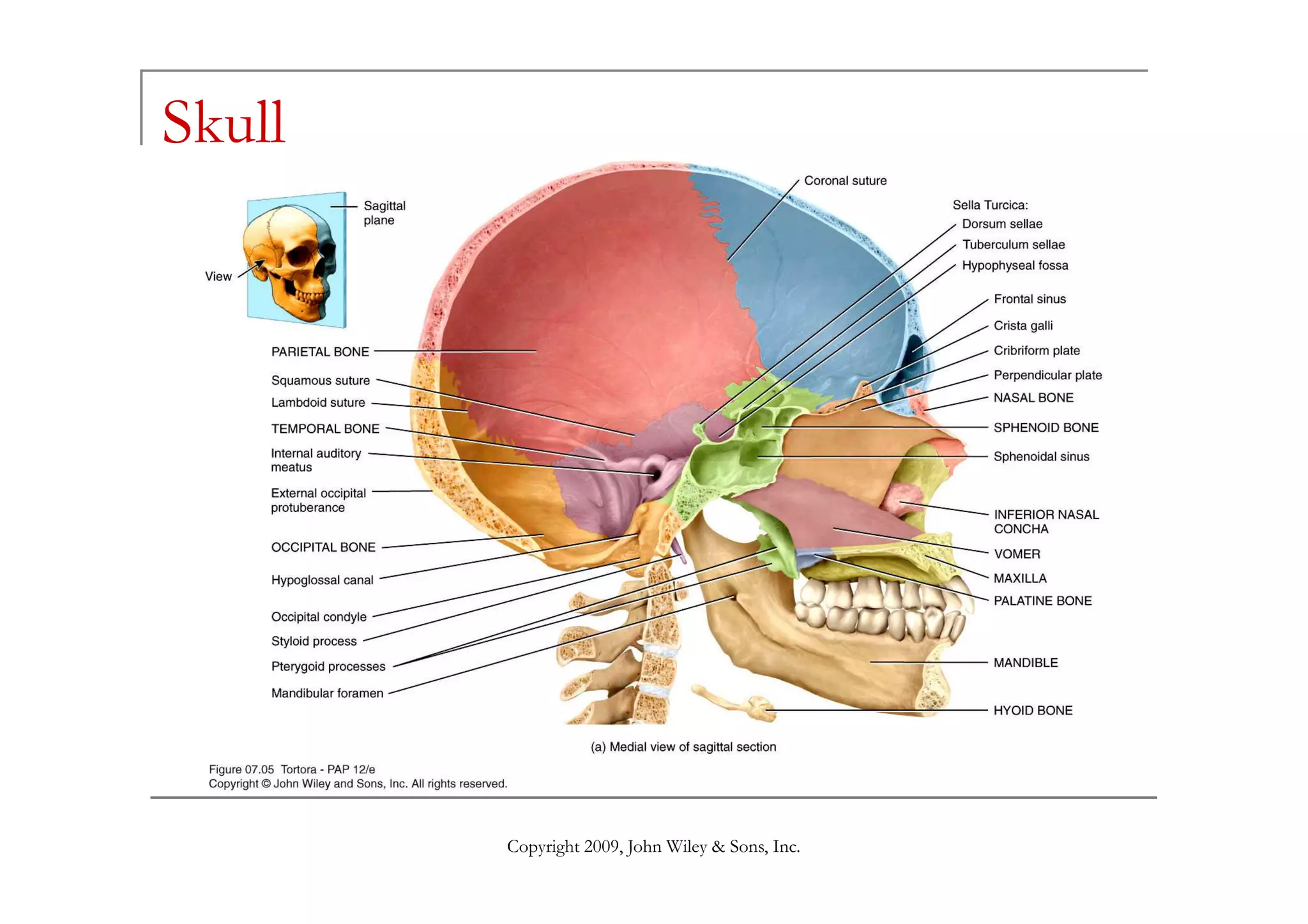
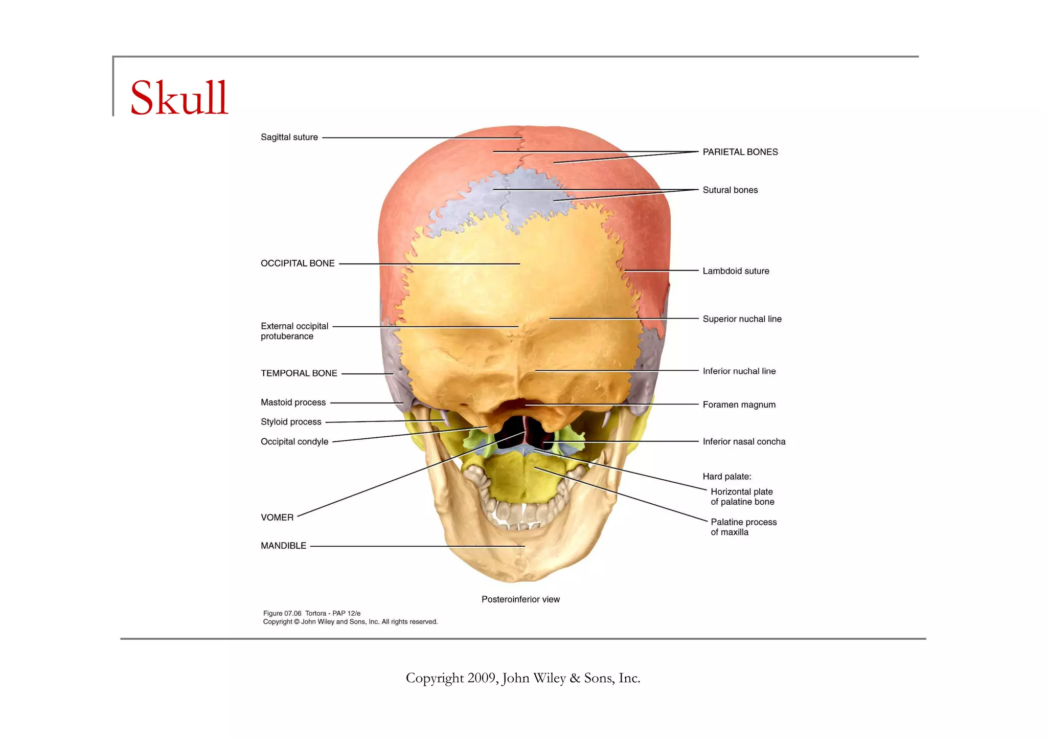
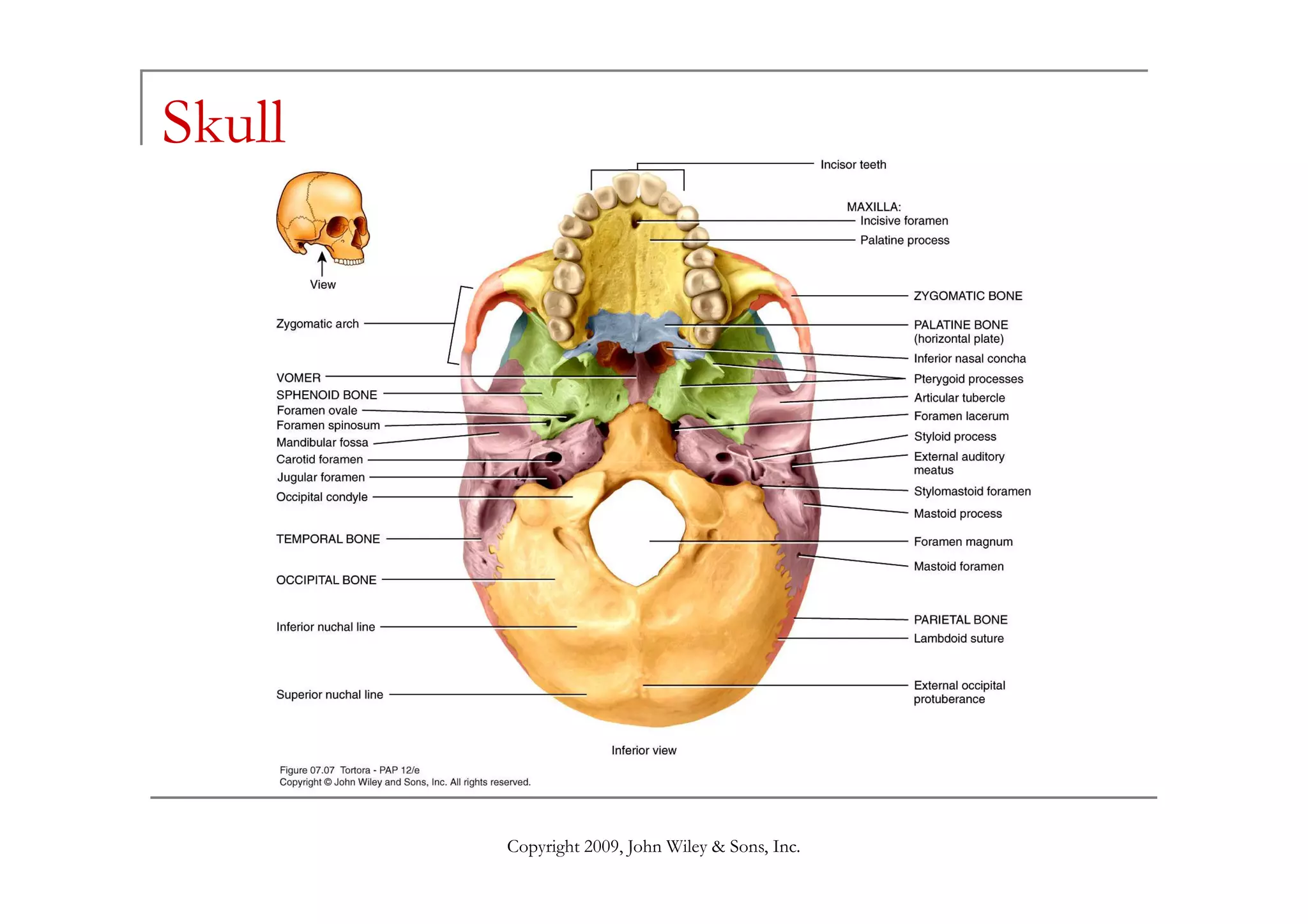
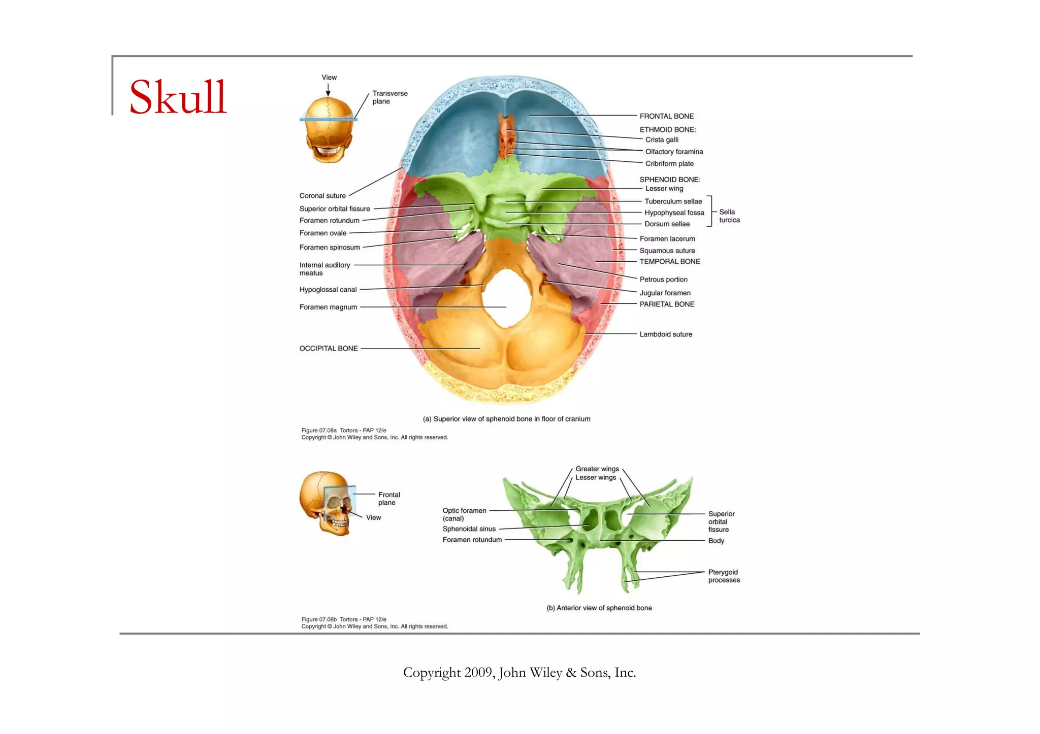
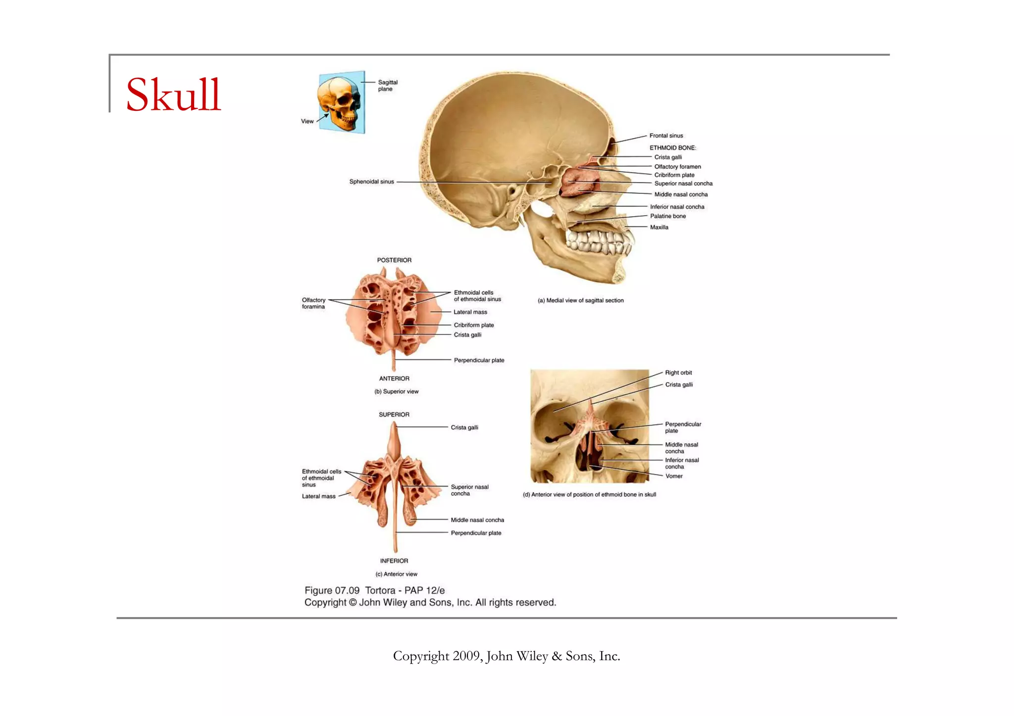
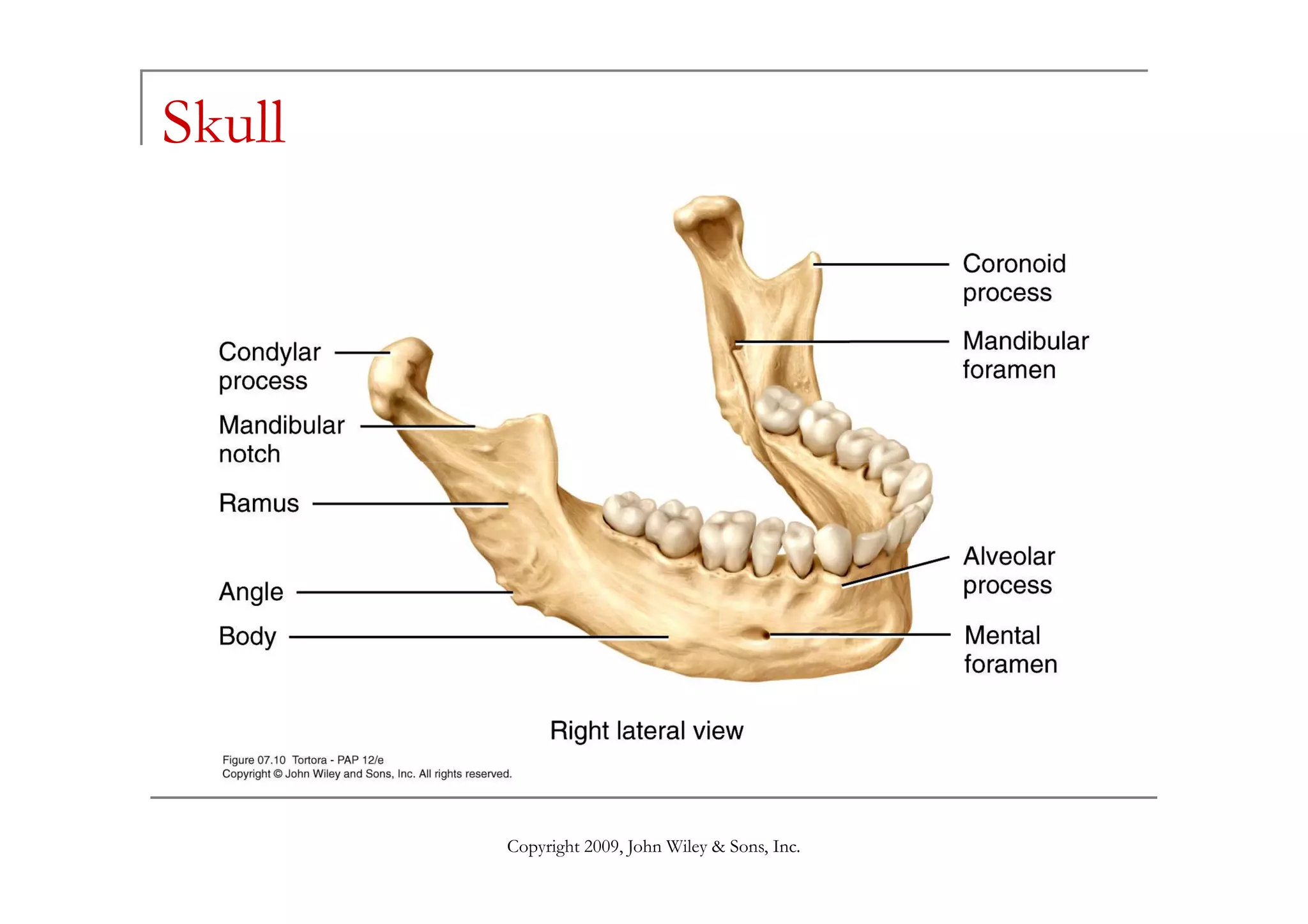
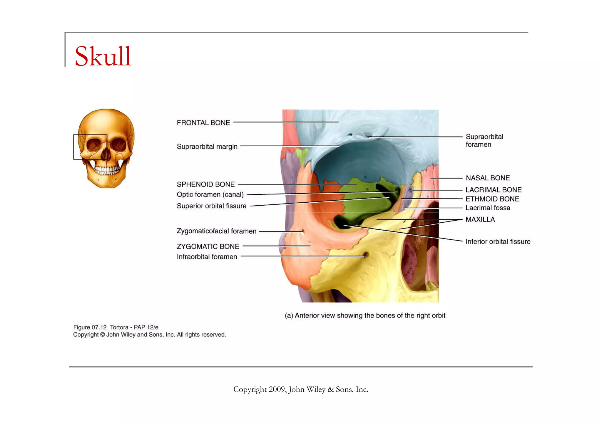
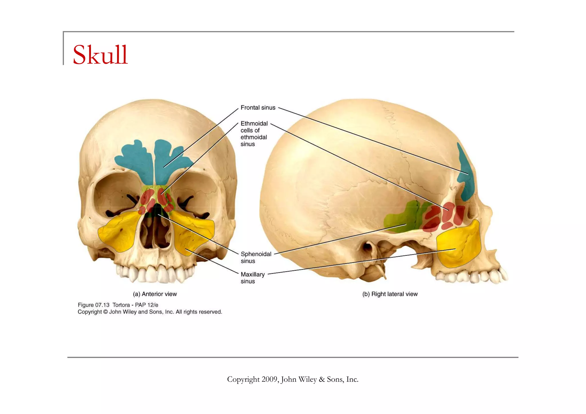
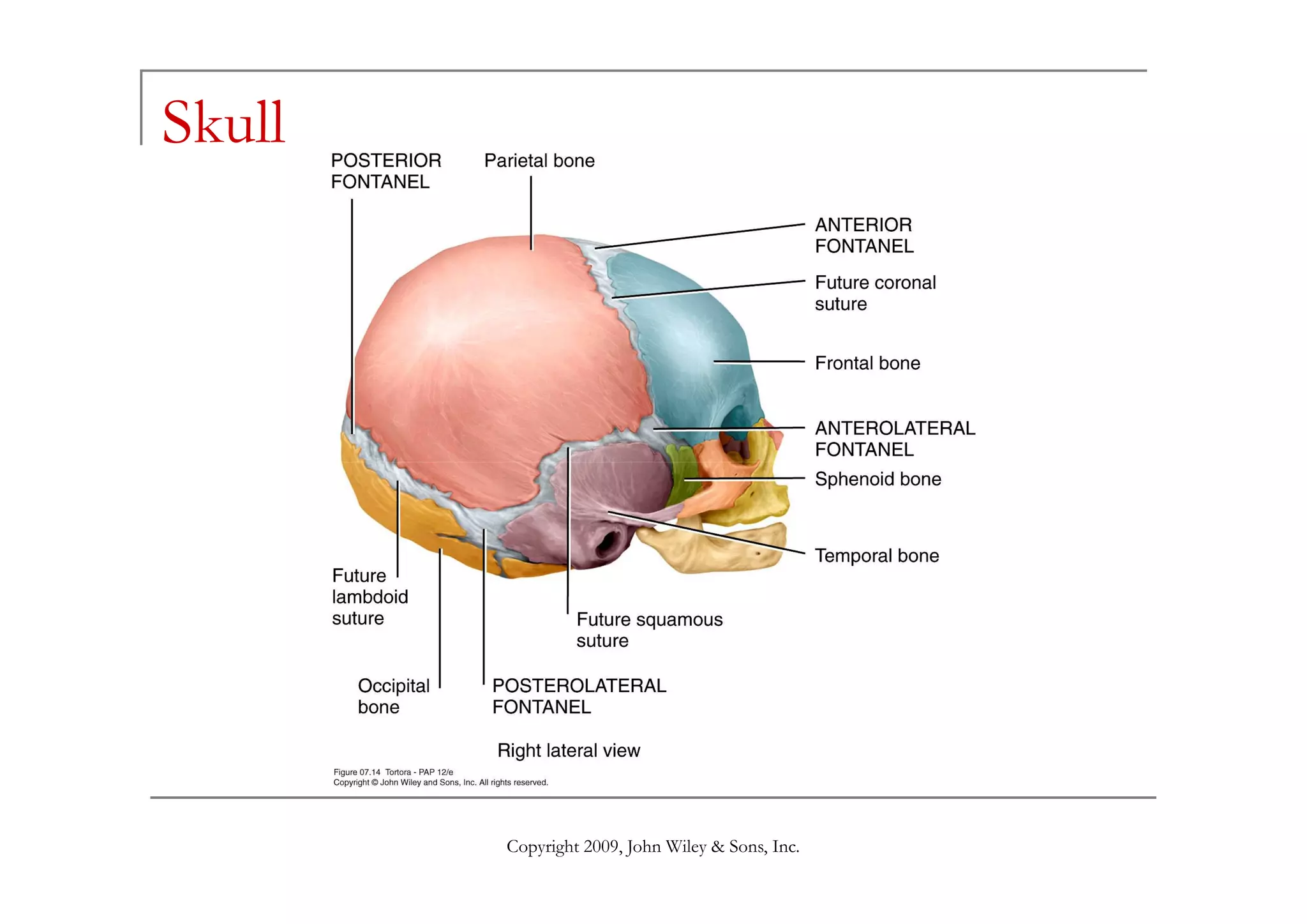
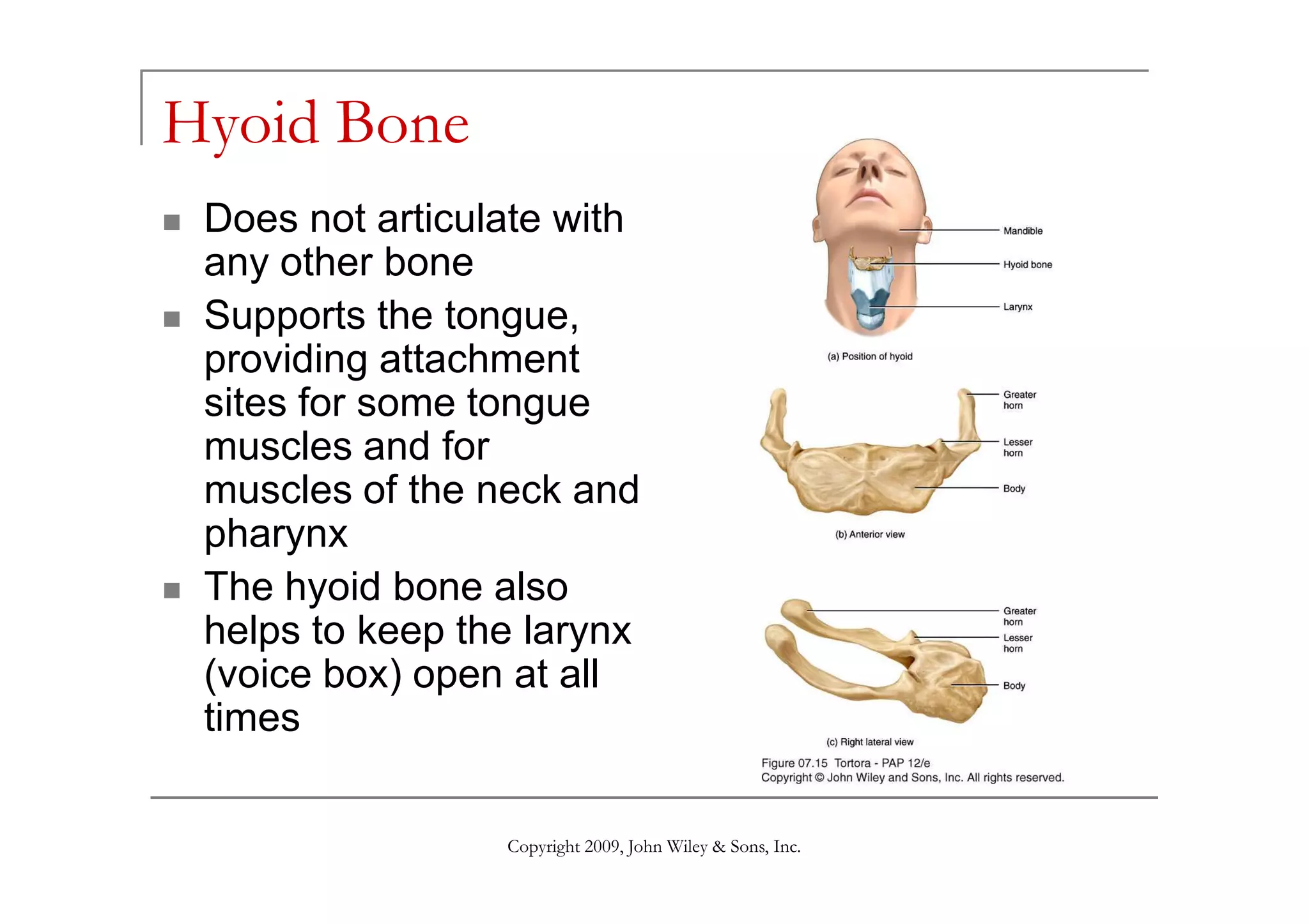
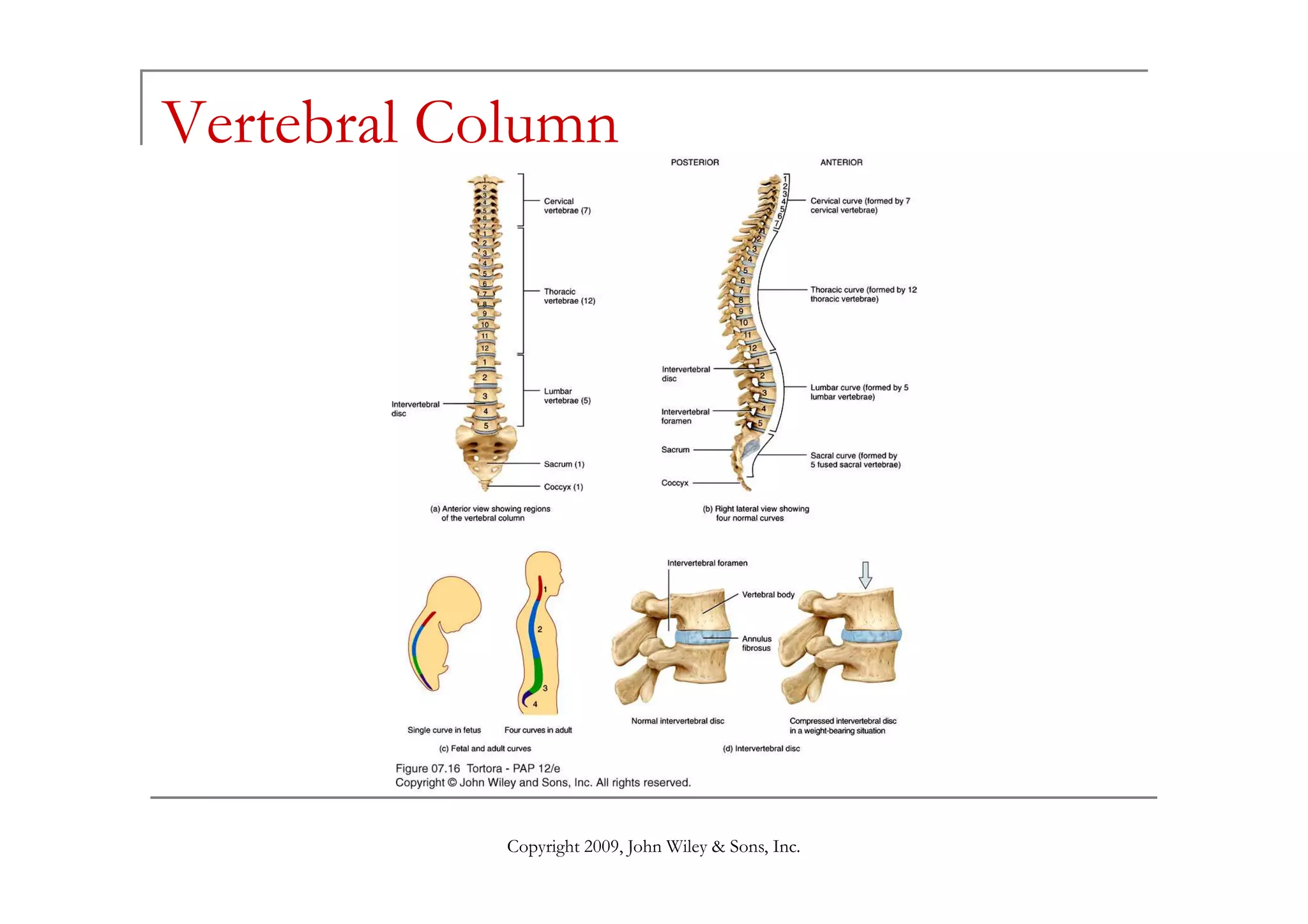
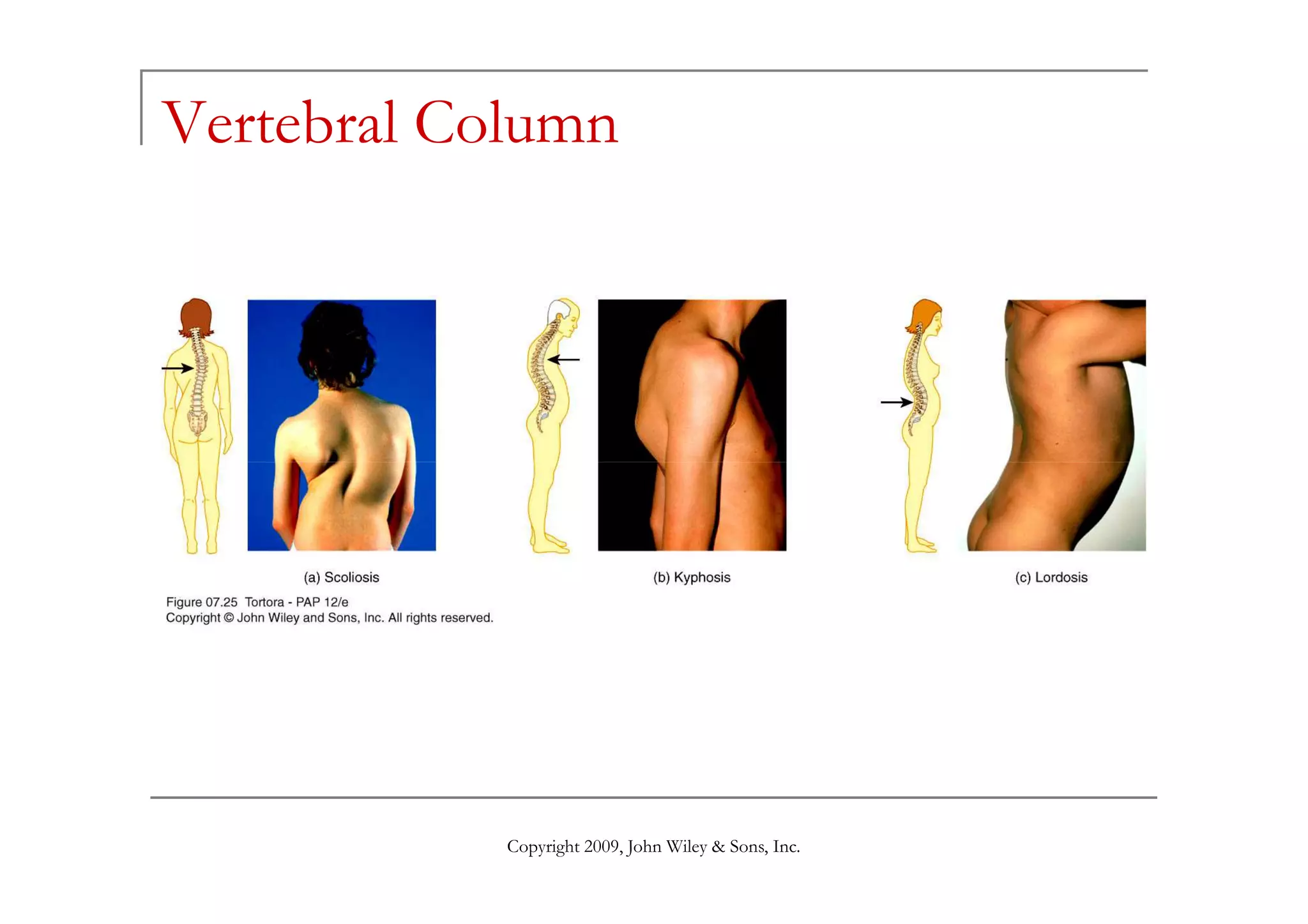
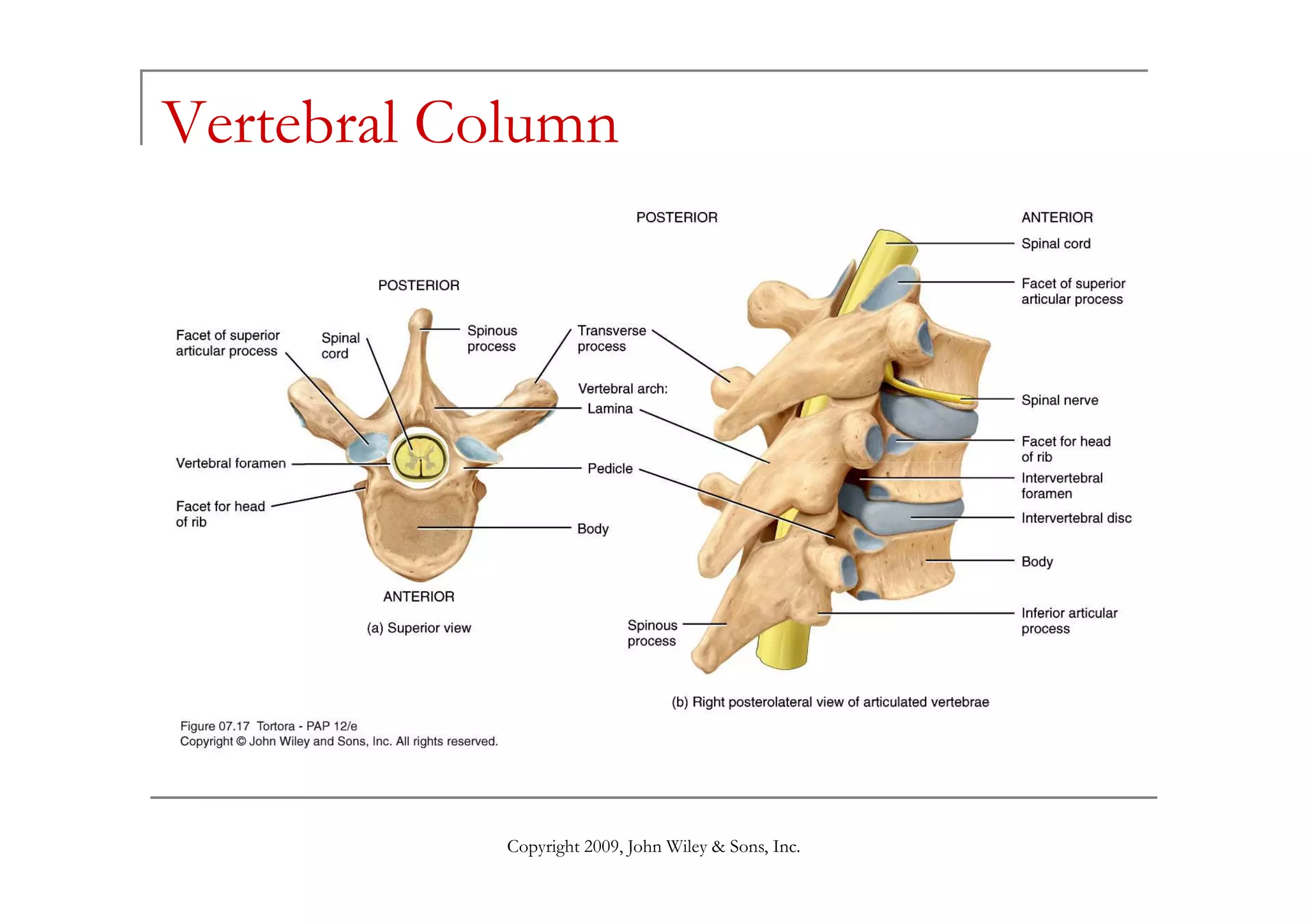
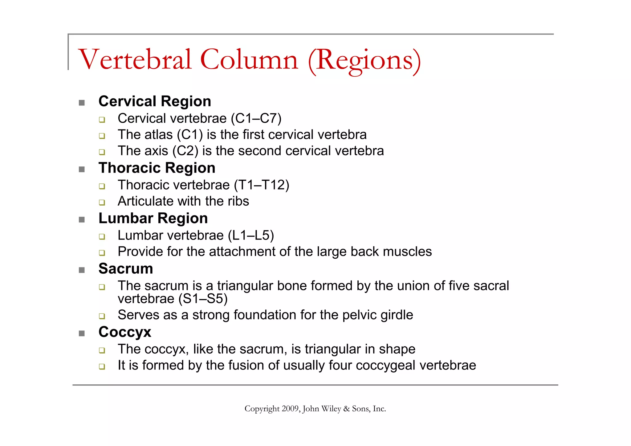
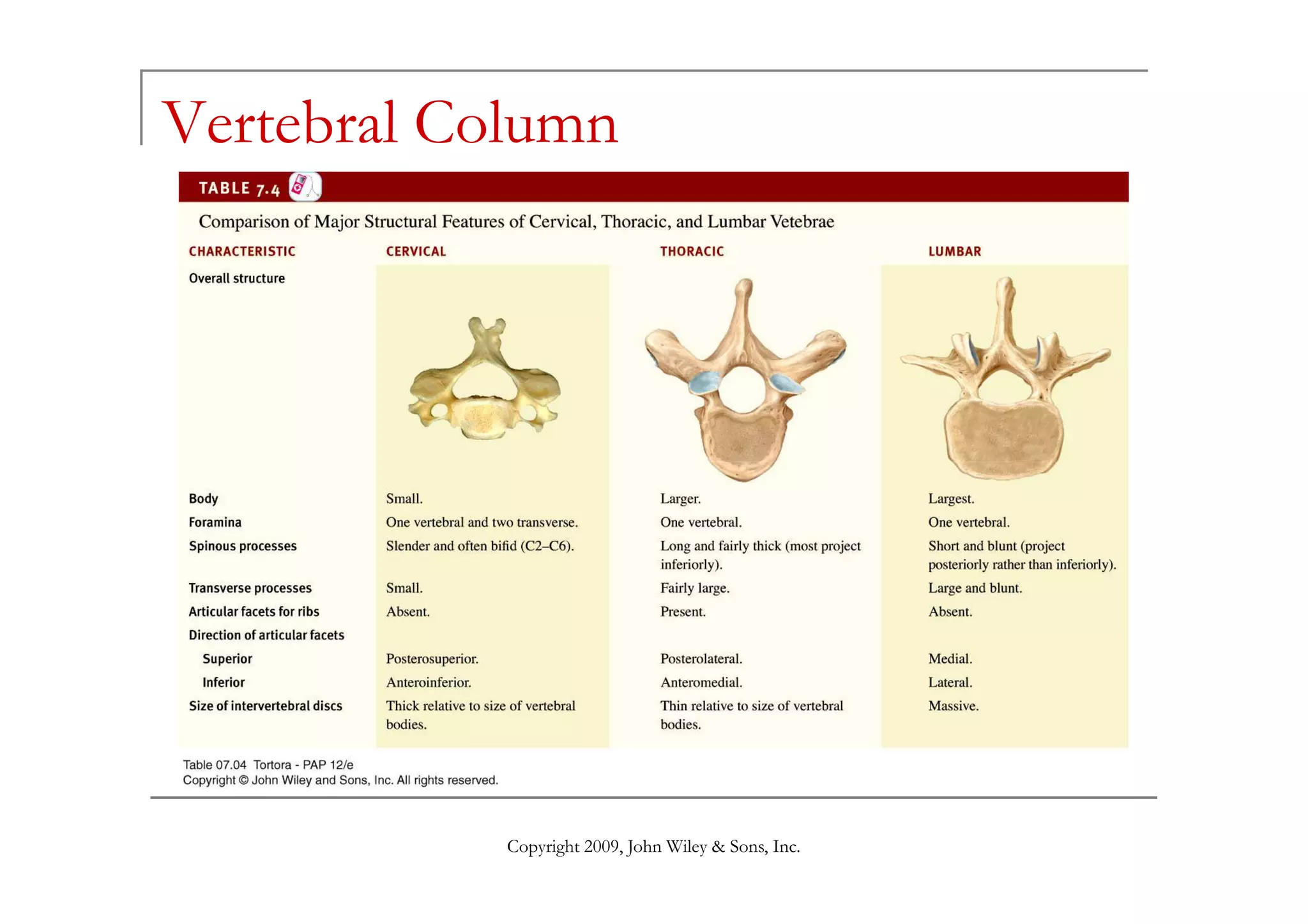
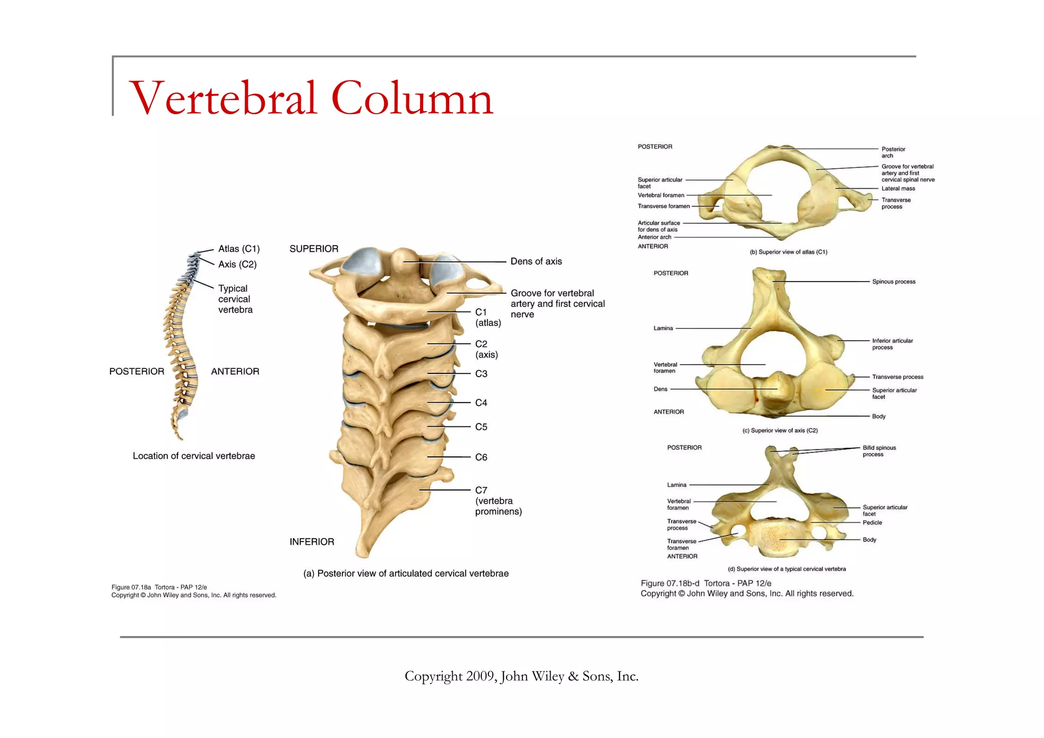
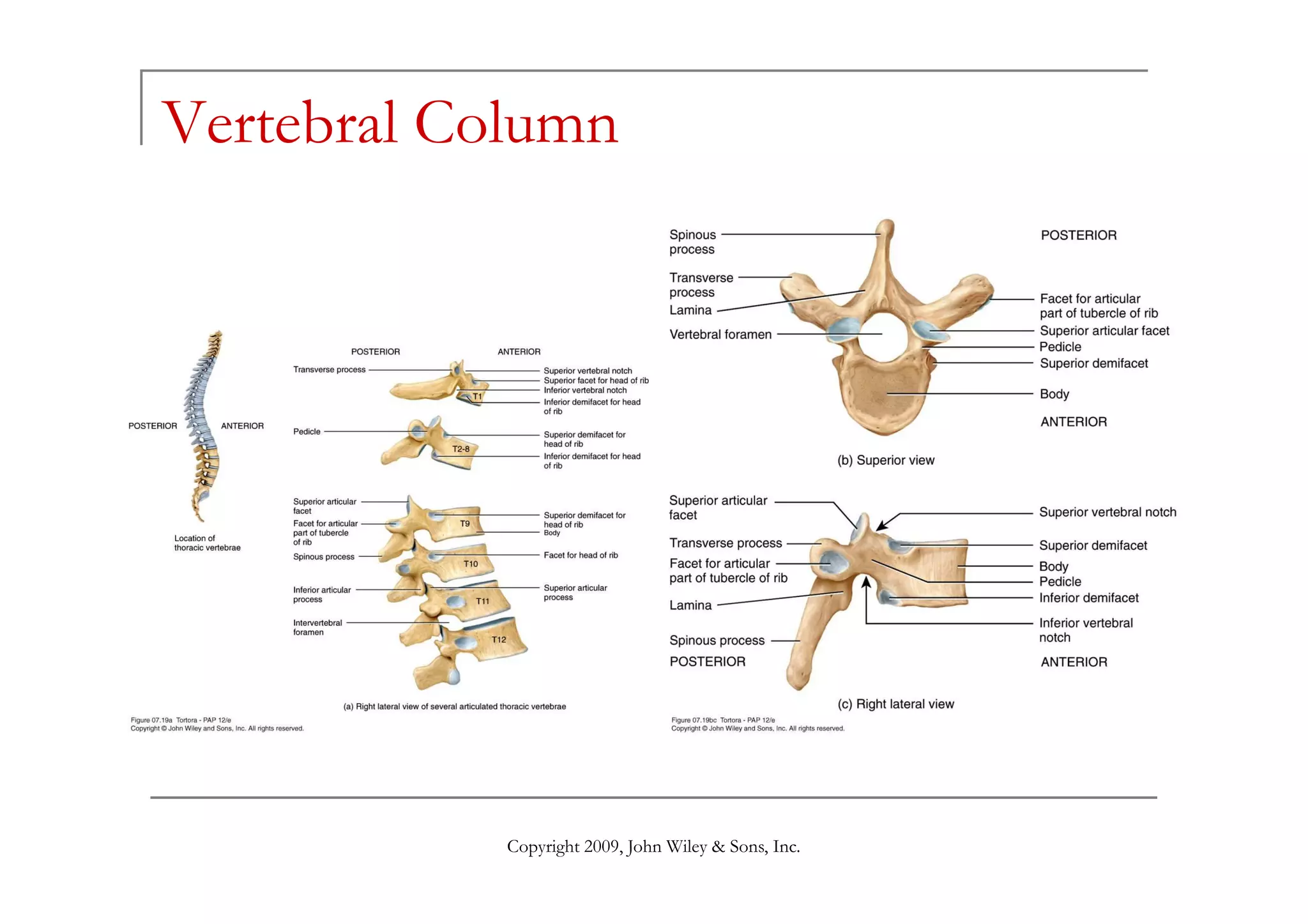
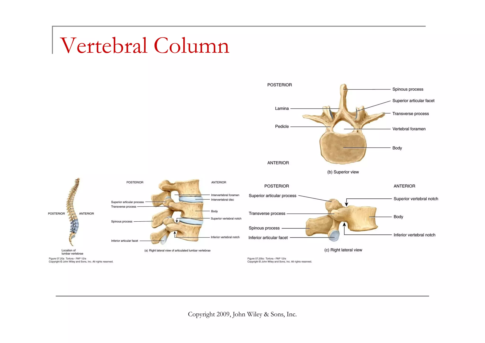
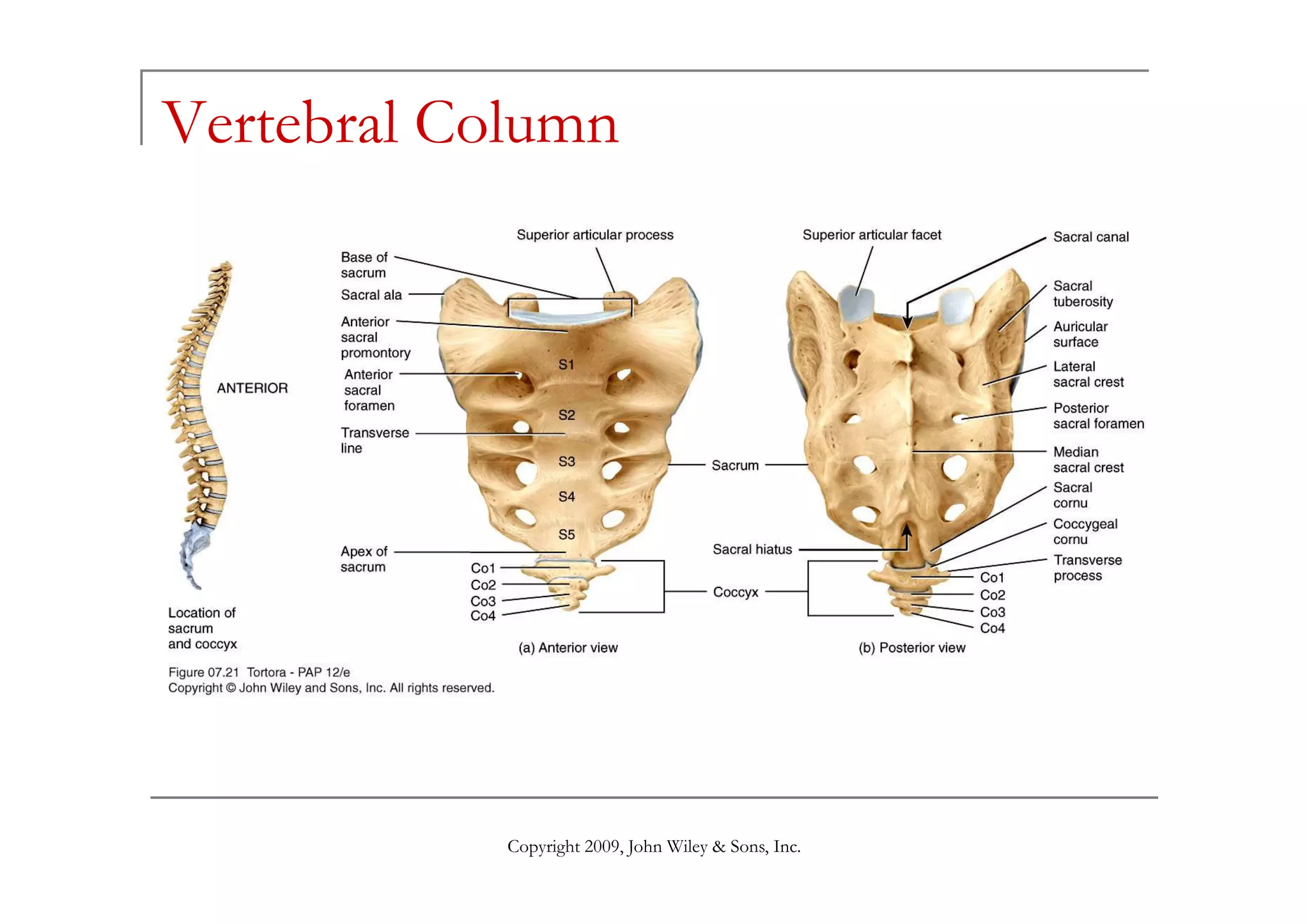
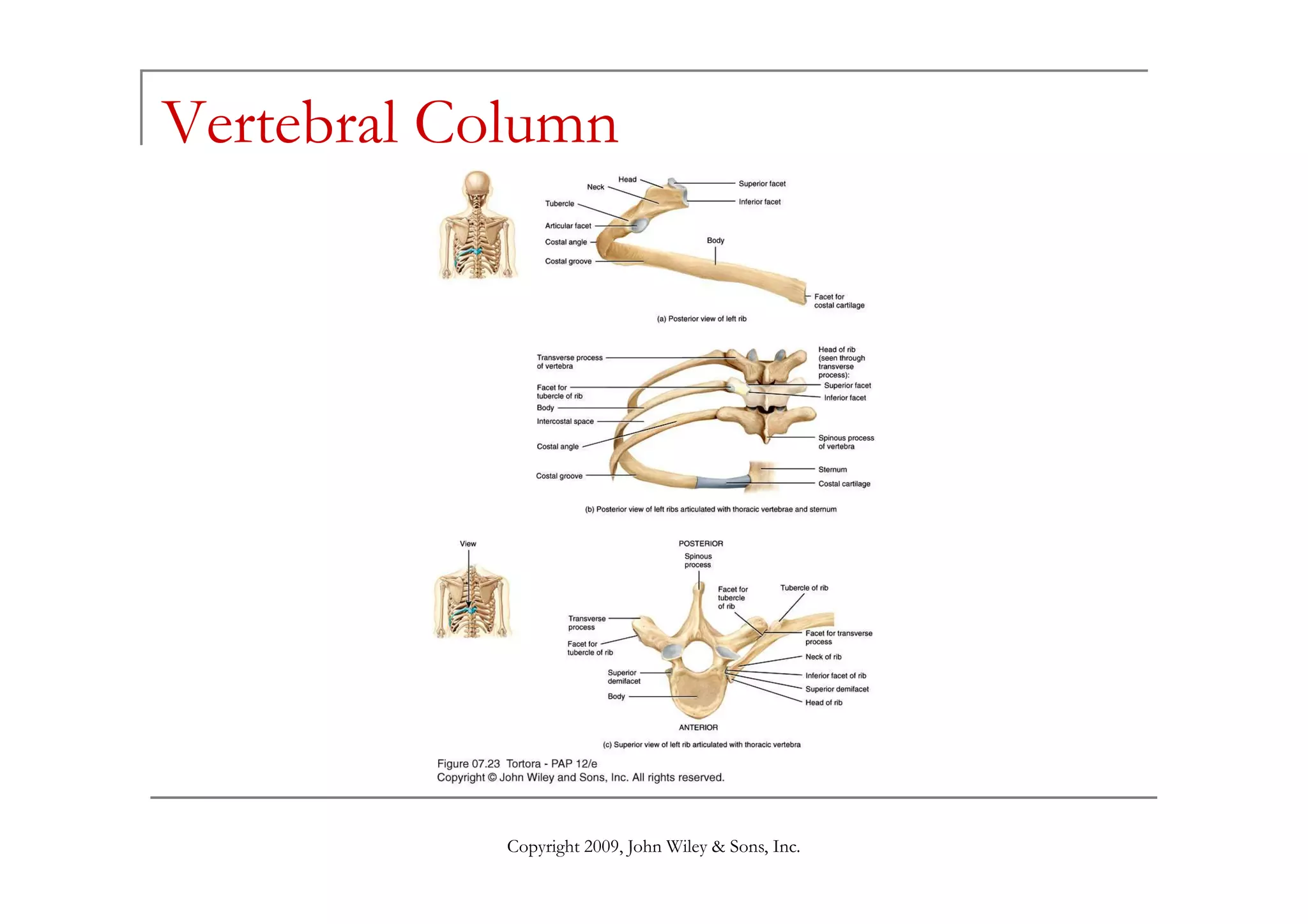
(A Project of National Medical Centre Karachi)
THE SKELETAL SYSTEM
BY:
Shahzad Bashir
Lecturer, NMC ION.
The Skeletal System:
The Axial Skeleton
The Appendicular Skeleton
Shahzad Bashir
The Skeletal System: The Axial Skeleton
Divisions of the Skeletal System
Types of Bones
Bone Surface Markings
Skull
Shahzad Bashir
Skull
Hyoid Bone
Vertebral Column
Thorax
Divisions of the Skeletal System
The human skeleton consists of 206 named bones
Bones of the skeleton are grouped into two principal
divisions:
Axial skeleton
Consists of the bones that lie around the longitudinal axis of
the human body
Shahzad Bashir
the human body
Skull bones, auditory ossicles (ear bones), hyoid bone, ribs,
sternum (breastbone), and bones of the vertebral column
Appendicular skeleton
Consists of the bones of the upper and lower limbs
(extremities), plus the bones forming the girdles that connect
the limbs to the axial skeleton
Divisions of the Skeletal System
Shahzad Bashir
Divisions of the Skeletal System
Shahzad Bashir
Types of Bones
Bones can be classified into five types based on
shape:
Long
Short
Flat
Shahzad Bashir
Flat
Irregular
Sesamoid
Types of Bones
Long Bones
Greater length than width and are slightly curved for strength
Femur, tibia, fibula, humerus, ulna, radius, phalanges
Short bones
Cube-shaped and are nearly equal in length and width
Carpal, tarsal
Flat bones
Thin and composed of two nearly parallel plates of compact bone tissue enclosing
a layer of spongy bone tissue
Shahzad Bashir
a layer of spongy bone tissue
Cranial, sternum, ribs, scapulae
Irregular bones
Complex shapes and cannot be grouped into any of the previous categories
Vertebrae, hip bones, some facial bones, calcaneus
Sesamoid bones
Protect tendons from excessive wear and tear
Patellae, foot, hand
Sutural bones
Small bones located in sutures of cranial bones
Bone Surface Markings
Bones have characteristic surface markings
Structural features adapted for specific functions
There are two major types of surface markings:
1) Depressions and openings
Allow the passage of blood vessels and nerves or form
Shahzad Bashir
Allow the passage of blood vessels and nerves or form
joints
2) Processes
Projections or outgrowths that form joints or serve as
attachment points for ligaments and tendons
Bone Surface Markings
Shahzad Bashir
Skull
Skull (cranium)
Consists of 22 bones
Bones of the skull are grouped into two
categories:
Cranial bones
Shahzad Bashir
Eight cranial bones form the cranial cavity
Frontal bone, two parietal bones, two temporal bones, the
occipital bone, the sphenoid bone, ethmoid bone
Facial bones
Fourteen facial bones form the face
Two nasal bones, two maxillae, two zygomatic bones, the
mandible, two lacrimal bones, two palatine bones, two inferior
nasal conchae, vomer
Skull
Shahzad Bashir
Skull
The cranial and facial bones protect and support
special sense organs and the brain
Besides forming the large cranial cavity, the skull
also forms several smaller cavities
Nasal cavity
Copyright 2009, John Wiley & Sons, Inc.
Nasal cavity
Orbits (eye sockets)
Paranasal sinuses
Small cavities which house organs involved in hearing
and equilibrium
Skull
Immovable joints called sutures fuse most of the
skull bones together
The skull provides large areas of attachment for
muscles that move various parts of the head
Skull and facial bones provide attachment for
Copyright 2009, John Wiley & Sons, Inc.
muscles that produce facial expressions
The facial bones form the framework of the face
and provide support for the entrances to the
digestive and respiratory systems
Skull (Cranial Bones)
Frontal Bone
Forms the forehead
Parietal Bones
Form the sides and roof of the cranial cavity
Temporal Bones
Form the lateral aspects and floor of the cranium
Occipital Bone
Forms the posterior part and most of the base of the cranium
Copyright 2009, John Wiley & Sons, Inc.
Forms the posterior part and most of the base of the cranium
Sphenoid Bone
Lies at the middle part of the base of the skull
Ethmoid Bone
Located on the midline in the anterior part of the cranial floor medial to
the orbits
A major superior supporting structure of the nasal cavity
Contain thin projections called conchae which are lined by mucous
membranes
Increased surface area in the nasal cavity helps to humidify inhaled air
trapping inhaled particles
Skull
Copyright 2009, John Wiley & Sons, Inc.
Skull
Copyright 2009, John Wiley & Sons, Inc.
Skull
Copyright 2009, John Wiley & Sons, Inc.
Skull (Facial Bones)
Nasal Bones
Form the bridge of the nose
Maxillae
Form the upper jawbone
Form most of the hard palate
Separates the nasal cavity from the oral cavity
Zygomatic Bones
Copyright 2009, John Wiley & Sons, Inc.
Zygomatic Bones
commonly called cheekbones, form the prominences of the
cheeks
Lacrimal Bones
Form a part of the medial wall of each orbit
Palatine Bones
Form the posterior portion of the hard palate
Inferior Nasal Conchae
Form a part of the inferior lateral wall of the nasal cavity
Skull (Facial Bones)
Vomer
Forms the inferior portion of the nasal septum
Mandible
Lower jawbone
The largest, strongest facial bone
The only movable skull bone
Nasal Septum
Copyright 2009, John Wiley & Sons, Inc.
Nasal Septum
Divides the interior of the nasal cavity into right and left sides
“Broken nose,” in most cases, refers to septal damage rather
than the nasal bones themselves
Orbits
Eye socket
Foramina
Openings for blood vessels , nerves , or ligaments of the skull
Skull
Copyright 2009, John Wiley & Sons, Inc.
Skull
Copyright 2009, John Wiley & Sons, Inc.
Skull
Copyright 2009, John Wiley & Sons, Inc.
Skull
Copyright 2009, John Wiley & Sons, Inc.
Skull
Copyright 2009, John Wiley & Sons, Inc.
Skull
Copyright 2009, John Wiley & Sons, Inc.
Skull
Unique Features of the Skull
Sutures, Paranasal sinuses, Fontanels
Sutures
an immovable joint that holds most skull bones together
Paranasal Sinuses
Cavities within cranial and facial bones near the nasal cavity
Secretions produced by the mucous membranes which line the sinuses,
drain into the nasal cavity
Copyright 2009, John Wiley & Sons, Inc.
drain into the nasal cavity
Serve as resonating chambers that intensify and prolong sounds
Fontanels
Areas of unossified tissue
At birth, unossified tissue spaces, commonly called “soft spots” link the
cranial bones
Eventually, they are replaced with bone to become sutures
Provide flexibility to the fetal skull, allowing the skull to change shape as
it passes through the birth canal
Skull
Copyright 2009, John Wiley & Sons, Inc.
Skull
Copyright 2009, John Wiley & Sons, Inc.
Hyoid Bone
Does not articulate with
any other bone
Supports the tongue,
providing attachment
sites for some tongue
muscles and for
Copyright 2009, John Wiley & Sons, Inc.
muscles and for
muscles of the neck and
pharynx
The hyoid bone also
helps to keep the larynx
(voice box) open at all
times
Vertebral Column
Also called the spine, backbone, or spinal
column
Functions to:
Protect the spinal cord
Support the head
Serve as a point of attachment for the ribs, pelvic
girdle, and muscles
Copyright 2009, John Wiley & Sons, Inc.
Serve as a point of attachment for the ribs, pelvic
girdle, and muscles
The vertebral column is curved to varying
degrees in different locations
Curves increase the column strength
Help maintain balance in the upright position
Absorb shocks during walking, and help protect the
vertebrae from fracture
Vertebral Column
Copyright 2009, John Wiley & Sons, Inc.
Vertebral Column
Various conditions may exaggerate the normal
curves of the vertebral column
Kyphosis
Lordosis
Scoliosis
Composed of a series of bones called vertebrae
(Adult=26)
Copyright 2009, John Wiley & Sons, Inc.
(Adult=26)
7 cervical are in the neck region
12 thoracic are posterior to the thoracic cavity
5 lumbar support the lower back
1 sacrum consists of five fused sacral vertebrae
1 coccyx consists of four fused coccygeal vertebrae
Vertebral Column
Copyright 2009, John Wiley & Sons, Inc.
Vertebral Column (Intervertebral Discs)
Found between the bodies of adjacent
vertebrae
Functions to:
Form strong joints
Permit various movements of the vertebral
column
Copyright 2009, John Wiley & Sons, Inc.
column
Absorb vertical shock
Vertebrae typically consist of:
A Body (weight bearing)
A vertebral arch (surrounds the spinal cord)
Several processes (points of attachment for
muscles)
Vertebral Column
Copyright 2009, John Wiley & Sons, Inc.
Vertebral Column (Regions)
Cervical Region
Cervical vertebrae (C1–C7)
The atlas (C1) is the first cervical vertebra
The axis (C2) is the second cervical vertebra
Thoracic Region
Thoracic vertebrae (T1–T12)
Articulate with the ribs
Lumbar Region
Copyright 2009, John Wiley & Sons, Inc.
Lumbar Region
Lumbar vertebrae (L1–L5)
Provide for the attachment of the large back muscles
Sacrum
The sacrum is a triangular bone formed by the union of five sacral
vertebrae (S1–S5)
Serves as a strong foundation for the pelvic girdle
Coccyx
The coccyx, like the sacrum, is triangular in shape
It is formed by the fusion of usually four coccygeal vertebrae
Vertebral Column
Copyright 2009, John Wiley & Sons, Inc.
Vertebral Column
Copyright 2009, John Wiley & Sons, Inc.
Vertebral Column
Copyright 2009, John Wiley & Sons, Inc.
Vertebral Column
Copyright 2009, John Wiley & Sons, Inc.
Vertebral Column
Copyright 2009, John Wiley & Sons, Inc.
Thorax
Thoracic cage is formed by the:
Sternum
Ribs
Costal cartilages
Thoracic vertebrae
Functions to:
Copyright 2009, John Wiley & Sons, Inc.
Functions to:
Enclose and protect the organs in the thoracic
and abdominal cavities
Provide support for the bones of the upper
limbs
Play a role in breathing
Thorax
Sternum
“Breastbone” located in the center of the
thoracic wall
Consists of the manubrium, body, xiphoid
process
Ribs
Copyright 2009, John Wiley & Sons, Inc.
Ribs
Twelve pairs of ribs give structural support to
the sides of the thoracic cavity
Costal cartilages
Costal cartilages contribute to the elasticity of
the thoracic cage
Vertebral Column
Copyright 2009, John Wiley & Sons, Inc.
Vertebral Column
Copyright 2009, John Wiley & Sons, Inc.
End of Chapter 7
Copyright 2009 John Wiley & Sons, Inc.
All rights reserved. Reproduction or translation of this work
beyond that permitted in section 117 of the 1976 United
States Copyright Act without express permission of the
copyright owner is unlawful. Request for further information
should be addressed to the Permission Department, John
Copyright 2009, John Wiley & Sons, Inc.
should be addressed to the Permission Department, John
Wiley & Sons, Inc. The purchaser may make back-up copies
for his/her own use only and not for distribution or resale.
The Publishers assumes no responsibility for errors,
omissions, or damages caused by the use of theses
programs or from the use of the information herein.

Introductiontoskeletalsystem 130406022426-phpapp02

  • 1.
    (A Project ofNational Medical Centre Karachi) THE SKELETAL SYSTEM BY: Shahzad Bashir Lecturer, NMC ION.
  • 2.
    The Skeletal System: TheAxial Skeleton The Appendicular Skeleton Shahzad Bashir
  • 3.
    The Skeletal System:The Axial Skeleton Divisions of the Skeletal System Types of Bones Bone Surface Markings Skull Shahzad Bashir Skull Hyoid Bone Vertebral Column Thorax
  • 4.
    Divisions of theSkeletal System The human skeleton consists of 206 named bones Bones of the skeleton are grouped into two principal divisions: Axial skeleton Consists of the bones that lie around the longitudinal axis of the human body Shahzad Bashir the human body Skull bones, auditory ossicles (ear bones), hyoid bone, ribs, sternum (breastbone), and bones of the vertebral column Appendicular skeleton Consists of the bones of the upper and lower limbs (extremities), plus the bones forming the girdles that connect the limbs to the axial skeleton
  • 5.
    Divisions of theSkeletal System Shahzad Bashir
  • 6.
    Divisions of theSkeletal System Shahzad Bashir
  • 7.
    Types of Bones Bonescan be classified into five types based on shape: Long Short Flat Shahzad Bashir Flat Irregular Sesamoid
  • 8.
    Types of Bones LongBones Greater length than width and are slightly curved for strength Femur, tibia, fibula, humerus, ulna, radius, phalanges Short bones Cube-shaped and are nearly equal in length and width Carpal, tarsal Flat bones Thin and composed of two nearly parallel plates of compact bone tissue enclosing a layer of spongy bone tissue Shahzad Bashir a layer of spongy bone tissue Cranial, sternum, ribs, scapulae Irregular bones Complex shapes and cannot be grouped into any of the previous categories Vertebrae, hip bones, some facial bones, calcaneus Sesamoid bones Protect tendons from excessive wear and tear Patellae, foot, hand Sutural bones Small bones located in sutures of cranial bones
  • 9.
    Bone Surface Markings Boneshave characteristic surface markings Structural features adapted for specific functions There are two major types of surface markings: 1) Depressions and openings Allow the passage of blood vessels and nerves or form Shahzad Bashir Allow the passage of blood vessels and nerves or form joints 2) Processes Projections or outgrowths that form joints or serve as attachment points for ligaments and tendons
  • 10.
  • 11.
    Skull Skull (cranium) Consists of22 bones Bones of the skull are grouped into two categories: Cranial bones Shahzad Bashir Eight cranial bones form the cranial cavity Frontal bone, two parietal bones, two temporal bones, the occipital bone, the sphenoid bone, ethmoid bone Facial bones Fourteen facial bones form the face Two nasal bones, two maxillae, two zygomatic bones, the mandible, two lacrimal bones, two palatine bones, two inferior nasal conchae, vomer
  • 12.
  • 13.
    Skull The cranial andfacial bones protect and support special sense organs and the brain Besides forming the large cranial cavity, the skull also forms several smaller cavities Nasal cavity Copyright 2009, John Wiley & Sons, Inc. Nasal cavity Orbits (eye sockets) Paranasal sinuses Small cavities which house organs involved in hearing and equilibrium
  • 14.
    Skull Immovable joints calledsutures fuse most of the skull bones together The skull provides large areas of attachment for muscles that move various parts of the head Skull and facial bones provide attachment for Copyright 2009, John Wiley & Sons, Inc. muscles that produce facial expressions The facial bones form the framework of the face and provide support for the entrances to the digestive and respiratory systems
  • 15.
    Skull (Cranial Bones) FrontalBone Forms the forehead Parietal Bones Form the sides and roof of the cranial cavity Temporal Bones Form the lateral aspects and floor of the cranium Occipital Bone Forms the posterior part and most of the base of the cranium Copyright 2009, John Wiley & Sons, Inc. Forms the posterior part and most of the base of the cranium Sphenoid Bone Lies at the middle part of the base of the skull Ethmoid Bone Located on the midline in the anterior part of the cranial floor medial to the orbits A major superior supporting structure of the nasal cavity Contain thin projections called conchae which are lined by mucous membranes Increased surface area in the nasal cavity helps to humidify inhaled air trapping inhaled particles
  • 16.
    Skull Copyright 2009, JohnWiley & Sons, Inc.
  • 17.
    Skull Copyright 2009, JohnWiley & Sons, Inc.
  • 18.
    Skull Copyright 2009, JohnWiley & Sons, Inc.
  • 19.
    Skull (Facial Bones) NasalBones Form the bridge of the nose Maxillae Form the upper jawbone Form most of the hard palate Separates the nasal cavity from the oral cavity Zygomatic Bones Copyright 2009, John Wiley & Sons, Inc. Zygomatic Bones commonly called cheekbones, form the prominences of the cheeks Lacrimal Bones Form a part of the medial wall of each orbit Palatine Bones Form the posterior portion of the hard palate Inferior Nasal Conchae Form a part of the inferior lateral wall of the nasal cavity
  • 20.
    Skull (Facial Bones) Vomer Formsthe inferior portion of the nasal septum Mandible Lower jawbone The largest, strongest facial bone The only movable skull bone Nasal Septum Copyright 2009, John Wiley & Sons, Inc. Nasal Septum Divides the interior of the nasal cavity into right and left sides “Broken nose,” in most cases, refers to septal damage rather than the nasal bones themselves Orbits Eye socket Foramina Openings for blood vessels , nerves , or ligaments of the skull
  • 21.
    Skull Copyright 2009, JohnWiley & Sons, Inc.
  • 22.
    Skull Copyright 2009, JohnWiley & Sons, Inc.
  • 23.
    Skull Copyright 2009, JohnWiley & Sons, Inc.
  • 24.
    Skull Copyright 2009, JohnWiley & Sons, Inc.
  • 25.
    Skull Copyright 2009, JohnWiley & Sons, Inc.
  • 26.
    Skull Copyright 2009, JohnWiley & Sons, Inc.
  • 27.
    Skull Unique Features ofthe Skull Sutures, Paranasal sinuses, Fontanels Sutures an immovable joint that holds most skull bones together Paranasal Sinuses Cavities within cranial and facial bones near the nasal cavity Secretions produced by the mucous membranes which line the sinuses, drain into the nasal cavity Copyright 2009, John Wiley & Sons, Inc. drain into the nasal cavity Serve as resonating chambers that intensify and prolong sounds Fontanels Areas of unossified tissue At birth, unossified tissue spaces, commonly called “soft spots” link the cranial bones Eventually, they are replaced with bone to become sutures Provide flexibility to the fetal skull, allowing the skull to change shape as it passes through the birth canal
  • 28.
    Skull Copyright 2009, JohnWiley & Sons, Inc.
  • 29.
    Skull Copyright 2009, JohnWiley & Sons, Inc.
  • 30.
    Hyoid Bone Does notarticulate with any other bone Supports the tongue, providing attachment sites for some tongue muscles and for Copyright 2009, John Wiley & Sons, Inc. muscles and for muscles of the neck and pharynx The hyoid bone also helps to keep the larynx (voice box) open at all times
  • 31.
    Vertebral Column Also calledthe spine, backbone, or spinal column Functions to: Protect the spinal cord Support the head Serve as a point of attachment for the ribs, pelvic girdle, and muscles Copyright 2009, John Wiley & Sons, Inc. Serve as a point of attachment for the ribs, pelvic girdle, and muscles The vertebral column is curved to varying degrees in different locations Curves increase the column strength Help maintain balance in the upright position Absorb shocks during walking, and help protect the vertebrae from fracture
  • 32.
    Vertebral Column Copyright 2009,John Wiley & Sons, Inc.
  • 33.
    Vertebral Column Various conditionsmay exaggerate the normal curves of the vertebral column Kyphosis Lordosis Scoliosis Composed of a series of bones called vertebrae (Adult=26) Copyright 2009, John Wiley & Sons, Inc. (Adult=26) 7 cervical are in the neck region 12 thoracic are posterior to the thoracic cavity 5 lumbar support the lower back 1 sacrum consists of five fused sacral vertebrae 1 coccyx consists of four fused coccygeal vertebrae
  • 34.
    Vertebral Column Copyright 2009,John Wiley & Sons, Inc.
  • 35.
    Vertebral Column (IntervertebralDiscs) Found between the bodies of adjacent vertebrae Functions to: Form strong joints Permit various movements of the vertebral column Copyright 2009, John Wiley & Sons, Inc. column Absorb vertical shock Vertebrae typically consist of: A Body (weight bearing) A vertebral arch (surrounds the spinal cord) Several processes (points of attachment for muscles)
  • 36.
    Vertebral Column Copyright 2009,John Wiley & Sons, Inc.
  • 37.
    Vertebral Column (Regions) CervicalRegion Cervical vertebrae (C1–C7) The atlas (C1) is the first cervical vertebra The axis (C2) is the second cervical vertebra Thoracic Region Thoracic vertebrae (T1–T12) Articulate with the ribs Lumbar Region Copyright 2009, John Wiley & Sons, Inc. Lumbar Region Lumbar vertebrae (L1–L5) Provide for the attachment of the large back muscles Sacrum The sacrum is a triangular bone formed by the union of five sacral vertebrae (S1–S5) Serves as a strong foundation for the pelvic girdle Coccyx The coccyx, like the sacrum, is triangular in shape It is formed by the fusion of usually four coccygeal vertebrae
  • 38.
    Vertebral Column Copyright 2009,John Wiley & Sons, Inc.
  • 39.
    Vertebral Column Copyright 2009,John Wiley & Sons, Inc.
  • 40.
    Vertebral Column Copyright 2009,John Wiley & Sons, Inc.
  • 41.
    Vertebral Column Copyright 2009,John Wiley & Sons, Inc.
  • 42.
    Vertebral Column Copyright 2009,John Wiley & Sons, Inc.
  • 43.
    Thorax Thoracic cage isformed by the: Sternum Ribs Costal cartilages Thoracic vertebrae Functions to: Copyright 2009, John Wiley & Sons, Inc. Functions to: Enclose and protect the organs in the thoracic and abdominal cavities Provide support for the bones of the upper limbs Play a role in breathing
  • 44.
    Thorax Sternum “Breastbone” located inthe center of the thoracic wall Consists of the manubrium, body, xiphoid process Ribs Copyright 2009, John Wiley & Sons, Inc. Ribs Twelve pairs of ribs give structural support to the sides of the thoracic cavity Costal cartilages Costal cartilages contribute to the elasticity of the thoracic cage
  • 45.
    Vertebral Column Copyright 2009,John Wiley & Sons, Inc.
  • 46.
    Vertebral Column Copyright 2009,John Wiley & Sons, Inc.
  • 47.
    End of Chapter7 Copyright 2009 John Wiley & Sons, Inc. All rights reserved. Reproduction or translation of this work beyond that permitted in section 117 of the 1976 United States Copyright Act without express permission of the copyright owner is unlawful. Request for further information should be addressed to the Permission Department, John Copyright 2009, John Wiley & Sons, Inc. should be addressed to the Permission Department, John Wiley & Sons, Inc. The purchaser may make back-up copies for his/her own use only and not for distribution or resale. The Publishers assumes no responsibility for errors, omissions, or damages caused by the use of theses programs or from the use of the information herein.