The human skeleton is the internal framework of the human body. It is composed of around 270 bones at birth – this total decreases to around 206 bones by adulthood after some bones get fused together. The bone mass in the skeleton reaches maximum density around age 21. The skeletal system includes all of the bones and joints in the body. Each bone is a complex living organ that is made up of many cells, protein fibers, and minerals. The skeleton acts as a scaffold by providing support and protection for the soft tissues that make up the rest of the body. this is brief study on skeletal system ,that i prepared for my academic purpose .
please comment
thank u
Anatomy of skeleton system full/Human all BonesMdMehbubAlam
All about human skeleton system
Full Skeleton system
Appendicular Skeleton
Axial Skeleton
Rib cage
Skull bones
Facial bones
Ear bones
Humerus bone
Femur bone
Tarsals
Carpals
Falanges
Bones of the Foot: Tarsals, Metatarsals and Phalanges · The Femur · The Patella · The Tibia · The Fibula.
The femur is the only bone in the thigh and the longest bone in the body.
It acts as the site of origin and attachment of many muscles and ligaments, and can be divided into three parts; proximal, shaft and distal.
Facial bones anatomy, Anatomic position of fascial bone, axial skeleton, arti...Dr Shahid Alam
Facial bones anatomy, Anatomic position of fascial bone, axial skeleton, articulation of fascial bone, maxillary sinuses, paranasal sinuses frontanelles, foramina and fissure of skull from BD chaurasia and Snell's anatomy lecture by dr shahid alam, dr shahid, dr alam, shahid, shahid alam
The lymphatic system is part of the immune system. It also maintains fluid balance and plays a role in absorbing fats and fat-soluble nutrients.
The lymphatic or lymph system involves an extensive network of vessels that passes through almost all our tissues to allow for the movement of a fluid called lymph. Lymph circulates through the body in a similar way to blood.
There are about 600 lymph nodes in the body. These nodes swell in response to infection, due to a build-up of lymph fluid, bacteria, or other organisms and immune system cells.
A person with a throat infection, for example, may feel that their "glands" are swollen. Swollen glands can be felt especially under the jaw, in the armpits, or in the groin area. These are, in fact, not glands but lymph nodes.
A detail account of Bones, their histological features, classification, composition, Formation, blood and nerve supply, functions, plus some interesting facts about bones.
Introduction in human anatomy
2. Anatomy • Definition - anatome = up (ana) + cutting (tome) • Disciplines of anatomy – Macroscopic – Microscopic – Developmental – Neuroanatomy • Approach to study of gross anatomy Upper extremity Back Head and neck Thorax Abdomen Pelvis and perineum Lower extremity
3. Basis for Terminology • Terms informative • Nomina anatomica • Use of eponyms Use correct terminology on exams; avoid nonspecific, general terms, like
The details description is given in the ppt about the axial skeleton. We include the topics which are mentions below :-
What is skeleton
Skull
Hyoid bone
Vertebrae
Thoracic cage
Skeleton system- bones and their number with detailed description.bhartisharma175
this ppt consist of skeleton system and its types. it consist of every individual bone and their number in adult human being. easy to understand for students as well as for teachers.
The skeletal system includes all of the bones and joints in the body. Each bone is a complex living organ that is made up of many cells, protein fibers, and minerals. The skeleton acts as a scaffold by providing support and protection for the soft tissues that make up the rest of the body. The skeletal system also provides attachment points for muscles to allow movements at the joints. New blood cells are produced by the red bone marrow inside of our bones.
this is brief study describes the aspects of iv cannulation for students and aspirants , this slide briefly comprises all the major aspects of cannulation .......................
thank you
martinsuja369@gmail.com
Wilsons disease- A brief medical study. martinshaji
this brief study describes all the basic aspects of Wilsons disease , including management. as this is a congenital abnormality associated with severe complications on the future proper diagnosis management , and lifestyle modifications , …..surgical options are also needed ,if necessary .
thank you
martinsuja369@gmail.com
Communicable diseases and causative agents- a detailed chart martinshaji
A communicable disease is one that is spread from one person to another through a variety of ways that include contact with blood and bodily fluids; breathing in an airborne virus or by being bitten by an insect................................................................this chart provides a clear idea regarding almost all communicable disease and their causative agents
thank you ...............
martinsuja369@gmail.com
TDM of drugs used in organ transplantation-detailed studymartinshaji
Therapeutic drug monitoring (TDM) is testing that measures the amount of certain medicines in your blood. It is done to make sure the amount of medicine you are taking is both safe and effective. Most medicines can be dosed correctly without special testing. the slide explain all the tdm aspects of the drug in detail / Therapeutic drug monitoring (TDM) is testing that measures the amount of certain medicines in your blood. It is done to make sure the amount of medicine you are taking is both safe and effective. Most medicines can be dosed correctly without special testing.
martinsuja369@gmail.com
inbox for queries and more explanatory study materials
Organophosphate poisoning - a brief toxicological study martinshaji
this is a brief study on organophosphate poisoning , as it being more common problem in the health sector and emergency medicine now a days , this will be much helpful among health professionals .........text me for more topics
martinsuaj369@gmail.com
thank you
this case study describes about maxillofacial trauma , which details about the treatment, management , diagnosis, surgical options, patient counselling, pharmacist interventions & discussions are followed in this case .
please comment
thank u
martinsuja369@gmail.com
Sleep disorders - a brief medical study martinshaji
A sleep disorder is any condition that involves difficulty experienced when sleep , such disorders involve daytime fatigue causing severe distress and impairment to work.
SD also have an impact upon social and personal functioning
this is a brief study on all aspects of this ...............
please comment
thank you
for any detailed suggestions and for any medical study materials connect with me
martinsuja369@gmail.com
than you
A case study on Pangastritis with pancreatitis martinshaji
this case study describes about Pangastritis with pancreatitis , which details about the treatment, management , diagnosis, patient counselling, pharmacist interventions & discussions are followed in this case .
please comment
thank u
martinsuja369@gmail.com
Blood transfusion-MANAGEMENT FOR PEOPLE LIVING WITH SICKLE CELL DISORDER/DISE...martinshaji
Sickle cell disease is caused by a genetic mutation that leads to the production of abnormal hemoglobin known as sickle hemoglobin . Blood transfusion is the transfer of blood from one individual to another ..
Red blood cell transfusions help lessen anemia and reduce the blood’s viscosity, allowing it to flow more freely and ease disease symptoms.
this is a brief study
please comment
thank you
martinsuja369@gmail.com
Compartment syndrome,- a detailed medical study .martinshaji
Compartment syndrome is a condition that occurs when injury causes generalized painful swelling and increased pressure within a compartment to the point that blood cannot supply the muscles and nerves with oxygen and nutrients. Muscles in the forearm, lower leg and other body areas are surrounded by fibrous bands of tissues. This creates distinct compartments. The fibrous tissue is very inflexible and cannot stretch to accommodate the generalized swelling. If left untreated, muscles and nerves fail and may eventually die.
hence this is a medical emergency needed fast and great medical supervision , his study provides a detailed information regarding compartment syndrome
please comment
thank you
martinsuja369@gamil.com
Muscles -A LONG CHART ON IMAGES OF DIFFERENT MUSCLES- Myology|, kinesiology- ...martinshaji
this is a long chart on different types of muscles of human body with its images , this will be helpful for medical academics and better understanding , along with its names
please comment
thank you
martinsuja369@gamil.com
P h value- a long chart on different ph. values martinshaji
pH is a measure of how acidic/basic water is. The range goes from 0 - 14, with 7 being neutral. pHs of less than 7 indicate acidity, whereas a pH of greater than 7 indicates a base. pH is really a measure of the relative amount of free hydrogen and hydroxyl ions in the water .
this is a long chart on ph value of different substances
please comment
thank you
martinsuja369@gamil.com
12 basic facts about tetracyclines - medical information martinshaji
Tetracyclines are a class of broad-spectrum antibiotics used in the management and treatment of a variety of infectious diseases. Naturally occurring drugs in this class are tetracycline, chlortetracycline, oxytetracycline, and demeclocycline. used to treat infections caused by susceptible microorganisms such as gram positive and gram negative bacteria, chlamydiae, mycoplasmata, protozoans, or rickettsiae.
please comment
thank you
martinsuja369@gamil.com
8 signs and symptoms of breast cancer you - medical information martinshaji
After skin cancer, breast cancer is the most common cancer diagnosed in women in the United States. hence it is necessary to know its major symptoms which will help you to avoid a life threatening condition easily .....
please comment
thank you
How to have a Clear Skin....HEALTH INFORMATION martinshaji
Clear skin is possible for everyone if you follow a basic good skincare routine for your unique skin type. these are some common methods for having good clear skin on considering medical aspects ,
please comment
thank you ........
Liver failure is a life-threatening condition that demands urgent medical care.
Alcoholic liver disease (ALD) is a leading cause of cirrhosis, liver cancer, and acute and chronic liver failure and as such causes significant morbidity and mortality.
this is a brief study on liver failure and associated liver conditions and stages of conditions ,
please comment
thank you ........
martinsuja369@gmail.com
Natural ways to build healthy bones - health information martinshaji
Bone health is important throughout life. While you can never regain the bone density you had in your youth, you can help prevent rapidly thinning bones, even after your diagnosis. Calcium is a crucial building block of bone tissue. Vitamin D helps the body absorb and process calcium. Together, these two nutrients are the cornerstone of healthy bones.
this is a brief study for healthy bones .................................................
for any doubts or questions you can mail me @
martinsuja369@gmail.com
please comment
thank you
Sleep According To your Age-health information | images martinshaji
Sleep plays an important role in your physical health. For example, sleep is involved in healing and repair of your heart and blood vessels.
please comment
thank you.....
Doctors once thought bony growths called heel spurs brought on the pain. Now they believe that heel spurs are the result -- not the cause -- of plantar fasciitis.
Plantar fasciitis is inflammation of the thick band of tissue (also called a fascia) at the bottom of your foot that runs from your heel to your toes.
this is a brief study on plantar fasciitis
please comment
thank you
Title: Sense of Smell
Presenter: Dr. Faiza, Assistant Professor of Physiology
Qualifications:
MBBS (Best Graduate, AIMC Lahore)
FCPS Physiology
ICMT, CHPE, DHPE (STMU)
MPH (GC University, Faisalabad)
MBA (Virtual University of Pakistan)
Learning Objectives:
Describe the primary categories of smells and the concept of odor blindness.
Explain the structure and location of the olfactory membrane and mucosa, including the types and roles of cells involved in olfaction.
Describe the pathway and mechanisms of olfactory signal transmission from the olfactory receptors to the brain.
Illustrate the biochemical cascade triggered by odorant binding to olfactory receptors, including the role of G-proteins and second messengers in generating an action potential.
Identify different types of olfactory disorders such as anosmia, hyposmia, hyperosmia, and dysosmia, including their potential causes.
Key Topics:
Olfactory Genes:
3% of the human genome accounts for olfactory genes.
400 genes for odorant receptors.
Olfactory Membrane:
Located in the superior part of the nasal cavity.
Medially: Folds downward along the superior septum.
Laterally: Folds over the superior turbinate and upper surface of the middle turbinate.
Total surface area: 5-10 square centimeters.
Olfactory Mucosa:
Olfactory Cells: Bipolar nerve cells derived from the CNS (100 million), with 4-25 olfactory cilia per cell.
Sustentacular Cells: Produce mucus and maintain ionic and molecular environment.
Basal Cells: Replace worn-out olfactory cells with an average lifespan of 1-2 months.
Bowman’s Gland: Secretes mucus.
Stimulation of Olfactory Cells:
Odorant dissolves in mucus and attaches to receptors on olfactory cilia.
Involves a cascade effect through G-proteins and second messengers, leading to depolarization and action potential generation in the olfactory nerve.
Quality of a Good Odorant:
Small (3-20 Carbon atoms), volatile, water-soluble, and lipid-soluble.
Facilitated by odorant-binding proteins in mucus.
Membrane Potential and Action Potential:
Resting membrane potential: -55mV.
Action potential frequency in the olfactory nerve increases with odorant strength.
Adaptation Towards the Sense of Smell:
Rapid adaptation within the first second, with further slow adaptation.
Psychological adaptation greater than receptor adaptation, involving feedback inhibition from the central nervous system.
Primary Sensations of Smell:
Camphoraceous, Musky, Floral, Pepperminty, Ethereal, Pungent, Putrid.
Odor Detection Threshold:
Examples: Hydrogen sulfide (0.0005 ppm), Methyl-mercaptan (0.002 ppm).
Some toxic substances are odorless at lethal concentrations.
Characteristics of Smell:
Odor blindness for single substances due to lack of appropriate receptor protein.
Behavioral and emotional influences of smell.
Transmission of Olfactory Signals:
From olfactory cells to glomeruli in the olfactory bulb, involving lateral inhibition.
Primitive, less old, and new olfactory systems with different path
Explore natural remedies for syphilis treatment in Singapore. Discover alternative therapies, herbal remedies, and lifestyle changes that may complement conventional treatments. Learn about holistic approaches to managing syphilis symptoms and supporting overall health.
263778731218 Abortion Clinic /Pills In Harare ,sisternakatoto
263778731218 Abortion Clinic /Pills In Harare ,ABORTION WOMEN’S CLINIC +27730423979 IN women clinic we believe that every woman should be able to make choices in her pregnancy. Our job is to provide compassionate care, safety,affordable and confidential services. That’s why we have won the trust from all generations of women all over the world. we use non surgical method(Abortion pills) to terminate…Dr.LISA +27730423979women Clinic is committed to providing the highest quality of obstetrical and gynecological care to women of all ages. Our dedicated staff aim to treat each patient and her health concerns with compassion and respect.Our dedicated group ABORTION WOMEN’S CLINIC +27730423979 IN women clinic we believe that every woman should be able to make choices in her pregnancy. Our job is to provide compassionate care, safety,affordable and confidential services. That’s why we have won the trust from all generations of women all over the world. we use non surgical method(Abortion pills) to terminate…Dr.LISA +27730423979women Clinic is committed to providing the highest quality of obstetrical and gynecological care to women of all ages. Our dedicated staff aim to treat each patient and her health concerns with compassion and respect.Our dedicated group of receptionists, nurses, and physicians have worked together as a teamof receptionists, nurses, and physicians have worked together as a team wwww.lisywomensclinic.co.za/
ARTIFICIAL INTELLIGENCE IN HEALTHCARE.pdfAnujkumaranit
Artificial intelligence (AI) refers to the simulation of human intelligence processes by machines, especially computer systems. It encompasses tasks such as learning, reasoning, problem-solving, perception, and language understanding. AI technologies are revolutionizing various fields, from healthcare to finance, by enabling machines to perform tasks that typically require human intelligence.
NVBDCP.pptx Nation vector borne disease control programSapna Thakur
NVBDCP was launched in 2003-2004 . Vector-Borne Disease: Disease that results from an infection transmitted to humans and other animals by blood-feeding arthropods, such as mosquitoes, ticks, and fleas. Examples of vector-borne diseases include Dengue fever, West Nile Virus, Lyme disease, and malaria.
Knee anatomy and clinical tests 2024.pdfvimalpl1234
This includes all relevant anatomy and clinical tests compiled from standard textbooks, Campbell,netter etc..It is comprehensive and best suited for orthopaedicians and orthopaedic residents.
Acute scrotum is a general term referring to an emergency condition affecting the contents or the wall of the scrotum.
There are a number of conditions that present acutely, predominantly with pain and/or swelling
A careful and detailed history and examination, and in some cases, investigations allow differentiation between these diagnoses. A prompt diagnosis is essential as the patient may require urgent surgical intervention
Testicular torsion refers to twisting of the spermatic cord, causing ischaemia of the testicle.
Testicular torsion results from inadequate fixation of the testis to the tunica vaginalis producing ischemia from reduced arterial inflow and venous outflow obstruction.
The prevalence of testicular torsion in adult patients hospitalized with acute scrotal pain is approximately 25 to 50 percent
Pulmonary Thromboembolism - etilogy, types, medical- Surgical and nursing man...VarunMahajani
Disruption of blood supply to lung alveoli due to blockage of one or more pulmonary blood vessels is called as Pulmonary thromboembolism. In this presentation we will discuss its causes, types and its management in depth.
Flu Vaccine Alert in Bangalore Karnatakaaddon Scans
As flu season approaches, health officials in Bangalore, Karnataka, are urging residents to get their flu vaccinations. The seasonal flu, while common, can lead to severe health complications, particularly for vulnerable populations such as young children, the elderly, and those with underlying health conditions.
Dr. Vidisha Kumari, a leading epidemiologist in Bangalore, emphasizes the importance of getting vaccinated. "The flu vaccine is our best defense against the influenza virus. It not only protects individuals but also helps prevent the spread of the virus in our communities," he says.
This year, the flu season is expected to coincide with a potential increase in other respiratory illnesses. The Karnataka Health Department has launched an awareness campaign highlighting the significance of flu vaccinations. They have set up multiple vaccination centers across Bangalore, making it convenient for residents to receive their shots.
To encourage widespread vaccination, the government is also collaborating with local schools, workplaces, and community centers to facilitate vaccination drives. Special attention is being given to ensuring that the vaccine is accessible to all, including marginalized communities who may have limited access to healthcare.
Residents are reminded that the flu vaccine is safe and effective. Common side effects are mild and may include soreness at the injection site, mild fever, or muscle aches. These side effects are generally short-lived and far less severe than the flu itself.
Healthcare providers are also stressing the importance of continuing COVID-19 precautions. Wearing masks, practicing good hand hygiene, and maintaining social distancing are still crucial, especially in crowded places.
Protect yourself and your loved ones by getting vaccinated. Together, we can help keep Bangalore healthy and safe this flu season. For more information on vaccination centers and schedules, residents can visit the Karnataka Health Department’s official website or follow their social media pages.
Stay informed, stay safe, and get your flu shot today!
Title: Sense of Taste
Presenter: Dr. Faiza, Assistant Professor of Physiology
Qualifications:
MBBS (Best Graduate, AIMC Lahore)
FCPS Physiology
ICMT, CHPE, DHPE (STMU)
MPH (GC University, Faisalabad)
MBA (Virtual University of Pakistan)
Learning Objectives:
Describe the structure and function of taste buds.
Describe the relationship between the taste threshold and taste index of common substances.
Explain the chemical basis and signal transduction of taste perception for each type of primary taste sensation.
Recognize different abnormalities of taste perception and their causes.
Key Topics:
Significance of Taste Sensation:
Differentiation between pleasant and harmful food
Influence on behavior
Selection of food based on metabolic needs
Receptors of Taste:
Taste buds on the tongue
Influence of sense of smell, texture of food, and pain stimulation (e.g., by pepper)
Primary and Secondary Taste Sensations:
Primary taste sensations: Sweet, Sour, Salty, Bitter, Umami
Chemical basis and signal transduction mechanisms for each taste
Taste Threshold and Index:
Taste threshold values for Sweet (sucrose), Salty (NaCl), Sour (HCl), and Bitter (Quinine)
Taste index relationship: Inversely proportional to taste threshold
Taste Blindness:
Inability to taste certain substances, particularly thiourea compounds
Example: Phenylthiocarbamide
Structure and Function of Taste Buds:
Composition: Epithelial cells, Sustentacular/Supporting cells, Taste cells, Basal cells
Features: Taste pores, Taste hairs/microvilli, and Taste nerve fibers
Location of Taste Buds:
Found in papillae of the tongue (Fungiform, Circumvallate, Foliate)
Also present on the palate, tonsillar pillars, epiglottis, and proximal esophagus
Mechanism of Taste Stimulation:
Interaction of taste substances with receptors on microvilli
Signal transduction pathways for Umami, Sweet, Bitter, Sour, and Salty tastes
Taste Sensitivity and Adaptation:
Decrease in sensitivity with age
Rapid adaptation of taste sensation
Role of Saliva in Taste:
Dissolution of tastants to reach receptors
Washing away the stimulus
Taste Preferences and Aversions:
Mechanisms behind taste preference and aversion
Influence of receptors and neural pathways
Impact of Sensory Nerve Damage:
Degeneration of taste buds if the sensory nerve fiber is cut
Abnormalities of Taste Detection:
Conditions: Ageusia, Hypogeusia, Dysgeusia (parageusia)
Causes: Nerve damage, neurological disorders, infections, poor oral hygiene, adverse drug effects, deficiencies, aging, tobacco use, altered neurotransmitter levels
Neurotransmitters and Taste Threshold:
Effects of serotonin (5-HT) and norepinephrine (NE) on taste sensitivity
Supertasters:
25% of the population with heightened sensitivity to taste, especially bitterness
Increased number of fungiform papillae
micro teaching on communication m.sc nursing.pdfAnurag Sharma
Microteaching is a unique model of practice teaching. It is a viable instrument for the. desired change in the teaching behavior or the behavior potential which, in specified types of real. classroom situations, tends to facilitate the achievement of specified types of objectives.
The prostate is an exocrine gland of the male mammalian reproductive system
It is a walnut-sized gland that forms part of the male reproductive system and is located in front of the rectum and just below the urinary bladder
Function is to store and secrete a clear, slightly alkaline fluid that constitutes 10-30% of the volume of the seminal fluid that along with the spermatozoa, constitutes semen
A healthy human prostate measures (4cm-vertical, by 3cm-horizontal, 2cm ant-post ).
It surrounds the urethra just below the urinary bladder. It has anterior, median, posterior and two lateral lobes
It’s work is regulated by androgens which are responsible for male sex characteristics
Generalised disease of the prostate due to hormonal derangement which leads to non malignant enlargement of the gland (increase in the number of epithelial cells and stromal tissue)to cause compression of the urethra leading to symptoms (LUTS
How STIs Influence the Development of Pelvic Inflammatory Disease.pptx
Skeletal system- a brief study
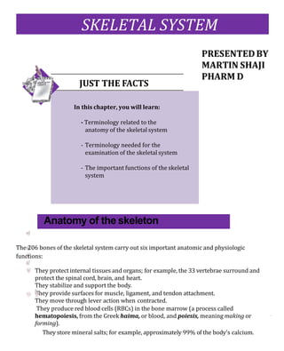
1. The 206 bones of the skeletal system carry out six important anatomic and physiologic
functions:
They protect internal tissues and organs; for example, the 33 vertebrae surround and
protect the spinal cord, brain, and heart.
They stabilize and support the body.
They provide surfaces for muscle, ligament, and tendon attachment.
They move through lever action when contracted.
They produce red blood cells (RBCs) in the bone marrow (a process called
hematopoiesis, from the Greek haima, or blood, and poiesis, meaning making or
forming).
They store mineral salts; for example, approximately 99% of the body’s calcium.
SKELETAL SYSTEM
Anatomy of the skeleton
JUST THE FACTS
In this chapter, you will learn:
- Terminology related to the
anatomy of the skeletal system
- Terminology needed for the
examination of the skeletal system
- The important functions of the skeletal
system
2. Bones-r-us
Theskeletonisdividedintotwoparts:theaxial(from theLatinaxis, meaningaxle
or wheel) and appendicular (from the Latin appendare, meaning to add or
append). The axialskeleton forms the body’sverticalaxis andcontains74bonesin
theheadandtorso;italsoincludes6bonesofthemiddleear,foratotalof 80bones.
(See the body’sbones.)
Below is a list of key terms, along with the
correct way to pronouncethem.
Calcaneus Kal-kay-nee-uhs
Coccyx Kok-siks
Hematopoiesis Hee-muh-toe-poy-ee-sis
Occipital Ok-sip-uh-tuhl
Periosteum Per-ee-os-tee-uhm
Xiphoid process Zeye-foyd Prah-sess
4. The appendicular skeleton contains 126 bones and
includes the body’s appendages, or upper and lower
extremities
The axial skeleton
The axial skeleton forms the long axis of the body and
includes bones of the skull, vertebral column, and rib
cage.
The skull
The skull contains 28 irregular bones in two major
areas:
the brain case, or cranium (from the Greek kranion,
meaning upper part of the head), and the face. Eight
bones form the cranium, 14 bones make up the face, and
the inner ears contain 6 ossicles (from the Latin
ossiculum, meaning bone), or 3 small bones in each ear. The
jaw bone, or mandible (from the Latin mandibula, meaning
jaw) is the only movable bone in the skull. (See Bones of the
skull.)
Getting it together
Sutures are immobile joints that hold the skull bones together.
The coronal suture unites the frontal boneand
the two parietal bones. In infants, this suture isn’t closed,
leaving a diamond-shaped area (called the anterior
fontanel), which is covered only by a membrane. This
soft spot closes between ages 10 and 18 months. At the
back of the head of infants, the posterior fontanel closes
by age 2 months.
A real airhead
Sinuses are air-filled spaces within the skull that lessen
the bone weight, moisten incoming air, and act as resonating
chambers for the voice.
Up front
The sinuses, the forehead, and the area directly behind it
are part of the frontal bone. This bone also forms the
orbits (eye sockets) and the front part of the cranial
floor.
Fontanel, also spelled
fontanelle,derives from
French and means little
fountain. Itcanalsorefer
to any membrane-
covered area between
twobones.
5. Take it from thetop
The main part of the skull consists of a number of bones
sutured together:
• The coronal suture connects the frontal bone with the
parietal bones.
• Two parietal bones crown the head, forming the roof
and the upper part of each side of the skull.
• The squamous suture connects the parietal bones
with the temporal bones.
• Temporal bones form the lower part of the sides of
the skull and part of its floor. They contain structures of the middle and inner
ear and the mastoid sinuses.
Anatomicallyspeaking
Bonesoftheskull
The skull is a complexbonystructure. It’sformedby twosetsof
bones, the cranial bonesand thefacial bones
6. • The lambdoid suture connects the parietal bones to
the occipital bone.
• The occipital bone forms the rear portion and the
base of the skull and forms a movable joint with the
first cervical vertebra.
• A large opening at the base of the occipital bone, called
the foramen magnum (meaning large hole), allows the
spinal cord to pass from the encephalon into the spine.
A bat in the belfry
The sphenoid bone looks like a bat with outstretched
wings and legs extended to the back. Located in the cranial
floor, this bone is an anchor for the frontal, parietal,
occipital, and ethmoid bones. It also supports part of the
eye sockets and forms the lateral walls of the skull. The
sphenoid sinuses are large air-filled spaces within the
sphenoid bone.
Facial bones
The bones of the face include:
• two maxillary bones that form the upper jaw, nose,
orbits, and roof of the mouth as well as the maxillary sinuses
• the cheekbones, called zygomatic or malar bones,
that attach to chewing muscles
•two nasal bones that form the upper part of the bridge
of the nose (cartilage forms the lower part)
• the mandible that forms the lower jaw
•two lacrymal bones that contain the lacrymal bag
(part of the conduit through which tears drain in the
nasal cannula)
• the vomer that’s part of the nasal septum
•two palatine bones that form the posterior portion of
the hard palate, lateral side of the nasal cavity, and small
part of the orbit.
jogger
As a way to
remember
the bones of the
skull,useyourhead
and think “part of
man”:
PARietal
Occipital
Frontal
MAlar
Nasal.
Jointsbetween
the vertebrae
allow forward,
backward, and
sideways
movement. Not
all at once,
though!
7. The spinal column
The flexible spinal column contains 24 vertebrae (plural
of vertebra), the sacrum, and the coccyx. (See Some
thorny words of the spine.)
Joints between the vertebrae allow forward, backward,
and sideways movement. The spinal column supports
the head while suspending the ribs and organs in
Spine comesfromtheLatinwordspina,whichmeansthorn,
and is related to spike as well. Latin writers likened the
thorn to the prickly bones in animals and fish and, thus, the
word alsobe- camethedesignationforthevertebralcolumn.
AlsofromLatin,vertebraderivesfromaverbmeaningtoturn.
Therefore,itformerlyconnotedanyjoint—notjustthoseofthe
spine. A Greek word, spondylos,has thesame meaning as
verte- bra.Itshowsup inwordslikespondylitis,which is an
inflammationofthevertebrae.
Sacrum and coccyxbringing up therear
Thesacrumwasformerlyknown astheossacrum,literally
the holybone, so called because it was thought to be a
particularly choice bit and so was offered to the gods in
sacrifice. The coc- cyx derives its name from the Greek
word for the cuckoo, kokkyx.The Greek anatomist Galen
thought this triangularbone resembledthe shape of the
bird’sbill.
8. front. It also anchors the pelvic girdle and provides
attachment points for many important muscles. The
spinal column contains:
•seven cervical (neck) vertebrae, which support the
skull and rotate
•twelve thoracic (chest) vertebrae, which attach to the
ribs
•five lumbar (lower back) vertebrae, which support the
small of the back
• the sacrum, a single bone that results from the fusion
of five vertebrae and attaches to the pelvic girdle
•the coccyx, or tailbone, which is located at the bottom
tip of the spinal column and is a single bone formed from
the fusion of four or five vertebrae.
The spinal column is curved to increase its strength
and make balance possible in an upright position. The
vertebrae are cushioned by intervertebral disks composed
of cartilage.
9. The33 vertebrae of thespinalcolumn
surroundandprotectthe spinalcord.They’re
divided into fivesections: cervical vertebrae,
thoracic vertebrae, lumbarvertebrae,
sacrum, andcoccyx. Yep. I have 33
vertebrae----and
they’re all perfect
specimens, if I do say
so myself
10. Sternum
Located in thecenter of thechest, thesternum is a flat, sword-shapedbonethat’s
attached totheclavicles (collar- bones) andtheinnermostpart ofthefirsttwopairs
ofribs.
Cagedin
Thesternum,ribs,andthoracicvertebraeformaprotectiveenclosurearoundthe
vitalorgans.Knownasthe thoracic cage, or thorax, this flexible structure
protects theheartandlungsandallowsthelungstoexpandduring respiration.
Ribs
Theflat,curvedbonesattachedtothethoracicportion
of thespinalcolumnarecalledribs.
Ribs—true or false?
Thetermcostalrefers toribs. Thefirstsevenpairs of
ribs are attached tothesternum by costal cartilage;
they’re called true ribs.Theremainingfivepairs of
ribsare called falseribs becausetheyaren’tattached
directly to the sternum.All ribsare independently
attached to the spinalcolumn.
Appendicularskeleton
Theappendicular skeletonincludestheupper andlower
extremities.
Theupperextremities
The clavicles, or collarbones, are twoflat bones at-
tachedtothesternumontheiranteriorsideandtothe
scapulae(shoulderblades)laterally.Thisformsthe
sternoclavicular joint.
Thescapulaeareapairoflarge,triangularbonesthat
arelocatedatthebackofthethorax.Thesebones,plus
the clavicles, form theshoulder girdles.
Armed and dangerous
Thehumerus,orupperarmbone,isalongbonewitha
shaftandtwobulbousends.Thetwolongbonesofthe
lower armaretheulna,located on thelittlefinger side
of thehumerus,andtheradius,onthethumbside.
These
The ulna andthe
radius articulate
with the
humerus toform
the elbow joint
11. bones articulate with the humerus to form the elbow
joint. The wrists are composed of eight small, irregular
carpal bones aligned in two rows. Ligaments bind the
carpals together.
Anatomically speaking
Bones of the hand
A view of the right hand, illustrating the positions of the
carpals, metacarpals, and phalanges.
12. A handful of terms
Thebones ofthehandare comprisedofmetacarpal
bonesandphalanges.(SeeBones ofthe hand.)
Thewaythesebonescometogetherenablesmove-
mentofthehand
• Fivesmalllongmetacarpalbonesattachtothecarpals
andformthepalmofthehand.
Phalanges, or fingerbones,are miniaturelong bones.
Eachfingerhas threephalanges, whilethethumbhastwo.
• Thethumbmetacarpalhasafreelymovablejoint,allow-
inga widerange ofmovementbetweenthethumbmeta-
carpalandthetrapezium,thecarpalatthebaseofthe
thumb.
Lowerextremities
Thelowerextremitiescontainbonesofthehip,thigh,leg,
ankle,and foot.
Girdlewords
Three pairs of bones fuseduringchildhood to form the
pelvic girdle, the broadest bone in the body. This bone
supportsthe trunk, protectsthe abdominalorgans within
its basin,andattaches thelowerextremities to thebody.
The three pairs of fusedbones include the ilium, which
isthelargestanduppermostofthethree;theischium,the
lowerandstrongestsetofbones;and thepubis,apairof
anteriorbonesthatmeetatthesymphysispubis—acar -
tilaginousjoint.
Give’emaleg up
Thetwofemurs,orupperlegbones,arethelongestand
heaviestbonesinthebody.Theyconnectattheproximal
endwith thehip,articulating with theacetabulum, or
hipsocket.Thefemurs connectwiththetibiaatthedis-
talend.Thekneecap, orpatella,isasmall,flatbonethat
protects the kneejoint andoverlaps thedistalend of the
femurandtheproximalendofthetibia.
Below the knee
Thetibia,sometimescalledtheshinbone,isthelargest
andstrongestof thelowerlegbones. Itarticulates with
thefemurattheproximalendandmeetsthefibulaand
Phalanges is the plural
of theGreekword pha-
lange, or phalanx. The
latter termwas applied
toGreekandRoman
noted for their closely
joinedandunified ma-
neuvers.
It says here that
the word patella,
for kneecap, is a
Latin word that
means a small, flat
dish—just what
the kneecap looks
like
13. the talus at the distal end. The fibula connects with the
tibia at its proximal and distal ends. The fibula’s distal end
also articulates with the talus. The articulation of the
fibula, tibia, and talus bones creates the bony prominence
on the outside of the ankle, called the lateral malleolus.
Now, fleetly, to the foot
The foot bones form a strong, stable arch with lengthwise
and crosswise support. Strong ligaments and tendons of
the leg muscles help the foot bones maintain their arched
position:
•Seven short tarsal bones structurally resemble the
wrist, and they articulate with the tibia and fibula:
– The talus bone (astragalus) forms part of the ankle
joint.
–The heel, called the calcaneus, is the largest tarsal
bone.
– The scaphoid bone is also called the navicular because
of its boat shape.
–The cuneiforms (the lateral, intermediate, and medial)
are three wedge-shaped bones that form the arch of
the foot.
–The cuboid bone articulates in the front with the
metatarsal bones.
• Five metatarsal bones form the foot and articulate
with the tarsal bone and the phalanges.
•The fourteen phalanges (toes) are similar to fingers,
with three bones in each toe except the great toe, which,
like the thumb, contains only two bones.
Bones are classified according to their shape:
•Long bones are the main bones of the limbs, except
the patella, and those of the wrists and ankles.
• Short bones are the bones of the wrists and ankles.
•Flat bones include the sternum, scapulae, and cranium,
among others.
• Irregular bones include the vertebrae and hip bones.
Boning up on bone material
All bones consist of two types of bone material: an outer
layer of dense, smooth compact bone and an inner layer
Anatomy of bones
Words will
never hurt
me. But let’s
keep sticks
out of it!
14. of spongy, cancellous (porous) bone. Compact bone is
found especially in the shaft of long bones and in the outer
layers of short, flat, and irregular bones. Cancellous
bone fills the central regions of the epiphysis (the end of
a long bone where bone formation takes place) and the
inner portions of short, flat, and irregular bones.
Asosteoblastsaddnewtissuetotheoutsideofa bone,large phagocytic
cellscalled osteoclasts eataway bonytissuein themedullarycavityto
keep thebonefrom becomingtoothick.Ahealthyboneisconstantly
broken down,resorbed,andrepairedlongafteritstopsgrowing in size.
Duringadulthood,boneformation (or ossifica- tion) and bone
resorption balance one another so that eachboneremainsaconstant
size.Duringchildhoodand adolescence, ossificationis faster than
resorptionand bonesgrowlarger.
Cartilage
Bones andjointsneedsupportas wellas shock absorp-
tion.Cartilageis adenseconnectivetissuethathas
thesecapabilities.It consistsof fibersembedded ina
strong,gel-likesubstance.Unlikerigidbone,cartilagehas
theflexibility offirm plastic.
Cartilagesupportsandshapesvariousstructures,such
astheauditorycanalandtheintervertebraldisks.
It alsocushionsandabsorbs shock.Cartilagehasnoblood
or nerve supply.
Osteon,Greekforbone,providesa keyword-formingrootformedicaltermsrelatingtobones,oste- orosteo-.Osteoblast isa
com- poundofosteo-and-blast; thelatterisanother commonmedicalrootderivedfromaGreekwordthatmeansabud ora
shootof a developing organism. An osteoblast is thus a cellthat buds forth new bone tissue. The Greek word clast, on the
other hand,means tobreakorfragment.Therefore,anosteoclast isa cellthat breaksdownbone.
TheRomanshadanameforit
Anotherverycommonrootforformingwordsisthe Latinword os,oross-,alsomeaningbone.Thisroot iscontained inwords like
ossify,meaningto change ortobecome bone, and ossification,theprocessofbecomingbone.
15. Typesofcartilage
Cartilagemaybe fibrous,hyaline, orelastic:
•Fibrouscartilage formsatthemeniscus andthe
inter- vertebraldisks.
•Hyalinecartilage coversarticularbonesurfaces
(where oneormorebones meet atajoint), connects
the ribs and sternum,andappears in the trachea,
bronchi,andnasalseptum.
•Elastic cartilageislocatedinthe auditorycanal,
ex- ternalear,andepiglottis.
Bonemovement
Bones are rigid structures that can’t bend without being
damaged, so individual bones move at joint sites, or
Articulations. Every bone in the body except the hyoid
bone, which anchors the tongue, is connected to another
bone by flexible connective tissue.
Howdoesitmove?
Classifying joints
Jointscanbeclassifiedbythetypeofmovementtheyal- lowand bytheirstructure.
Thethreeclasses ofjointsidentifiedby therangeof movement theyalloware:
• synarthrosis—immovable
• amphiarthrosis—slightly movable
• diarthrosis—freely movable.
What is it made of?
Bystructure,ajointmaybeclassifiedasfibrous,carti-
laginous,orsynovial.Infibrous joints,thearticularsur-
facesofthe twobones arebound closelyby fibrouscon-
nective tissueandlittle movement ispossible.Thecranial
suturesareexamplesoffibrousjoints.
In cartilaginous joints, cartilage connects one
bone to another; thesejoints allow slight movement. An
example is the symphysis pubis (the junction of the
pelvicbones).
Body surfaces in thesynovial joints arecovered
by articularcartilageandjoined by ligaments(dense,
strong,flexiblebandsoffibrousconnectivetissuethat
bindbonesto otherbones)linedwithsynovialmem-
brane. Freelymovable,synovial joints includemost
joints of thearms andlegs.Synovialjointsalsoinclude
an ar- ticularcapsule—a saclikeenvelope, whose
outerlayer
Our joints are
jumpin’! It must
be that
diarthrosis