Download as PDF, PPTX

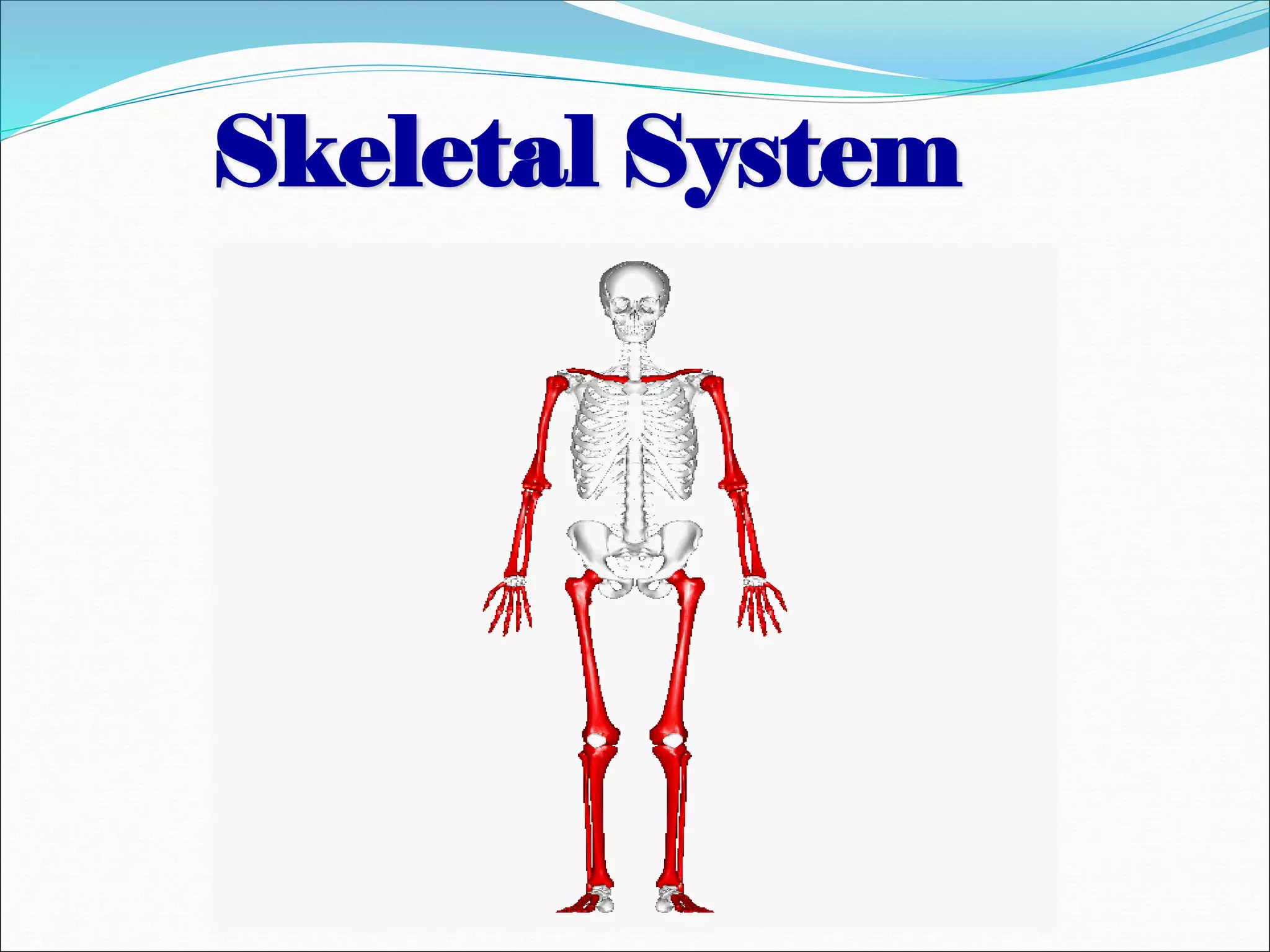
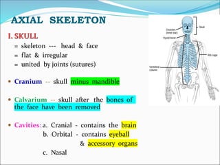
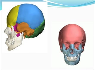
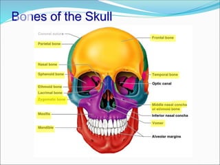
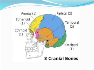
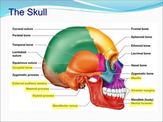
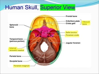
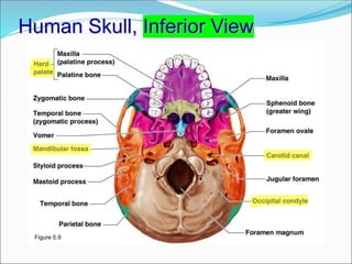
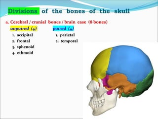
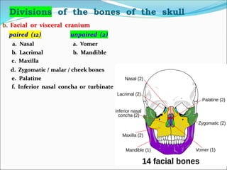
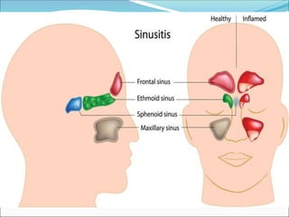
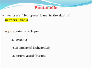
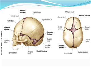
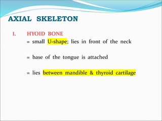
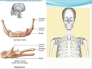
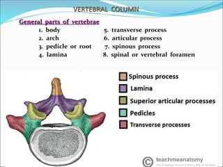
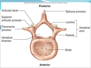
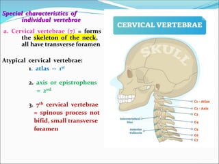
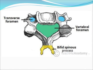
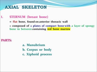
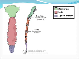
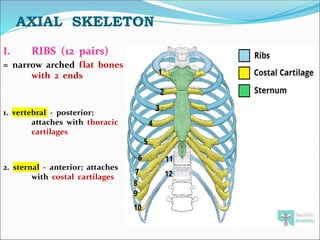
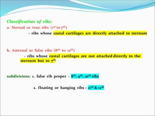
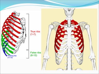
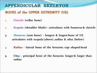
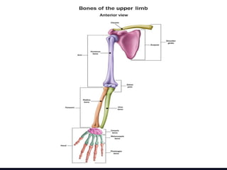
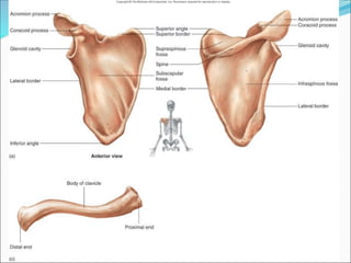
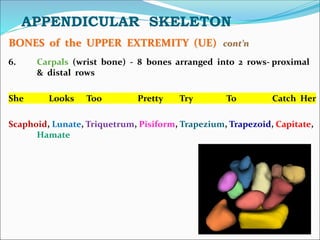
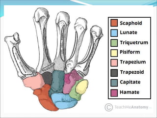
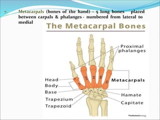
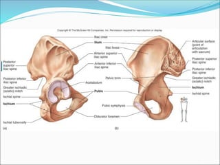
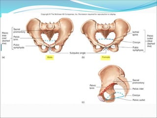
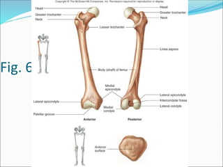
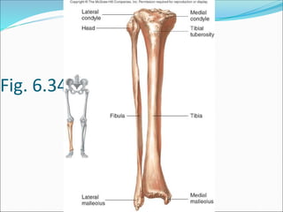
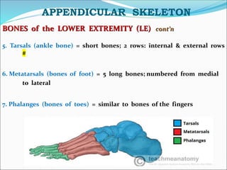
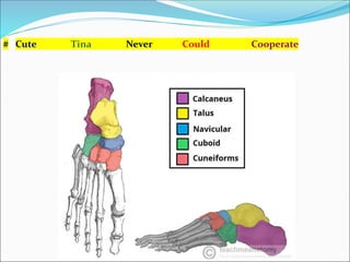
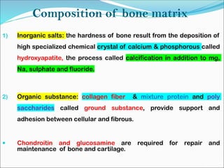
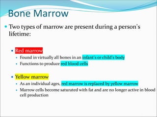

The document provides information about the skeletal system, including the axial skeleton and appendicular skeleton. It describes the bones that make up the skull, vertebral column, ribs, sternum, and bones of the upper and lower extremities. It also discusses the composition of bone, bone marrow, microscopic structure of compact bone, and several common diseases and conditions that affect the skeletal system such as arthritis, osteoporosis, scoliosis, and rickets.