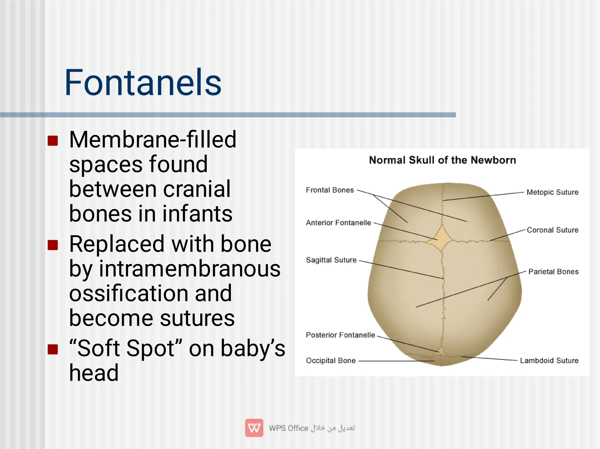
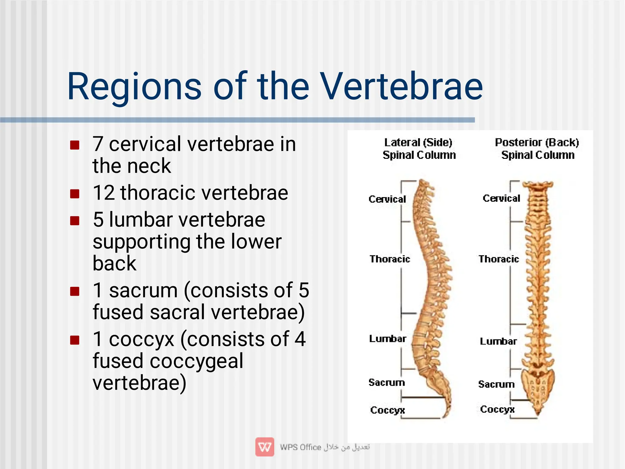
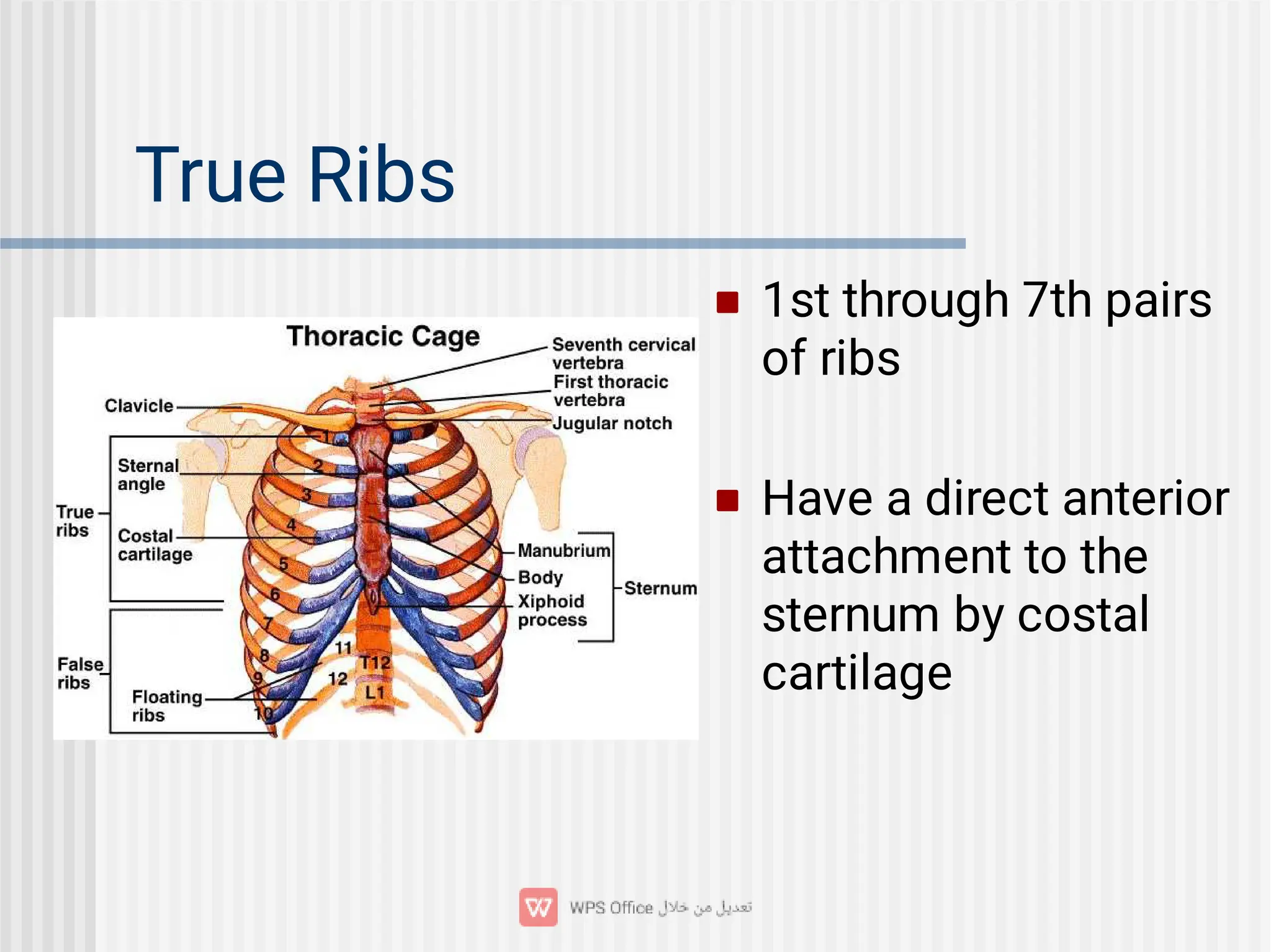
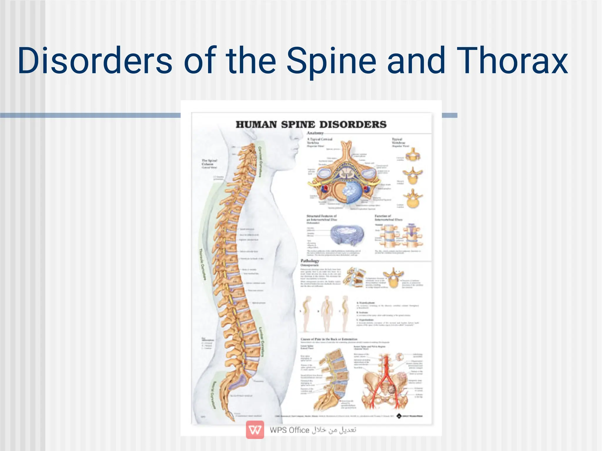
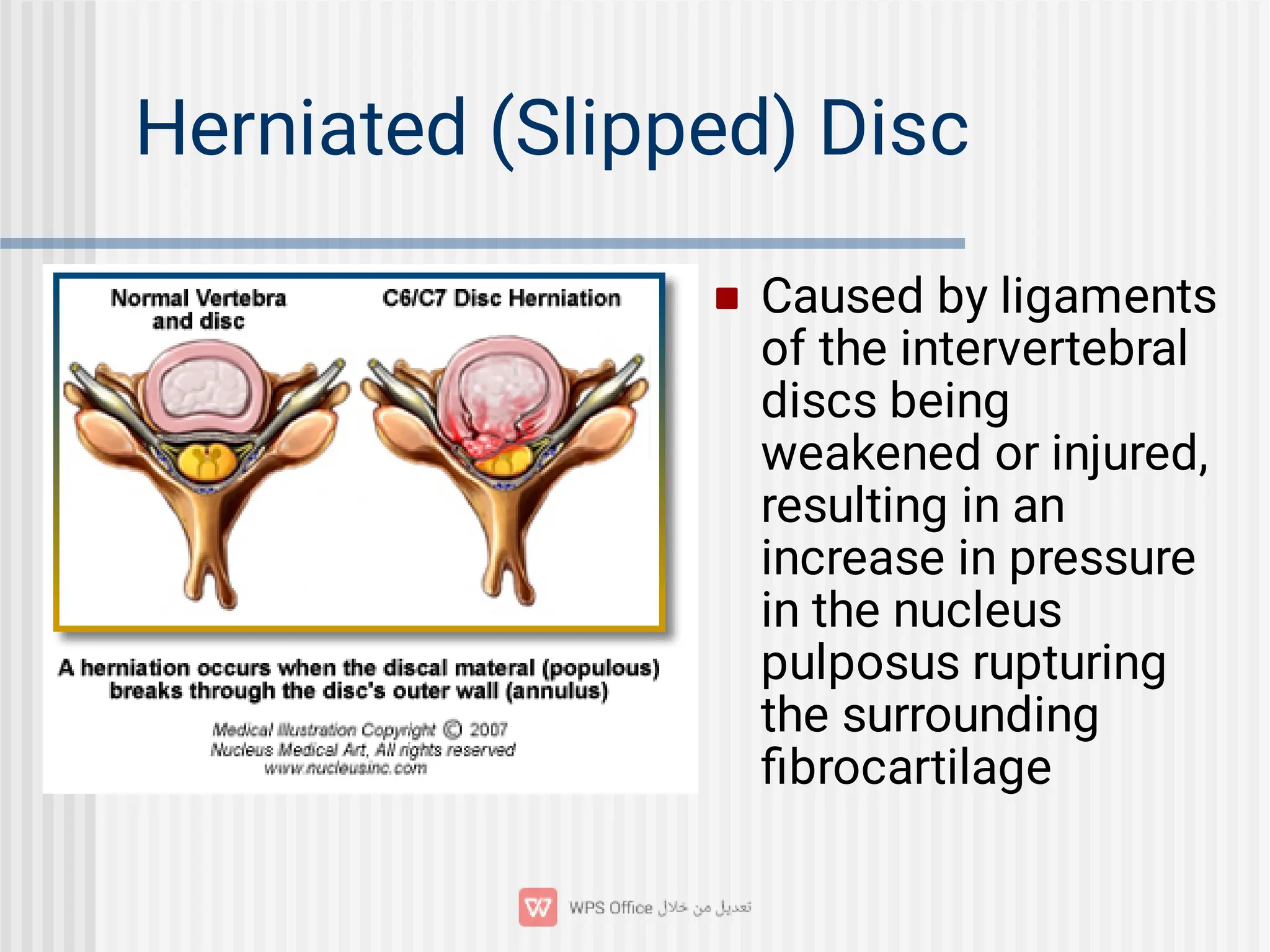
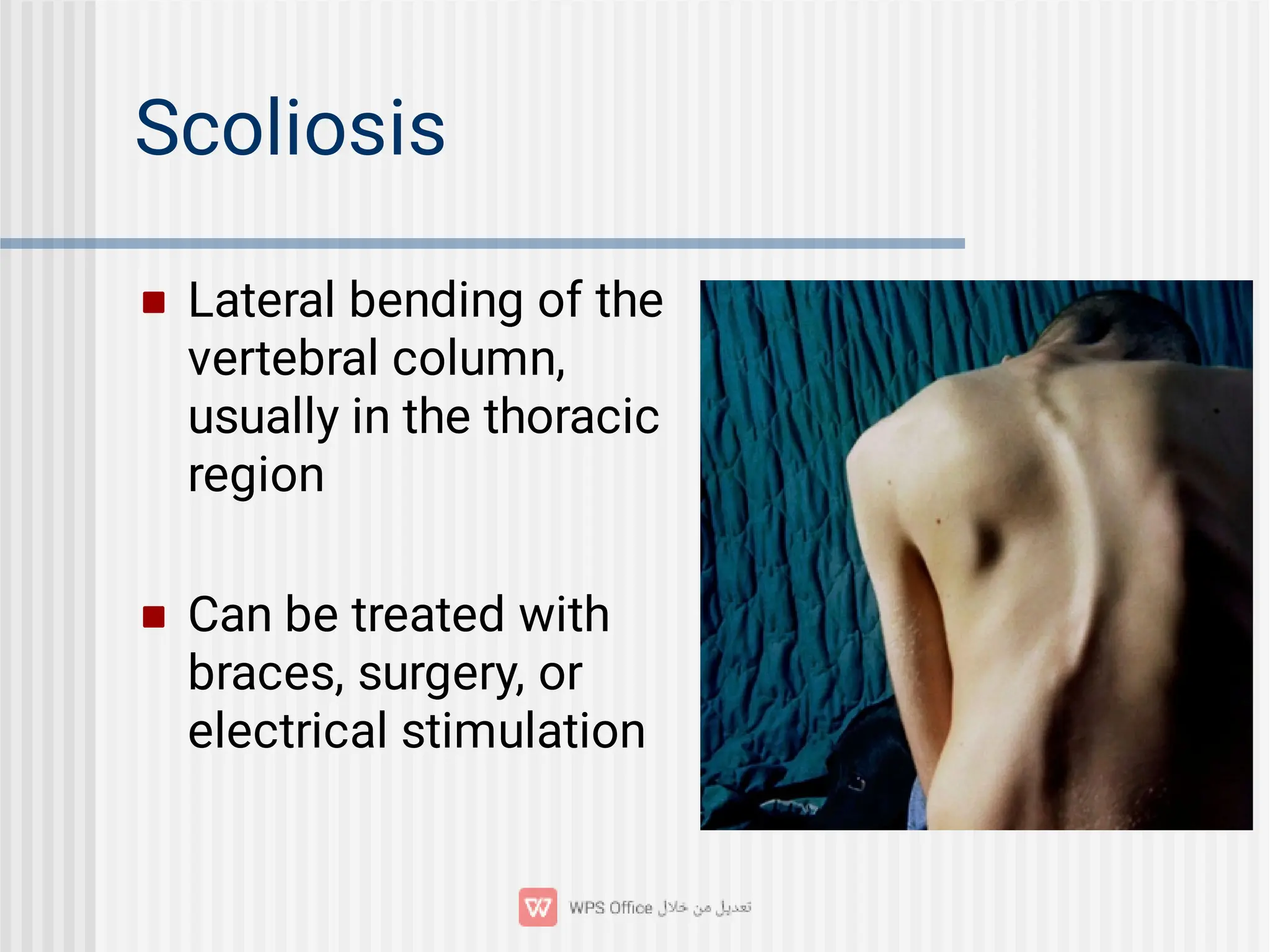
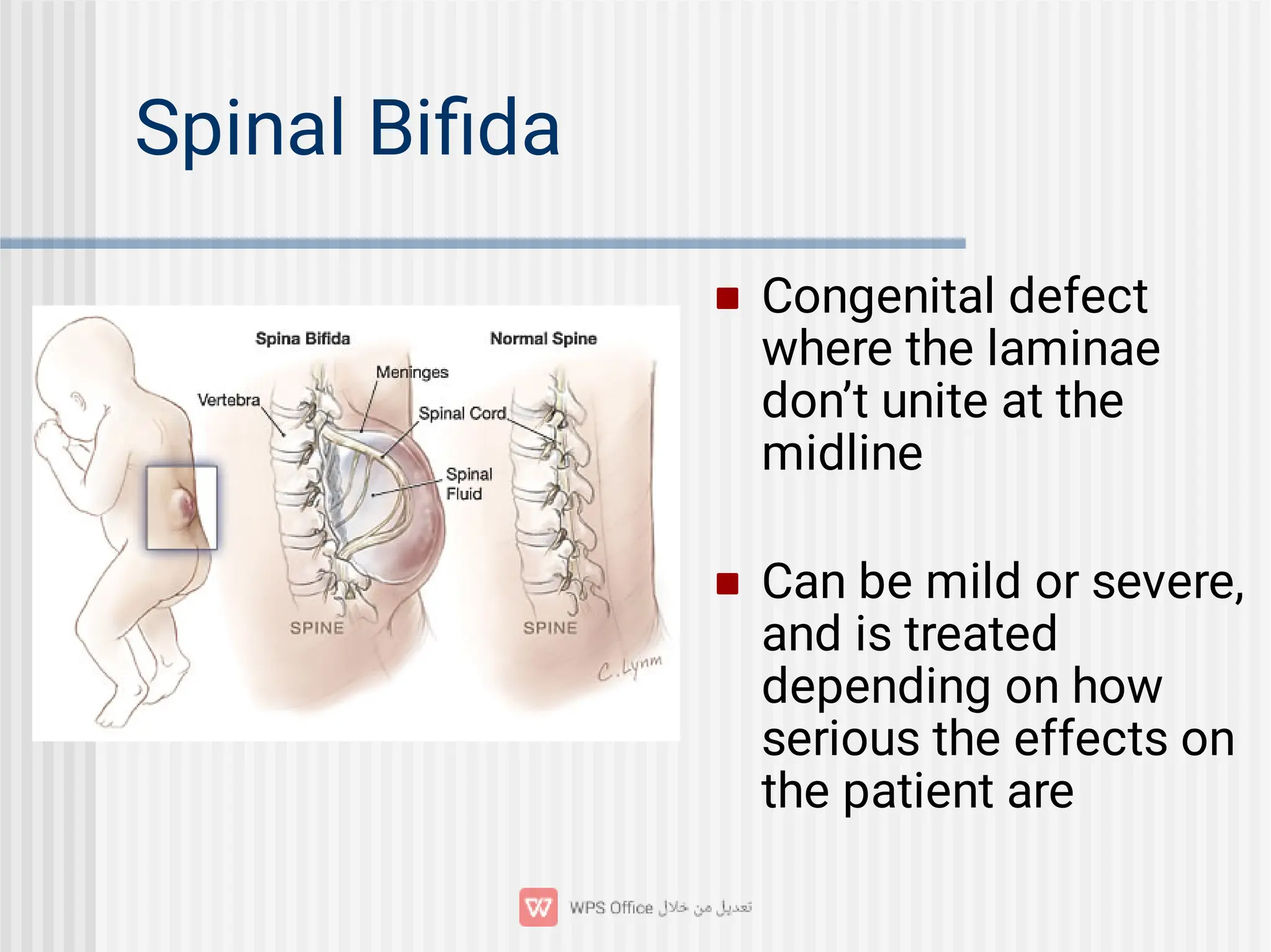
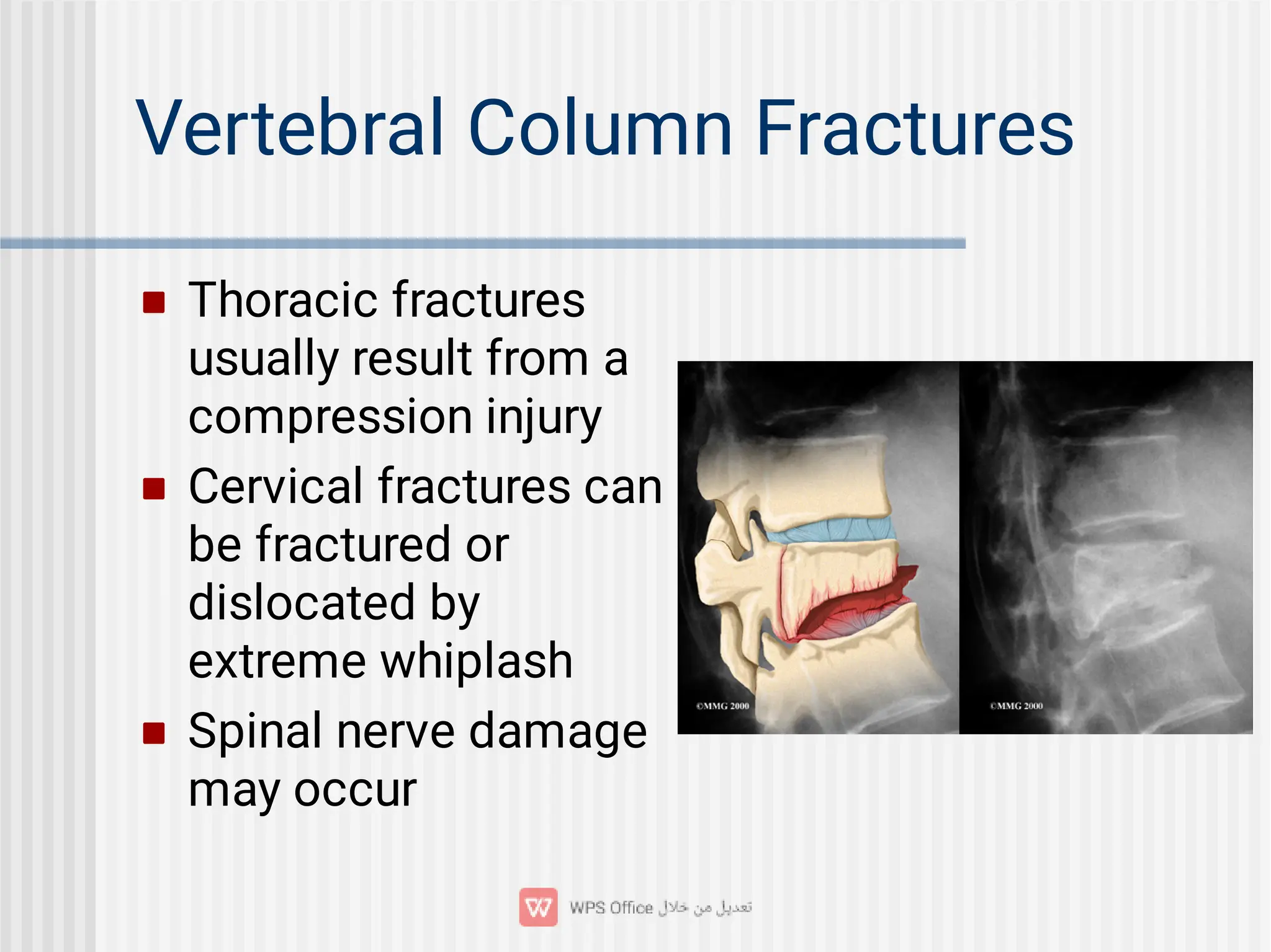
5. Frontal Bone
Forms forehead,
roofs of the eye
sockets (orbits), and
most of the front part
of the cranial floor
Frontal sinuses lie
deep within the bone
6. Parietal Bones
Form the sides and
roof of the cranial
cavity
Separated on top of
skull by the sagittal
suture
7. Temporal Bones
Form lower sides of
the cranium and part
of the cranial floor
External auditory
meatus (ear canal) is
located within these
bones
8.
Mastoid process is
behind external auditory
meatus and is a point of
neck muscle
attachment
Styloid process is point
of neck and tongue
muscle and ligament
attachment
Mandibular fossa forms
half of the
temporomandibular
joint with the mandible
(lower jaw bone)
9. Occipital Bone
Forms posterior part and
most of the cranial base
Foramen magnum
passes through this bone
Occipital condyles are on
either side of the
foramen magnum that
connect with the first
vertebrae
10. Sphenoid Bone
The “keystone” of the
cranial floor because
it holds together all of
the other cranial
bones
The hypophyseal
fossa is a depression
for the pituitary gland
11.
12. Ethmoid Bone
Light spongy bone in the
anterior part of cranial
floor between the eye
sockets
Houses the nasal cavity
Contains the nasal
conchae that cause
turbulence in inhaled air,
cleaning the air before it
passes into the rest of
the respiratory tract
15. Maxillae
Paried to form the
upper jawbone
Articulates with every
bone in the face
except the mandible
Forms the anterior
3/4 of the hard palate
16. Palatine Bones
Paired L-shaped
bones
Form the posterior
portion of the hard
palate, part of the
floor and lateral wall
of the nasal cavity,
and a small portion
of the eye sockets
17. Mandible
The lower jawbone
Largest and strongest
facial bone and only
movable skull bone
Condylar process
articulates with the
mandibular fossa of
the temporal bone to
form the TMJ
23. Cleft Palate
Occurs when fusion
of the left and right
maxillary bones is not
completed before
birth
Repaired between 12
and 18 months with
surgery
24. TMJ Syndrome
Caused by improperly
aligned teeth,
grinding or clenching
teeth, trauma, or
arthritis
Generally results in
pain around the ear
and jaw muscles
25. Deviated Nasal Septum
Nasal septum bends
sideways from the
middle of the nose
Can entirely block
nasal passage in
extreme cases
29.
Lambdoid Suture-
between the parietal
bones and occipital
bone
Squamous Suture-
between the parietal
bones and temporal
bones
30. Paranasal Sinuses
Paired cavities near
nasal cavity
Located in the frontal
bone, sphenoid bone,
ethmoid bone, and
maxillae
Lined with mucous
membranes
32. Hyoid Bone
Does not articulate
with or attach to any
other bone
Suspended from the
styloid processes by
ligaments and
muscles
Located in the neck
between the
mandible and larynx
34. Vertebral Column
Also called the spine or backbone
Composed of vertebrae
Functions as strong flexible rod that can rotate
and move in all directions
Encloses and protects spinal cord
Supports the skull
Point of attachment for ribs, pelvic girdle, and
back muscles
35. Regions of the Vertebrae
7 cervical vertebrae in
the neck
12 thoracic vertebrae
5 lumbar vertebrae
supporting the lower
back
1 sacrum (consists of 5
fused sacral vertebrae)
1 coccyx (consists of 4
fused coccygeal
vertebrae)
36. Intervertebral Discs
Lie in between the
vertebrae from the
2nd cervical
vertebrae to the
sacrum
Form strong joints,
permit movement,
and absorb vertical
shock
37. Vertebral Column Curvature
The spine curves like
a snake
Cervical and lumbar
curves are convex
(bulging out anteriorly)
Thoracic and sacral
curves are concave
(bulge out posteriorly)
40. Vertebral Arch
Extends backwards from
the body of the vertebra
Formed by two short,
thick processes (pedicles)
that unite with the flat
parts of the arch
(laminae), ending with a
single sharp, slender
projection (spinous
process)
41. Spinal Cord Openings
The vertebral foramen is
the space between the
vertebral arch and body
that contains the spinal
cord
All of the vertebral
foramen combined forms
the vertebral canal
The intervertebral
foramen is the opening
between adjoining
vertebrae on both sides
of the column contains a
single spinal nerve
43. Spinous Process
Projects from the
junction of the
laminae
Combined with the
two transverse
processes, these
three are points of
attachment for
muscles to the
vertebral column
44. Articular Processes
Superior Articular
Processes join with the
vertebra right above
them
Inferior Articular
Processes join with the
vertebra right below
them
The articulating surfaces
are called facets and are
lined with hyaline
cartilage
47. Atlas (C1 Vertebra)
Supports the head
Does not have a body
or spinous process
Upper surface
contains the superior
articular facets that
articulate with the
occipital bone
(allows you to nod
“yes”)
48. Axis (C2 Vertebra)
Does have a body and
spinous process
The dens, a tooth-
shaped process,
projects up through
the vertebral foramen
of the atlas and
serves as a pivot to
allow you to shake
your head “no”
49. Remaining Cervical Vertebrae
C3 - C6 all follow the
normal anatomy of
the typical vertebra
C7 is also called the
vertebra prominens; it
has a single, large
spinous process that
can be felt at the
base of the neck
50. Thoracic Vertebrae (T1 - T12)
Much larger and
stronger than cervical
vertebrae
Have facets for
articulating with the
ribs, which limits
movement of the
vertebrae
51. Lumbar Vertebrae (L1 - L5)
Largest and strongest of
the column
Projections are short and
thick
Spinous processes are
well adapted for the
attachment of large back
muscles
52. Sacrum
Triangular bone
formed by the fusion
of 5 sacral vertebrae
that occurs between
16-30 years old
Serves as strong
foundation for the
pelvic girdle
53.
Four sacral foramina on
the anterior and posterior
sides where nerves and
blood vessels pass
Sacral canal is a
continuation of the
vertebral canal
The lower entrance of
the canal is the sacral
hiatus
The sacral promontory is
a projection on the top
border
56. Thoracic Cage
Bony cage formed by the
sternum, costal
cartilages, ribs, and
bodies of the thoracic
vertebrae
Encloses and protects
the organs of the
thoracic cavity and upper
abdominal cavity
Provides support for the
bones of the shoulder
and upper limbs
58.
The manubrium is the upper part,
articulating with the clavicles and first
and second ribs.
The body is the largest and middle part,
articulating directly or indirectly with the
2nd-10th ribs.
The xiphoid process is the lowest and
smallest part that has some abdominal
muscles attached to it.
59. Ribs
Twelve pairs make up
the sides of the
thoracic cavity
Each rib articulates
posteriorly with its
corresponding
thoracic vertebra
60. True Ribs
1st through 7th pairs
of ribs
Have a direct anterior
attachment to the
sternum by costal
cartilage
61. False Ribs
8th through 12th pairs of
ribs
Costal cartilages either
attach indirectly to the
sternum or not at all
Cartilages of ribs pairs 8
-10 attach to each other
and the cartilages of the
7th pair of ribs
62. Floating Ribs
11th and 12th pair of
ribs are also called
floating ribs
The costal cartilage
at their anterior ends
does not attach to the
sternum at all
Attach only
posteriorly to the
thoracic vertebrae
64. Herniated (Slipped) Disc
Caused by ligaments
of the intervertebral
discs being
weakened or injured,
resulting in an
increase in pressure
in the nucleus
pulposus rupturing
the surrounding
fibrocartilage
65. Scoliosis
Lateral bending of the
vertebral column,
usually in the thoracic
region
Can be treated with
braces, surgery, or
electrical stimulation
66. Spinal Bifida
Congenital defect
where the laminae
don’t unite at the
midline
Can be mild or severe,
and is treated
depending on how
serious the effects on
the patient are
67. Vertebral Column Fractures
Thoracic fractures
usually result from a
compression injury
Cervical fractures can
be fractured or
dislocated by
extreme whiplash
Spinal nerve damage
may occur
68. Rib Fractures
Most common chest
injuries
Break at the point
where the greatest
force is applied or at
the weakest point on
the rib
Middle ribs are most
commonly broken
69. Checkpoint Questions
Describe the general features of the skull.
Define the following: suture, foramen, nasal
septum, paranasal sinus, and fontanel.
What are the functions of the vertebral column?
What are the main distinguishing characteristics
of the bones of the various regions of the
vertebral column?
What are the functions of the bones of the
thorax?
What are the parts of the sternum?